

3D bioprinting and the revolution in experimental cancer model systems-A review of developing new models and experiences with in vitro 3D bioprinted breast cancer tissue-mimetic structures
- PMID: 36843955
- PMCID: PMC9946983
- DOI: 10.3389/pore.2023.1610996
3D bioprinting and the revolution in experimental cancer model systems-A review of developing new models and experiences with in vitro 3D bioprinted breast cancer tissue-mimetic structures
Abstract
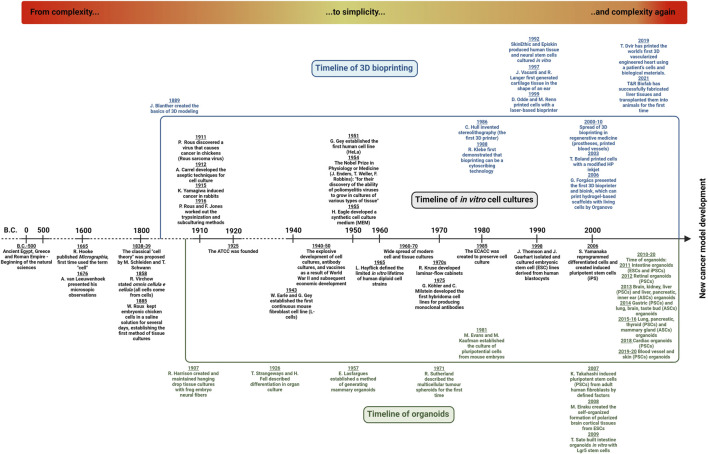
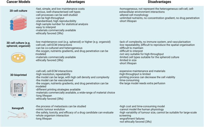
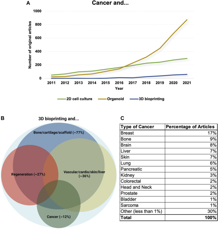
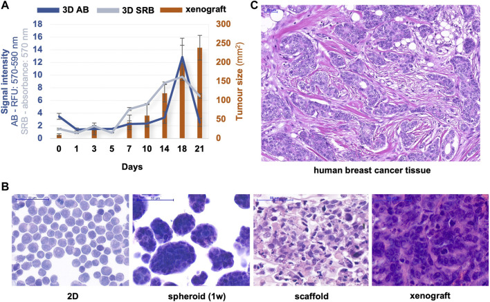
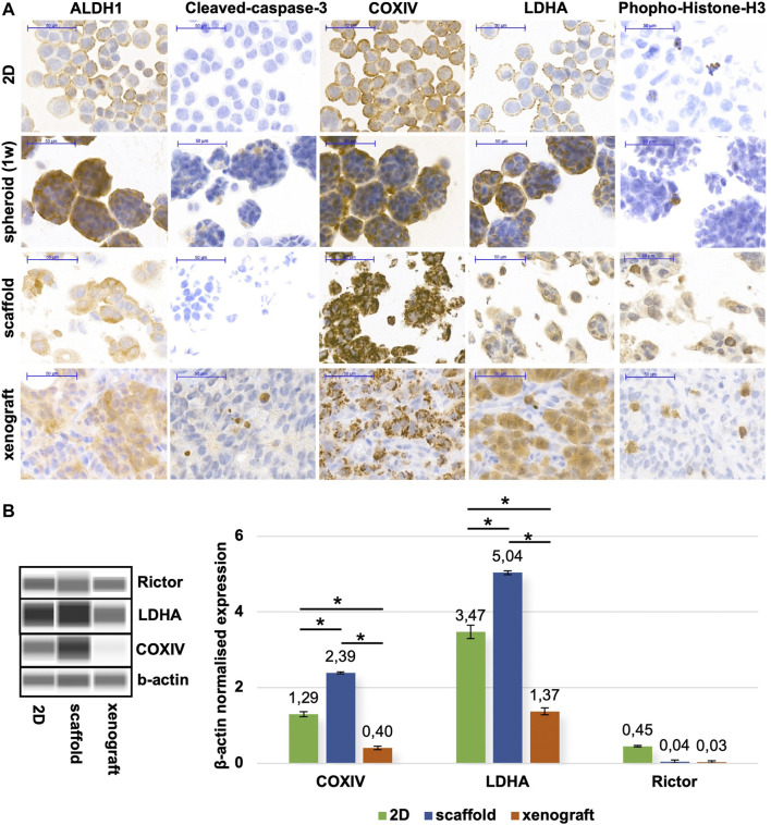
Growing evidence propagates those alternative technologies (relevant human cell-based-e.g., organ-on-chips or biofabricated models-or artificial intelligence-combined technologies) that could help in vitro test and predict human response and toxicity in medical research more accurately. In vitro disease model developments have great efforts to create and serve the need of reducing and replacing animal experiments and establishing human cell-based in vitro test systems for research use, innovations, and drug tests. We need human cell-based test systems for disease models and experimental cancer research; therefore, in vitro three-dimensional (3D) models have a renaissance, and the rediscovery and development of these technologies are growing ever faster. This recent paper summarises the early history of cell biology/cellular pathology, cell-, tissue culturing, and cancer research models. In addition, we highlight the results of the increasing use of 3D model systems and the 3D bioprinted/biofabricated model developments. Moreover, we present our newly established 3D bioprinted luminal B type breast cancer model system, and the advantages of in vitro 3D models, especially the bioprinted ones. Based on our results and the reviewed developments of in vitro breast cancer models, the heterogeneity and the real in vivo situation of cancer tissues can be represented better by using 3D bioprinted, biofabricated models. However, standardising the 3D bioprinting methods is necessary for future applications in different high-throughput drug tests and patient-derived tumour models. Applying these standardised new models can lead to the point that cancer drug developments will be more successful, efficient, and consequently cost-effective in the near future.
Keywords: 3D bioprinting; biofabrication; breast cancer; cancer; disease models.
Copyright © 2023 Sztankovics, Moldvai, Petővári, Gelencsér, Krencz, Raffay, Dankó and Sebestyén.
Conflict of interest statement
The authors declare that the research was conducted in the absence of any commercial or financial relationships that could be construed as a potential conflict of interest.
Figures
Similar articles
-
Characterisation of 3D Bioprinted Human Breast Cancer Model for In Vitro Drug and Metabolic Targeting.Int J Mol Sci. 2022 Jul 4;23(13):7444. doi: 10.3390/ijms23137444. Int J Mol Sci. 2022. PMID: 35806452 Free PMC article.
-
High Throughput Bioprinting Using Decellularized Adipose Tissue-Based Hydrogels for 3D Breast Cancer Modeling.Macromol Biosci. 2024 Aug;24(8):e2400035. doi: 10.1002/mabi.202400035. Epub 2024 Apr 29. Macromol Biosci. 2024. PMID: 38685795
-
3D bioprinting of engineered breast cancer constructs for personalized and targeted cancer therapy.J Control Release. 2021 May 10;333:91-106. doi: 10.1016/j.jconrel.2021.03.026. Epub 2021 Mar 25. J Control Release. 2021. PMID: 33774120 Review.
-
Strategies for 3D bioprinting of spheroids: A comprehensive review.Biomaterials. 2022 Dec;291:121881. doi: 10.1016/j.biomaterials.2022.121881. Epub 2022 Oct 28. Biomaterials. 2022. PMID: 36335718 Review.
-
Bioprinting technologies for disease modeling.Biotechnol Lett. 2017 Sep;39(9):1279-1290. doi: 10.1007/s10529-017-2360-z. Epub 2017 May 26. Biotechnol Lett. 2017. PMID: 28550360 Review.
Cited by
-
Recent Advances in Decellularized Extracellular Matrix-Based Bioinks for 3D Bioprinting in Tissue Engineering.Materials (Basel). 2023 Apr 18;16(8):3197. doi: 10.3390/ma16083197. Materials (Basel). 2023. PMID: 37110034 Free PMC article. Review.
-
Tumorigenic role of tacrolimus through mTORC1/C2 activation in post-transplant renal cell carcinomas.Br J Cancer. 2024 Apr;130(7):1119-1130. doi: 10.1038/s41416-024-02597-8. Epub 2024 Feb 10. Br J Cancer. 2024. PMID: 38341510 Free PMC article.
-
Deciphering Common Traits of Breast and Ovarian Cancer Stem Cells and Possible Therapeutic Approaches.Int J Mol Sci. 2023 Jun 26;24(13):10683. doi: 10.3390/ijms241310683. Int J Mol Sci. 2023. PMID: 37445860 Free PMC article. Review.
-
Preclinical Testing Techniques: Paving the Way for New Oncology Screening Approaches.Cancers (Basel). 2023 Sep 7;15(18):4466. doi: 10.3390/cancers15184466. Cancers (Basel). 2023. PMID: 37760435 Free PMC article. Review.
-
Emerging roles of 3D-culture systems in tackling tumor drug resistance.Cancer Drug Resist. 2023 Nov 21;6(4):788-804. doi: 10.20517/cdr.2023.93. eCollection 2023. Cancer Drug Resist. 2023. PMID: 38263982 Free PMC article. Review.
References
-
- Regulation of the European parliament and of the Council on cosmetic products, No 1223/2009 (2009). Available at: https://eur-lex.europa.eu/legal-content/EN/ALL/?uri=celex%3A32009R1223 (Accessed November 03, 2022).
Publication types
MeSH terms
LinkOut - more resources
Full Text Sources
Medical