

Pentad: A reproducible cytoarchitectonic protocol and its application to parcellation of the human hippocampus
- PMID: 36843959
- PMCID: PMC9947247
- DOI: 10.3389/fnana.2023.1114757
Pentad: A reproducible cytoarchitectonic protocol and its application to parcellation of the human hippocampus
Abstract
Introduction: The hippocampus is integral for learning and memory and is targeted by multiple diseases. Neuroimaging approaches frequently use hippocampal subfield volumes as a standard measure of neurodegeneration, thus making them an essential biomarker to study. Collectively, histologic parcellation studies contain various disagreements, discrepancies, and omissions. The present study aimed to advance the hippocampal subfield segmentation field by establishing the first histology based parcellation protocol, applied to n = 22 human hippocampal samples.
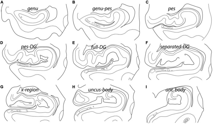
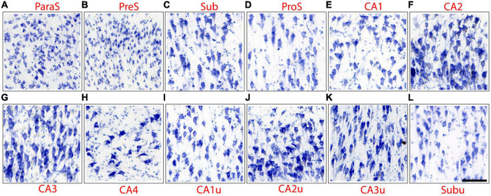
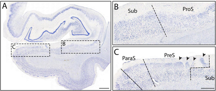
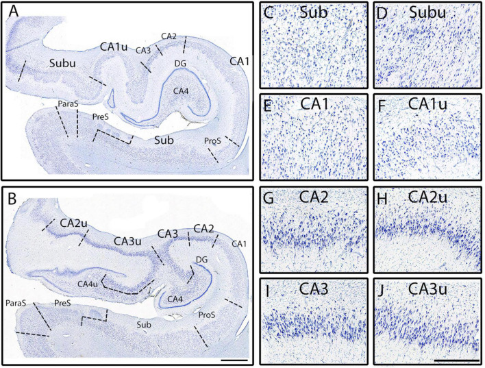
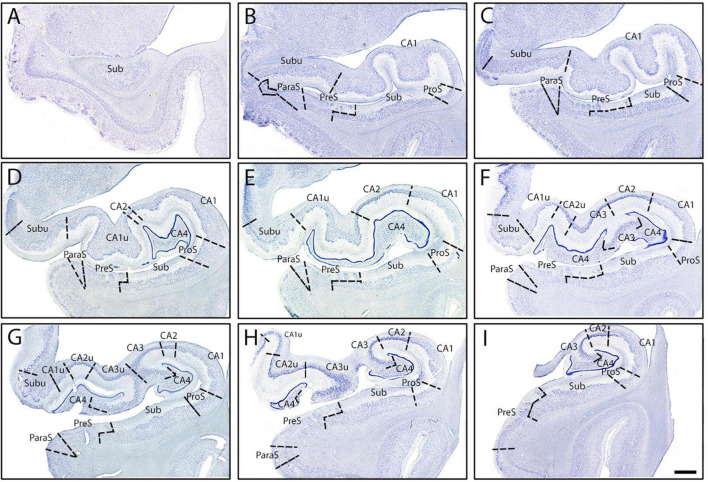
Methods: The protocol focuses on five cellular traits observed in the pyramidal layer of the human hippocampus. We coin this approach the pentad protocol. The traits were: chromophilia, neuron size, packing density, clustering, and collinearity. Subfields included were CA1, CA2, CA3, CA4, prosubiculum, subiculum, presubiculum, parasubiculum, as well as the medial (uncal) subfields Subu, CA1u, CA2u, CA3u, and CA4u. We also establish nine distinct anterior-posterior levels of the hippocampus in the coronal plane to document rostrocaudal differences.
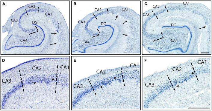
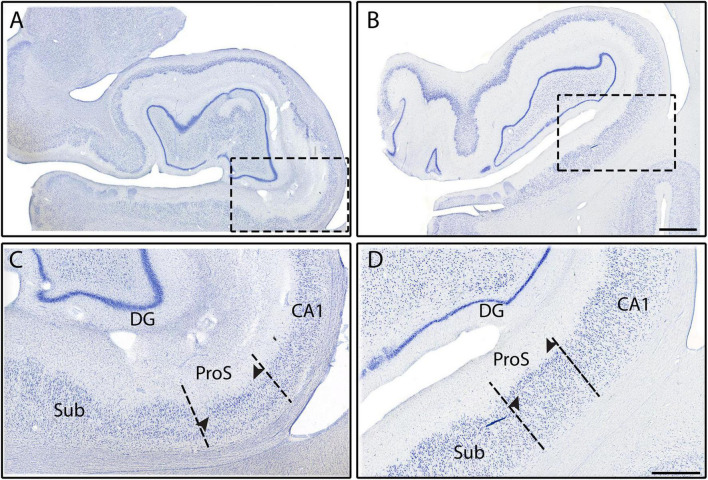
Results: Applying the pentad protocol, we parcellated 13 subfields at nine levels in 22 samples. We found that CA1 had the smallest neurons, CA2 showed high neuronal clustering, and CA3 displayed the most collinear neurons of the CA fields. The border between presubiculum and subiculum was staircase shaped, and parasubiculum had larger neurons than presubiculum. We also demonstrate cytoarchitectural evidence that CA4 and prosubiculum exist as individual subfields.
Discussion: This protocol is comprehensive, regimented and supplies a high number of samples, hippocampal subfields, and anterior-posterior coronal levels. The pentad protocol utilizes the gold standard approach for the human hippocampus subfield parcellation.
Keywords: CA1; CA2; CA3; hippocampal subfields; histology; pyramidal neurons; segmentation; subiculum.
Copyright © 2023 Williams, Rosenblum, Pihlstrom, Llamas-Rodríguez, Champion, Frosch and Augustinack.
Conflict of interest statement
The authors declare that the research was conducted in the absence of any commercial or financial relationships that could be construed as a potential conflict of interest.
Figures
Similar articles
-
Development of a histologically validated segmentation protocol for the hippocampal body.Neuroimage. 2017 Aug 15;157:219-232. doi: 10.1016/j.neuroimage.2017.06.008. Epub 2017 Jun 3. Neuroimage. 2017. PMID: 28587896
-
Organization and Detailed Parcellation of Human Hippocampal Head and Body Regions Based on a Combined Analysis of Cyto- and Chemoarchitecture.J Comp Neurol. 2015 Oct 15;523(15):2233-53. doi: 10.1002/cne.23786. Epub 2015 Jul 15. J Comp Neurol. 2015. PMID: 25872498
-
Hippocampal subfield CA2+3 exhibits accelerated aging in Alcohol Use Disorder: A preliminary study.Neuroimage Clin. 2019;22:101764. doi: 10.1016/j.nicl.2019.101764. Epub 2019 Mar 14. Neuroimage Clin. 2019. PMID: 30904825 Free PMC article.
-
The prosubiculum in the human hippocampus: A rostrocaudal, feature-driven, and systematic approach.J Comp Neurol. 2024 Mar;532(3):e25604. doi: 10.1002/cne.25604. J Comp Neurol. 2024. PMID: 38477395 Free PMC article.
-
Multimodal mapping and analysis of the cyto- and receptorarchitecture of the human hippocampus.Brain Struct Funct. 2020 Apr;225(3):881-907. doi: 10.1007/s00429-019-02022-4. Epub 2020 Jan 18. Brain Struct Funct. 2020. PMID: 31955294 Free PMC article.
Cited by
-
Comparison of histological delineations of medial temporal lobe cortices by four independent neuroanatomy laboratories.Hippocampus. 2024 May;34(5):241-260. doi: 10.1002/hipo.23602. Epub 2024 Feb 28. Hippocampus. 2024. PMID: 38415962 Free PMC article.
-
Diffusion MRI of the Hippocampus.J Neurosci. 2024 Jun 5;44(23):e1705232024. doi: 10.1523/JNEUROSCI.1705-23.2024. J Neurosci. 2024. PMID: 38839341 Free PMC article. Review.
-
Neuron collinearity differentiates human hippocampal subregions: a validated deep learning approach.Brain Commun. 2024 Sep 3;6(5):fcae296. doi: 10.1093/braincomms/fcae296. eCollection 2024. Brain Commun. 2024. PMID: 39262825 Free PMC article.
-
Stereology neuron counts correlate with deep learning estimates in the human hippocampal subregions.Sci Rep. 2023 Apr 11;13(1):5884. doi: 10.1038/s41598-023-32903-y. Sci Rep. 2023. PMID: 37041300 Free PMC article.
-
Immunohistochemical field parcellation of the human hippocampus along its antero-posterior axis.Brain Struct Funct. 2024 Mar;229(2):359-385. doi: 10.1007/s00429-023-02725-9. Epub 2024 Jan 5. Brain Struct Funct. 2024. PMID: 38180568 Free PMC article.
References
-
- Adler D. H., Wisse L. E. M., Ittyerah R., Pluta J. B., Ding S.-L., Xie L., et al. (2018). Characterizing the human hippocampus in aging and Alzheimer’s disease using a computational atlas derived from ex vivo MRI and histology. Proc. Natl. Acad. Sci. U. S. A. 115 4252–4257. 10.1073/pnas.1801093115 - DOI - PMC - PubMed
Grants and funding
LinkOut - more resources
Full Text Sources
Miscellaneous