FeS2-incorporated 3D PCL scaffold improves new bone formation and neovascularization in a rat calvarial defect model
- PMID: 36844239
- PMCID: PMC9947485
- DOI: 10.18063/ijb.v9i1.636
FeS2-incorporated 3D PCL scaffold improves new bone formation and neovascularization in a rat calvarial defect model
Abstract
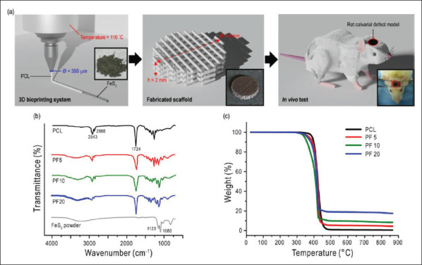
199Three-dimensional (3D) scaffolds composed of various biomaterials, including metals, ceramics, and synthetic polymers, have been widely used to regenerate bone defects. However, these materials possess clear downsides, which prevent bone regeneration. Therefore, composite scaffolds have been developed to compensate these disadvantages and achieve synergetic effects. In this study, a naturally occurring biomineral, FeS2, was incorporated in PCL scaffolds to enhance the mechanical properties, which would in turn influence the biological characteristics. The composite scaffolds consisting of different weight fractions of FeS2 were 3D printed and compared to pure PCL scaffold. The surface roughness (5.77-fold) and the compressive strength (3.38-fold) of the PCL scaffold was remarkably enhanced in a dose-dependent manner. The in vivo results showed that the group with PCL/ FeS2 scaffold implanted had increased neovascularization and bone formation (2.9-fold). These results demonstrated that the FeS2 incorporated PCL scaffold might be an effective bioimplant for bone tissue regeneration.
Keywords: 3D printed; Bone formation; FeS2; Mechanical properties; PCL.
Copyright: © 2022 Kang et al.
Figures


Similar articles
-
Stem Cell-Seeded 3D-Printed Scaffolds Combined with Self-Assembling Peptides for Bone Defect Repair.Tissue Eng Part A. 2022 Feb;28(3-4):111-124. doi: 10.1089/ten.TEA.2021.0055. Epub 2021 Dec 30. Tissue Eng Part A. 2022. PMID: 34157886
-
Incorporation of BMP-2 nanoparticles on the surface of a 3D-printed hydroxyapatite scaffold using an ε-polycaprolactone polymer emulsion coating method for bone tissue engineering.Colloids Surf B Biointerfaces. 2018 Oct 1;170:421-429. doi: 10.1016/j.colsurfb.2018.06.043. Epub 2018 Jun 20. Colloids Surf B Biointerfaces. 2018. PMID: 29957531
-
Evaluation of mechanical strength and bone regeneration ability of 3D printed kagome-structure scaffold using rabbit calvarial defect model.Mater Sci Eng C Mater Biol Appl. 2019 May;98:949-959. doi: 10.1016/j.msec.2019.01.050. Epub 2019 Jan 14. Mater Sci Eng C Mater Biol Appl. 2019. PMID: 30813102
-
3D-printed poly(Ɛ-caprolactone) scaffold with gradient mechanical properties according to force distribution in the mandible for mandibular bone tissue engineering.J Mech Behav Biomed Mater. 2020 Apr;104:103638. doi: 10.1016/j.jmbbm.2020.103638. Epub 2020 Jan 14. J Mech Behav Biomed Mater. 2020. PMID: 32174396
-
Recent advances on 3D-printed PCL-based composite scaffolds for bone tissue engineering.Front Bioeng Biotechnol. 2023 Jun 19;11:1168504. doi: 10.3389/fbioe.2023.1168504. eCollection 2023. Front Bioeng Biotechnol. 2023. PMID: 37469447 Free PMC article. Review.
Cited by
-
Bioinformatic analysis of the molecular mechanisms underlying the progression of bone defects.Front Med (Lausanne). 2023 Jun 8;10:1157099. doi: 10.3389/fmed.2023.1157099. eCollection 2023. Front Med (Lausanne). 2023. PMID: 37359021 Free PMC article.
-
A Multidisciplinary Evaluation of Three-Dimensional Polycaprolactone Bioactive Glass Scaffolds for Bone Tissue Engineering Purposes.Materials (Basel). 2024 May 17;17(10):2413. doi: 10.3390/ma17102413. Materials (Basel). 2024. PMID: 38793481 Free PMC article.
-
Neuro-bone tissue engineering: emerging mechanisms, potential strategies, and current challenges.Bone Res. 2023 Dec 20;11(1):65. doi: 10.1038/s41413-023-00302-8. Bone Res. 2023. PMID: 38123549 Free PMC article. Review.
References
-
- Datta H, Ng W, Walker J, et al. The cell biology of bone metabolism. J Clin Pathol . 2008;61(5):577–587. - PubMed
-
- Lanyon L. The success and failure of the adaptive response to functional load-bearing in averting bone fracture. Bone . 1992;13:S17–S21. - PubMed
-
- Phillips A. Overview of the fracture healing cascade. Injury . 2005;36(3):S5–S7. - PubMed
LinkOut - more resources
Full Text Sources
Miscellaneous