

A "T.E.S.T." hydrogel bioadhesive assisted by corneal cross-linking for in situ sutureless corneal repair
- PMID: 36844364
- PMCID: PMC9946819
- DOI: 10.1016/j.bioactmat.2023.02.006
A "T.E.S.T." hydrogel bioadhesive assisted by corneal cross-linking for in situ sutureless corneal repair
Abstract
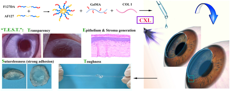
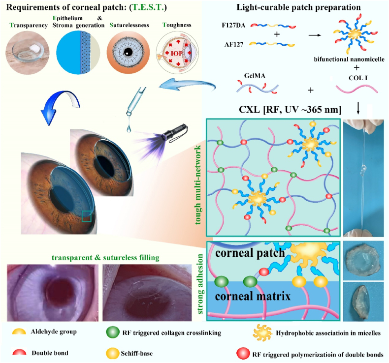
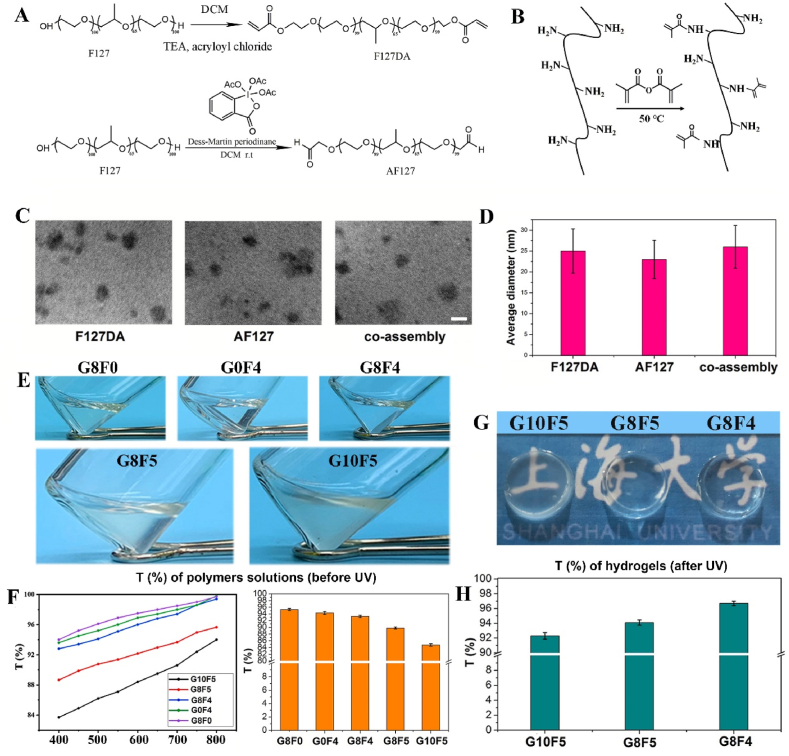
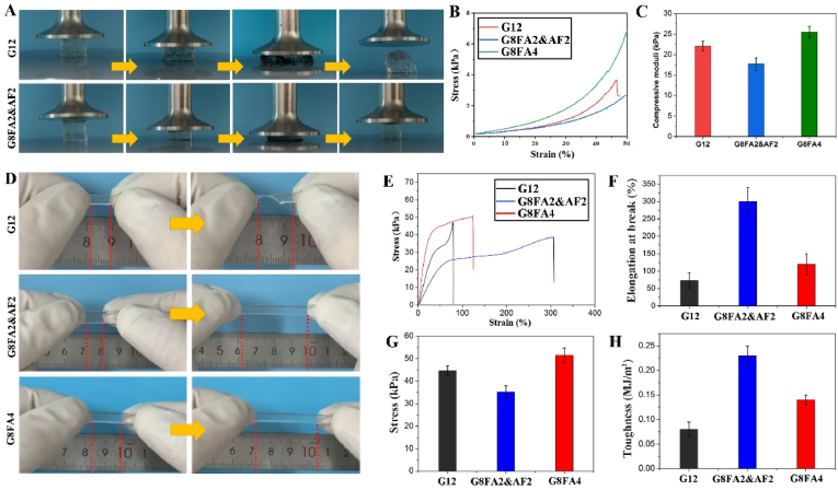
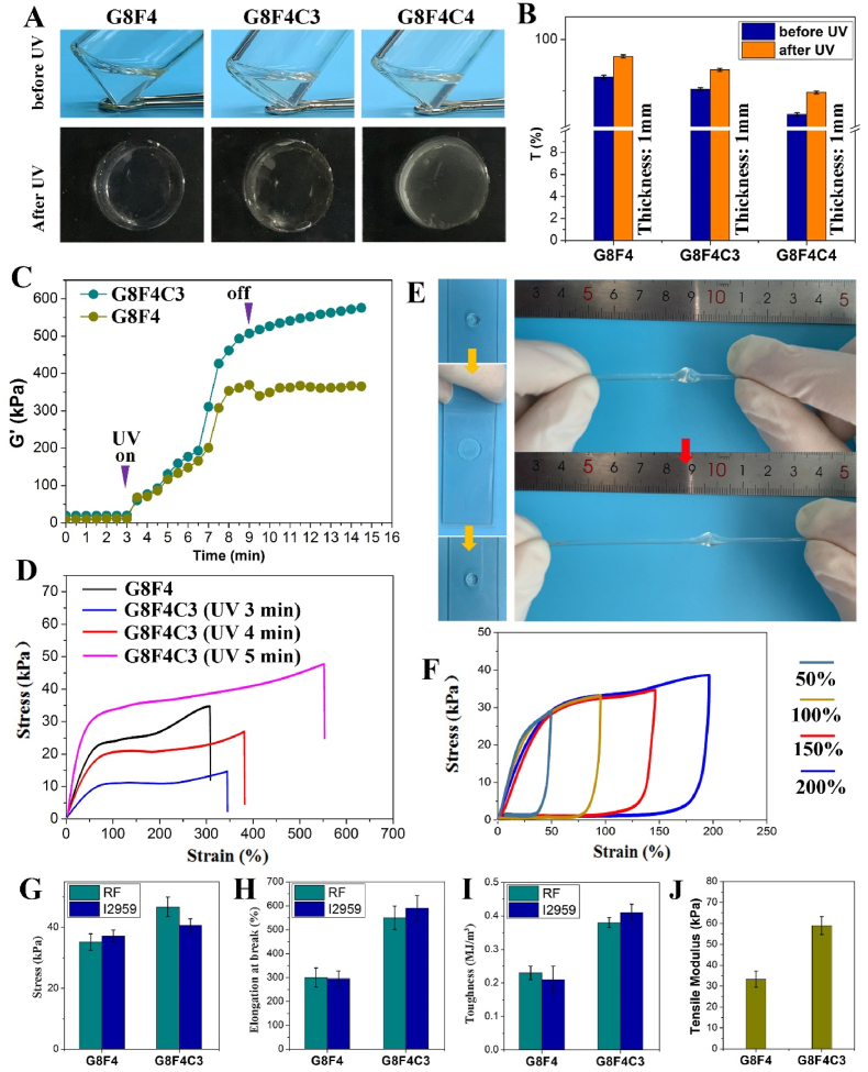
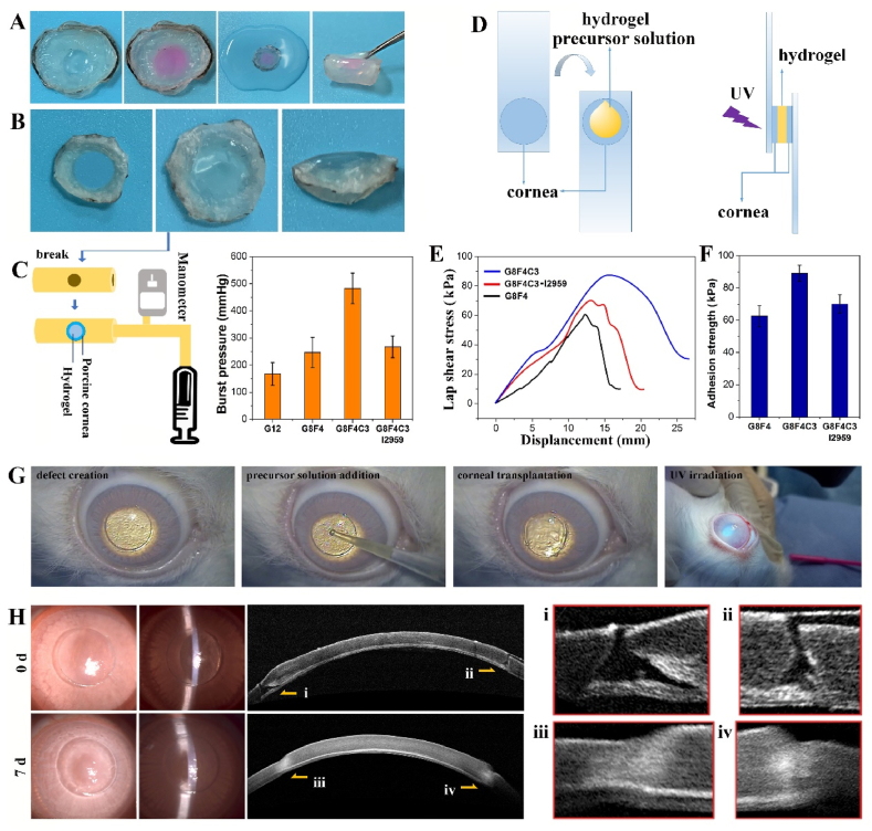
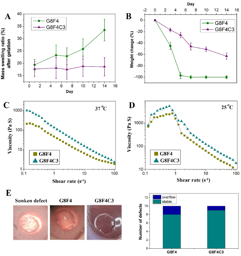
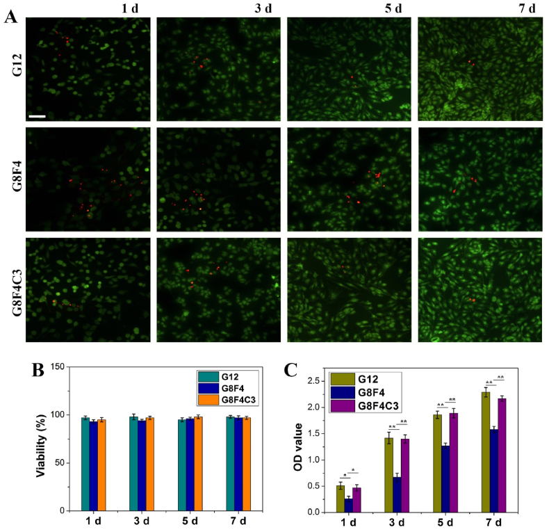
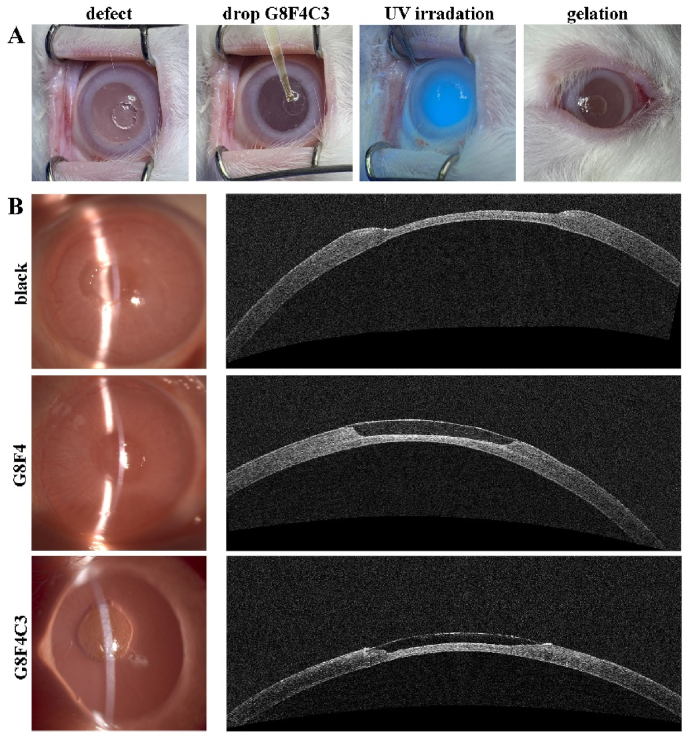
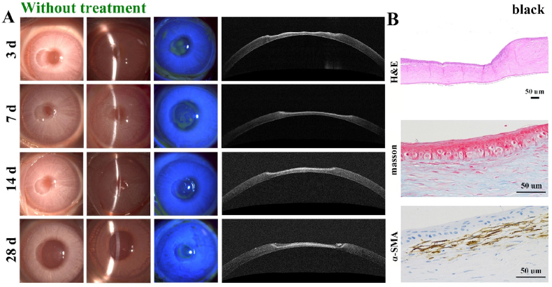
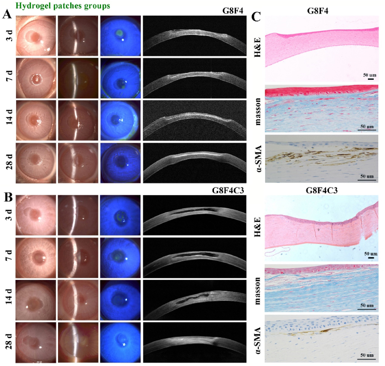
Corneal transplantation is an effective clinical treatment for corneal diseases, which, however, is limited by donor corneas. It is of great clinical value to develop bioadhesive corneal patches with functions of "Transparency" and "Epithelium & Stroma generation", as well as "Suturelessness" and "Toughness". To simultaneously meet the "T.E.S.T." requirements, a light-curable hydrogel is designed based on methacryloylated gelatin (GelMA), Pluronic F127 diacrylate (F127DA) & Aldehyded Pluronic F127 (AF127) co-assembled bi-functional micelles and collagen type I (COL I), combined with clinically applied corneal cross-linking (CXL) technology for repairing damaged cornea. The patch formed after 5 min of ultraviolet irradiation possesses transparent, highly tough, and strongly bio-adhesive performance. Multiple cross-linking makes the patch withstand deformation near 600% and exhibit a burst pressure larger than 400 mmHg, significantly higher than normal intraocular pressure (10-21 mmHg). Besides, the slower degradation than GelMA-F127DA&AF127 hydrogel without COL I makes hydrogel patch stable on stromal beds in vivo, supporting the regrowth of corneal epithelium and stroma. The hydrogel patch can replace deep corneal stromal defects and well bio-integrate into the corneal tissue in rabbit models within 4 weeks, showing great potential in surgeries for keratoconus and other corneal diseases by combining with CXL.
Keywords: AF127, Aldehyded Pluronic F127; AS-OCT, Anterior Segment Optical Coherence Tomography; Bioadhesives; CCK-8, Cell Counting Kit-8; COL I, Collagen Type I; CXL; CXL, Corneal Cross-linking; Corneal patch; DLS, Dynamic Light Scattering; DMEM, Dulbecco's Modified Eagle's Medium; ECM, Extracellular Matrix; F127DA, Pluronic F127 diacrylate; FBS, Fetal Bovine Serum; GelMA, Methacryloylated Gelatin; H&E, Hematoxylin and Eosin; IHC, Immunohistochemistry; IOP, Intraocular Pressure; PBS, Phosphate-buffered Saline; RF, Riboflavin-5-phosphate; ROS, Reactive Oxygen Species; SD, Standard Deviation; Sutureless repair; TEM, Transmission Electron Microscopy; Tough hydrogel; UV, Ultraviolet; α-SMA, Alpha Smooth Muscle Actin.
© 2023 The Authors.
Conflict of interest statement
The authors declare that they have no known competing financial interests or personal relationships that could have appeared to influence the work reported in this paper.
Figures
Similar articles
-
ECM-based bioadhesive hydrogel for sutureless repair of deep anterior corneal defects.Biomater Sci. 2024 Apr 30;12(9):2356-2368. doi: 10.1039/d4bm00129j. Biomater Sci. 2024. PMID: 38497791
-
3D bioprinting of in situ vascularized tissue engineered bone for repairing large segmental bone defects.Mater Today Bio. 2022 Aug 8;16:100382. doi: 10.1016/j.mtbio.2022.100382. eCollection 2022 Dec. Mater Today Bio. 2022. PMID: 36033373 Free PMC article.
-
Corneal stromal structure replicating humanized hydrogel patch for sutureless repair of deep anterior-corneal defect.Biomaterials. 2025 Feb;313:122754. doi: 10.1016/j.biomaterials.2024.122754. Epub 2024 Aug 14. Biomaterials. 2025. PMID: 39197237
-
[Principles of corneal cross-linking : Presentation based on the development of the various treatment protocols].Ophthalmologe. 2022 Apr;119(4):332-341. doi: 10.1007/s00347-021-01538-7. Epub 2021 Dec 9. Ophthalmologe. 2022. PMID: 34882268 Review. German.
-
[Transplantation of corneal endothelial cells].Nippon Ganka Gakkai Zasshi. 2002 Dec;106(12):805-35; discussion 836. Nippon Ganka Gakkai Zasshi. 2002. PMID: 12610838 Review. Japanese.
Cited by
-
Engineering a functionality-integrated artificial cornea stromal Substitute: Janus bio-adhesive implant with a collagen-based multi-scale biomimetic skeleton.Bioact Mater. 2025 Jun 24;51:740-757. doi: 10.1016/j.bioactmat.2025.06.030. eCollection 2025 Sep. Bioact Mater. 2025. PMID: 40641839 Free PMC article.
-
Advancements in Hydrogels for Corneal Healing and Tissue Engineering.Gels. 2024 Oct 16;10(10):662. doi: 10.3390/gels10100662. Gels. 2024. PMID: 39451315 Free PMC article. Review.
-
Harnessing Mechanical Stress with Viscoelastic Biomaterials for Periodontal Ligament Regeneration.Adv Sci (Weinh). 2024 May;11(18):e2309562. doi: 10.1002/advs.202309562. Epub 2024 Mar 9. Adv Sci (Weinh). 2024. PMID: 38460171 Free PMC article.
-
Biomaterials for Corneal Regeneration.Adv Sci (Weinh). 2025 Feb;12(6):e2408021. doi: 10.1002/advs.202408021. Epub 2024 Dec 31. Adv Sci (Weinh). 2025. PMID: 39739318 Free PMC article. Review.
-
Photothermal hydrogels for infection control and tissue regeneration.Front Bioeng Biotechnol. 2024 Mar 28;12:1389327. doi: 10.3389/fbioe.2024.1389327. eCollection 2024. Front Bioeng Biotechnol. 2024. PMID: 38605983 Free PMC article. Review.
References
-
- Krachmer J.H., Feder R.S., Belin M.W. Keratoconus and related noninflammatory corneal thinning disorders. Surv. Ophthalmol. 1984;28:293. - PubMed
-
- Gain P., Jullienne R., He Z., Aldossary M., Acquart S., Cognasse F., Thuret G. Global survey of corneal transplantation and eye banking. JAMA Ophthalmol. 2016;134:167. - PubMed
-
- Jeng B.H., Ahmad S. In pursuit of the elimination of corneal blindness: is establishing eye banks and training surgeons enough? Opthalmology. 2021;128:813–815. - PubMed
-
- Ahearne M., Fernández‐Pérez J., Masterton S., Madden P.W., Bhattacharjee P. Designing scaffolds for corneal regeneration. Adv. Funct. Mater. 2020;30
-
- Kharod-Dholakia B., Randleman J.B., Bromley J.G., Stulting R.D. Prevention and treatment of corneal graft rejection current practice patterns of the cornea society. Cornea. 2011;34(2015):609–614. - PubMed
LinkOut - more resources
Full Text Sources
Other Literature Sources
Research Materials
