. 2023 Jan 28;5(1):obad004.
doi: 10.1093/iob/obad004. eCollection 2023.

Vibroacoustic Response of the Tympanic Membrane to Hyoid-Borne Sound Generated during Echolocation in Bats

Affiliations

Vibroacoustic Response of the Tympanic Membrane to Hyoid-Borne Sound Generated during Echolocation in Bats

C C G Snipes et al. Integr Org Biol. .

Abstract

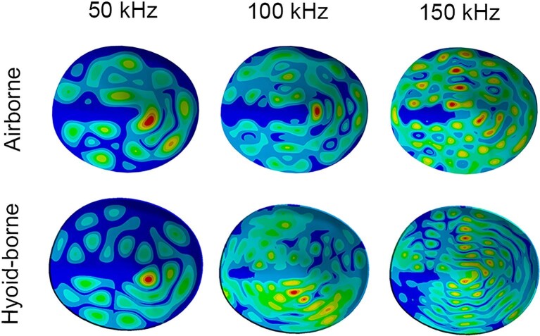
The hyoid apparatus in laryngeally echolocating bats is unique as it forms a mechanical connection between the larynx and auditory bullae, which has been hypothesized to transfer the outgoing echolocation call to the middle ear during call emission. Previous finite element modeling (FEM) found that hyoid-borne sound can reach the bulla at an amplitude likely heard by echolocating bats; however, that study did not model how or if the signal could reach the inner ear (or cochlea). One route that sound could take is via stimulation of the eardrum-similarly to that of air-conducted sound. We used micro computed tomography (μCT) data to build models of the hyoid apparatus and middle ear from six species of bats with variable morphology. Using FEM, we ran harmonic response analyses to measure the vibroacoustic response of the tympanic membrane due to hyoid-borne sound generated during echolocation and found that hyoid-borne sound in all six species stimulated the eardrum within a range likely heard by bats. Although there was variation in the efficiency between models, there are no obvious morphological patterns to account for it. This suggests that hyoid morphology in laryngeal echolocators is likely driven by other associated functions.


Figures

Fig. 1
Fig. 1
Volume-rendered lateral and ventral views of the cranium (brown), trachea/larynx (gray), and hyoid apparatus from R. ferrumequinum. The hyoid apparatus consists of the fused basihyal and thyrohyals (blue), hypohyal (green), ceratohyal (purple), and stylohyal (yellow) bone(s). The hypohyal, ceratohyal, and stylohyal bones are collectively referred to as the anterior cornu, and the bony segments are connected via cartilaginous joints (gray). In laryngeal echolocators, the stylohyal bones articulate with the auditory bullae (orange), which houses the TM and middle ear bones that transfer airborne sound to the cochleae (red).
Fig. 2
Fig. 2
Stylohyal-auditory bulla articulation from LDC/FM and HDC/NB echolocators. The stylohyal (yellow) articulates with the lateral rim of the auditory bulla (orange) in LDC/FM echolocators, whereas the stylohyal articulates with the medial rim of the auditory bulla in HDC/NB echolocators.
Fig. 3
Fig. 3
3D models/geometry used for the FE models. Ventral and lateral views of the hyoid apparatus and auditory bullae from (A) A. jamaicensis, (B) M. spasma, (C) R. ferrumequinum, (D) R. rouxi, (E) R. hildebrandtii, and (F) H. diadema. Bones are color coded as follows: fused basihyal and thyrohyals (blue), hypohyal (green), ceratohyal (purple), stylohyal (yellow), auditory bulla (orange), and intervening cartilaginous segments (gray).
Fig. 4
Fig. 4
Transverse slices through the auditory bulla and cochlea from a contrast enhanced μCT scan of A. jamaicensis (A) and a μCT scan of M. spasma (B). Due to the contrast between the lateral and medial sides of the TM, we were able to segment the negative space on the lateral side (i.e., air) to get the shape of the TM, which was used to construct the TM with NURBS surfaces (C and D). The medial side of the TM was likely filled with fluid as these specimens were stored in 70% ethanol, resulting in the contrast between the outer and middle ear cavities. Arrows indicate the manubrium of the malleus, which articulates with the medial side of the TM. Note that the slices are from different planes within the middle and inner ears.
Fig. 5
Fig. 5
Ventral (A) and dorso-lateral (B) views of the geometry from the A. jamaicensis FE model. All fixed points are indicated with red triangles (A) with: four fixed supports on the ventral surface of the basihyal to model muscle attachments, two fixed supports on the ends of the thyrohyals to model their attachment to the larynx, and five fixed supports on the surface of the auditory bullae that closely articulates with the skull. TM displacement data were generated in the axis orthogonal to the plane of the TM, indicated by the blue axis on the triad (B). Bones are color coded as follows: fused basihyal and thyrohyals (blue), hypohyal (green), ceratohyal (purple), stylohyal (yellow), auditory bulla (orange), and intervening cartilaginous segments (gray).
Fig. 6
Fig. 6
Geometry, including excitation surfaces and surfaces from which results data were generated, for the Harmonic response/modal superposition analyses on R. ferrumequinum. (A) To verify the TM geometry, the TM was excited with 100 dB sound/pressure on the lateral surface, and response data were generated from the same lateral surface of the TM. (B) To establish the displacement hearing threshold, the validated TM was then excited with 0 dB sound/pressure, and response data were generated from the same lateral surface of the TM. (C) To mimic an outgoing echolocation call, the laryngeal surface of the basihyal was excited with a 120 dB sound/pressure, and response data were generated from the lateral surface of the TM. Bones are color coded as follows: fused basihyal and thyrohyals (blue), hypohyal (green), ceratohyal (purple), stylohyal (yellow), and auditory bulla (orange). The intervening cartilage segments are gray.
Fig. 7
Fig. 7
TM displacements (m) in response to a 100 dB excitation on the lateral surface of the TM from H. diadema (solid blue line), R. ferrumequinum (dashed green line), R. rouxi (dotted green line), R. hildebrandtii (solid green line), M. spasma (solid yellow line), and A. jamaicensis (solid red line). Data were generated in the axis orthogonal to the plane of the TM and therefore in the direction that would set the ear ossicles into motion during airborne hearing. The experimental data from Manley et al. (1972) used to verify our TM models are indicated by the black horizontal lines at 6.5e–8 m (2.5 kHz) and 2.9e–10 m (100 kHz).
Fig. 8
Fig. 8
Displacement contour plots of the TM at 50, 100, and 150 kHz in response to airborne and hyoid-borne sound excitations. Warmer colors indicate areas of greater displacement (peaks), while cooler colors indicate areas with less displacement. TM displacements across a range of frequencies are characterized by fewer peaks at lower frequencies and an increase in the number of peaks as frequency increases. Our results show that this is the case in both air- and hyoid-borne excitation.
Fig. 9
Fig. 9
Average maximum TM displacements (m) from 0 to 150 kHz in response to 120 dB excitation on the laryngeal surface of the basihyal from H. diadema (solid blue line), R. ferrumequinum (dashed green line), R. rouxi (dotted green line), R. hildebrandtii (solid green line), M. spasma (solid yellow line), and A. jamaicensis (solid red line). Data were generated in the plane orthogonal to the plane of the TM and therefore in the direction that would set the ear ossicles into motion during airborne hearing. The average maximum TM displacement in response to a 0 dB excitation on the lateral side of the TM was measured from 0 to 150 kHz, and the average from each model was used to represent the average lowest hearing displacement threshold across species (solid black line).

References

    1. Brinkløv S, Kalko EK, Surlykke A. 2009. Intense echolocation calls from two “whispering” bats, Artibeus jamaicensis and Macrophyllum macrophyllum (Phyllostomidae). J Exp Biol 212:11–20. - PubMed
    1. Caminos L, Garcia-Manrique J, Lima-Rodriguez A, Gonzalez-Herrera A. 2018. Analysis of the mechanical properties of the human tympanic membrane and its influence on the dynamic behaviour of the human hearing system. Appl Bionics Biomech 2018:1736957. - PMC - PubMed
    1. Carter RT. 2020. Reinforcement of the larynx and trachea in echolocating and non-echolocating bats. J Anat 237: 495–503. - PMC - PubMed
    1. Carter RT, Shaw JB, Adams RA. 2014. Ontogeny of vocalization in Jamaican fruit bats with implications for the evolution of echolocation. J Zool 293:25–32.
    1. Cheng JT, Maftoon N, Guignard J, Ravicz ME, Rosowski J. 2019. Tympanic membrane surface motions in forward and reverse middle ear transmissions. J Acoust Soc Am 145:272. - PMC - PubMed

LinkOut - more resources