. 2023 Feb 17;26(2):106075.
doi: 10.1016/j.isci.2023.106075. Epub 2023 Feb 13.

Delta-Omicron recombinant escapes therapeutic antibody neutralization

Affiliations

Delta-Omicron recombinant escapes therapeutic antibody neutralization

Ralf Duerr et al. iScience. .

Abstract

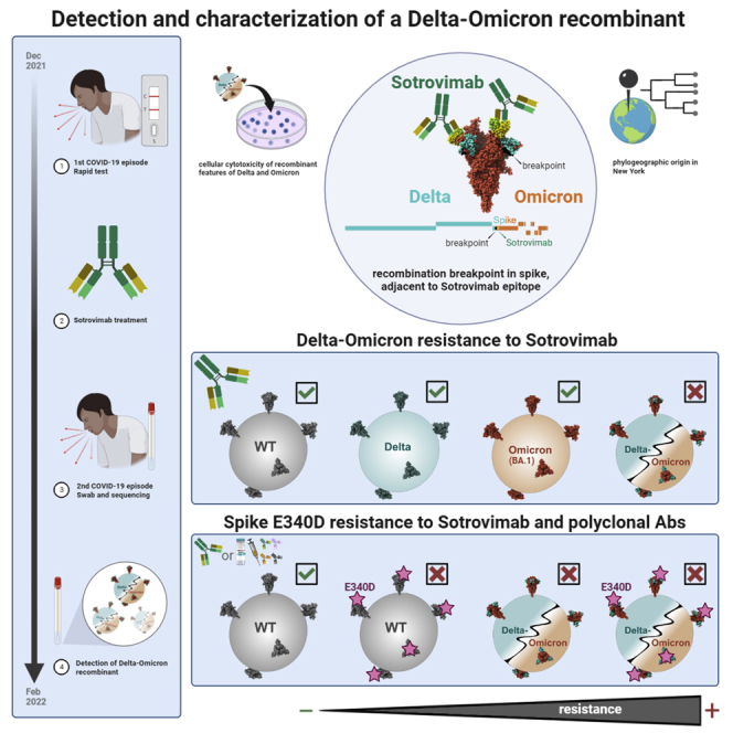
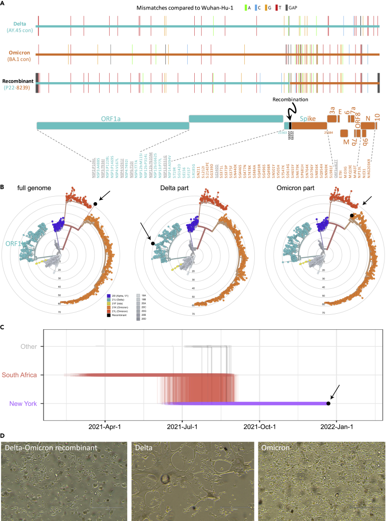
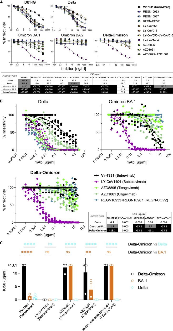
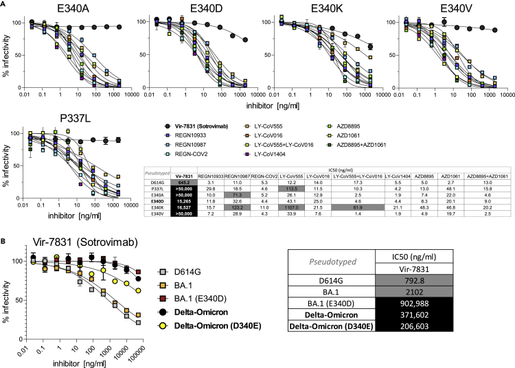
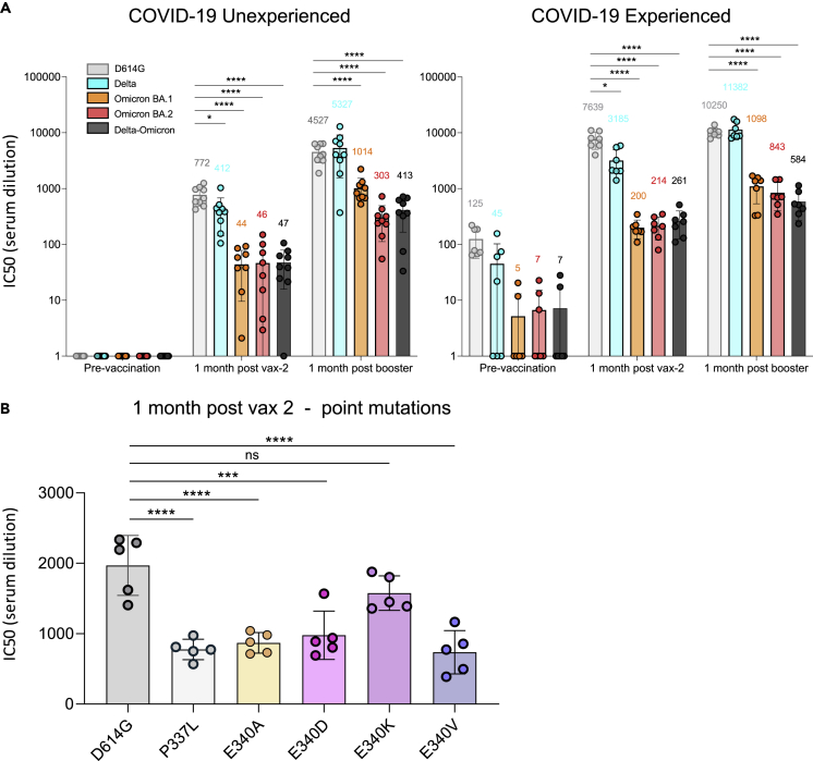
The emergence of recombinant viruses is a threat to public health, as recombination may integrate variant-specific features that together result in escape from treatment or immunity. The selective advantages of recombinant SARS-CoV-2 isolates over their parental lineages remain unknown. We identified a Delta-Omicron (AY.45-BA.1) recombinant in an immunosuppressed transplant recipient treated with monoclonal antibody Sotrovimab. The single recombination breakpoint is located in the spike N-terminal domain adjacent to the Sotrovimab binding site. While Delta and BA.1 are sensitive to Sotrovimab neutralization, the Delta-Omicron recombinant is highly resistant. To our knowledge, this is the first described instance of recombination between circulating SARS-CoV-2 variants as a functional mechanism of resistance to treatment and immune escape.

Keywords: biological sciences; immunity; immunology.


Conflict of interest statement

The authors declare no competing interests.

Figures

None
Graphical abstract
Figure 1
Figure 1
Identification of a Delta (AY.45)-Omicron (BA.1) recombinant virus with traces to New York and cytopathic effects (A) Full genome mutations of the Delta-Omicron recombinant in comparison with Delta AY.45 and Omicron BA.1 consensus (con) sequences. In the schematic of the Delta and Omicron portions, all non-synonymous mutations are listed and shown in teal (Delta), orange (Omicron), or gray (not Delta or Omicron). (B) Maximum likelihood phylogenies of 1557 global SARS-CoV-2 sequences based on a New York, New Jersey, and Connecticut-focused subsampling. The recombinant is shown in black. The phylogenetic analysis was done using the recombinant’s full genome (left) and Delta or Omicron parts only (middle and right). Concentrical rings indicate the number of mutations compared to Wuhan-Hu-1 (root). (C) Markov jump trajectory plot depicting the ancestral location history of the AY.45 segment of the recombinant sequence. Each individual trajectory corresponds to the Markov jumps in a single tree from the posterior distribution of 1,800 trees (Figure S2B). Horizontal lines represent the time maintained at an ancestral location and vertical lines represent a Markov jump between two locations. The first SARS-CoV-2 positive test of the patient is indicated in black. (D) Cytopathic effects observed in various lineages of SARS-CoV-2 on VeroE6/TMPRSS2 cells. Images were taken with an EVOS M5000 inverted microscope (ThermoFisher Scientific, Waltham, MA); 10X magnification. (see also Figures S1 and S2 and Tables S1 and S2).
Figure 2
Figure 2
Breakpoint region of the Delta-Omicron recombinant is adjacent to the Sotrovimab (S309) binding epitope in spike Trimer spike structure of the Delta-Omicron recombinant in the open, one RBD-up conformation with one Sotrovimab Fab molecule bound to the RBD in up-position. Sotrovimab is a slightly refined, engineered version of its precursor S309 that has been used in the structures shown here. The spike portions are shown as ribbons and the Sotrovimab epitopes as spheres. One spike protomer is shown in gray; the other two protomers are colored according to the legend. Lower right: schematic of the location of the Delta (teal) and Omicron (orange) spike portions as well as the recombinant breakpoint region (black) including the mutations related to Delta (teal, facing up) or Omicron (orange, facing down). The mutation E340D, which is not related to Delta or Omicron, is highlighted in pink. The recombinant spike structure is a homology model of the recombinant spike based on pdb: 7TO4 (1 RBD-up Omicron spike trimer). The S309 (Sotrovimab) molecule was added by structural overlay of a S309-bound spike co-structure in 1 RBD-up position (pdb: 7TM0). FP: fusion peptide, NTD: N-terminal domain, RBD: receptor-binding domain, SP: signal peptide, TM: transmembrane domain.
Figure 3
Figure 3
Delta and Omicron (BA.1) are sensitive to Sotrovimab, but Delta-Omicron is resistant (A) Neutralization of viruses with D614G, Delta, Omicron, and Delta-Omicron spike proteins by monoclonal antibodies (mAbs) Regeneron REGN10933 (Casirivamab), REGN10987 (Imdevimab), the REGN-COV-2 cocktail (Casirivimab + Imdevimab), LY-CoV555 (Bamlanivimab), LY-CoV016 (Etesevimab), the LY-CoV555+LY-CoV016 combination, LY-CoV1404 (Bebtelovimab), AZD8895 (Tixagevimab), AZD1061 (Cilgavimab), the AZD8895 + AZD1061 combination (Evusheld), and VIR-7831 (Sotrovimab). The heatmap table shows the half-maximal inhibitory concentrations (IC50) of the therapeutic mAbs calculated using the data from the antibody neutralization curves shown above. High resistance (IC50 > 3000 ng/mL) is highlighted in black, moderate resistance (50 ng/mL < IC50 < 3000 ng/mL) in gray, and no/low resistance (IC50 < 50 ng/mL) shown in white background. (B) Neutralization of infectious native Delta-Omicron virus in comparison with Delta and BA.1 by a selection of five mAbs used in A, including Vir-7831 (Sotrovimab). Inhibition was determined in plaque reduction neutralization tests using serially diluted mAb doses. The graphs display all data points (staggered) from two to four biological replicates with technical duplicates (Delta-Omicron: four biological replicates). Neutralization curves are shown as non-linear regression fits for each mAb. In the IC50 heatmap table, high resistance (IC50 > 10 μg/mL) is highlighted in black, moderate resistance (1 μg/mL < IC50 < 10 μg/mL) in gray, and no/low resistance (IC50 < 1 μg/mL) shown in white background. (C) Statistical comparison of IC50 values from all biological replicates in B (two-way ANOVA with Tukey’s multiple comparison test, ∗∗p < 0.005, ∗∗∗∗p < 0.0001, ns: not significant). Data in A and C are shown as mean with SD. (see also Table S3).
Figure 4
Figure 4
Delta-Omicron is resistant to Sotrovimab, irrespective of the E340D mutation in RBD (A) Neutralization of D614G (backbone) and P337L, E340A, E340D, E340K, and E340V point-mutated spike protein-pseudotyped viruses by the same set of mAbs as in Figure 3A. Experiments in Figures 3A and 4A were conducted simultaneously with D614G virus as a reference. (B) Neutralization of D614G (E340), BA.1 (E340), E340D point-mutated BA.1, Delta-Omicron (D340), and D340E point-mutated Delta-Omicron spike protein-pseudotyped viruses by VIR-7831 (Sotrovimab). The tables show the half-maximal inhibitory concentrations (IC50) of the therapeutic mAbs calculated using the data from the antibody neutralization curves shown above and to the left, respectively. High resistance (IC50 > 3000 ng/mL) is highlighted in black, moderate resistance (50 ng/mL < IC50 < 3000 ng/mL) in gray, and no/low resistance (IC50 < 50 ng/mL) shown in white background. Data points in A and B are shown as mean with SD. (see also Table S3).
Figure 5
Figure 5
Resistance of the Delta-Omicron recombinant and P337L or E340X-mutated viruses to vaccine-elicited polyclonal antibodies (A) Neutralizing antibody titers of participants with or without previous history of SARS-CoV-2 infection and/or vaccination were measured using pseudotyped viruses with D614G, Delta, Omicron, and Delta-Omicron spike. Sera were collected from study participants pre-vaccination, 1-month post-second vaccination with Pfizer BNT162b2, and 1-month post-boost (third vaccination). Study participants were without previous SARS-CoV-2 infection (unexperienced) (left) (n = 9) or previously infected (experienced) (right) (n = 7). COVID-19 history was determined by symptoms and a PCR-positive test or serology. Equivalent amounts of D614G, Delta, Omicron, and Delta-Omicron spike protein-pseudotyped viruses were mixed with a 2-fold serial dilution of donor serum and then applied to ACE2.293T cells. Luciferase activity was measured two days post-infection. Each serum dilution was measured in triplicate and the experiment was done twice with similar results. Half-maximal inhibitory concentrations (IC50) are shown for one representative experiment. Mean values for each group are shown above the bar. (B) Neutralizing antibody titers of sera from COVID-19 unexperienced individuals (n = 5) collected 1 month after second vaccination against D614G (as a backbone) and P337L, E340A, E340D, E340K, and E340V point mutated spike protein-pseudotyped viruses. Statistical significance was calculated by two-sided testing. (one-way ANOVA with Tukey’s multiple comparison test, ∗p ≤ 0.05, ∗∗∗p ≤ 0.001, ∗∗∗∗p ≤ 0.0001, ns: not significant). Bars are shown as mean with SD. (see also Tables S3 and S4).

Update of

References

    1. Simon-Loriere E., Holmes E.C. Why do RNA viruses recombine? Nat. Rev. Microbiol. 2011;9:617–626. doi: 10.1038/nrmicro2614. - DOI - PMC - PubMed
    1. Bean W.J., Jr., Cox N.J., Kendal A.P. Recombination of human influenza A viruses in nature. Nature. 1980;284:638–640. doi: 10.1038/284638a0. - DOI - PubMed
    1. Moradigaravand D., Kouyos R., Hinkley T., Haddad M., Petropoulos C.J., Engelstädter J., Bonhoeffer S. Recombination accelerates adaptation on a large-scale empirical fitness landscape in HIV-1. PLoS Genet. 2014;10:e1004439. doi: 10.1371/journal.pgen.1004439. - DOI - PMC - PubMed
    1. Zhu Z., Meng K., Meng G. Genomic recombination events may reveal the evolution of coronavirus and the origin of SARS-CoV-2. Sci. Rep. 2020;10:21617. doi: 10.1038/s41598-020-78703-6. - DOI - PMC - PubMed
    1. Pollett S., Conte M.A., Sanborn M., Jarman R.G., Lidl G.M., Modjarrad K., Maljkovic Berry I. A comparative recombination analysis of human coronaviruses and implications for the SARS-CoV-2 pandemic. Sci. Rep. 2021;11:17365. doi: 10.1038/s41598-021-96626-8. - DOI - PMC - PubMed

LinkOut - more resources