

Methods for Detecting Abnormal Ventilation in Children - the Case Study of 13-Years old Pitt-Hopkins Girl
- PMID: 36844470
- PMCID: PMC9944179
- DOI: 10.1177/2329048X231151361
Methods for Detecting Abnormal Ventilation in Children - the Case Study of 13-Years old Pitt-Hopkins Girl
Abstract
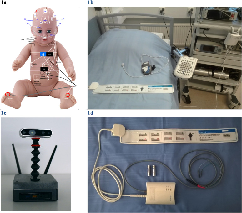
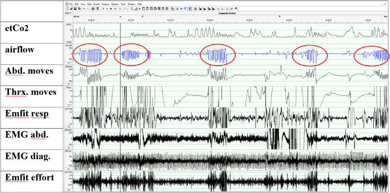
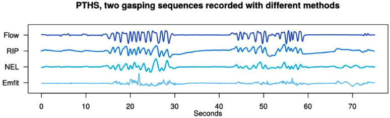
We present contactless technology measuring abnormal ventilation and compare it with polysomnography (PSG). A 13-years old girl with Pitt-Hopkins syndrome presented hyperpnoea periods with apneic spells. The PSG was conducted simultaneously with Emfit movement sensor (Emfit, Finland) and video camera with depth sensor (NEL, Finland). The respiratory efforts from PSG, Emfit sensor, and NEL were compared. In addition, we measured daytime breathing with tracheal microphone (PneaVox,France). The aim was to deepen the knowledge of daytime hyperpnoea periods and ensure that no upper airway obstruction was present during sleep. The signs of upper airway obstruction were not detected despite of minor sleep time. Monitoring respiratory effort with PSG is demanding in all patient groups. The used unobtrusive methods were capable to reveal breathing frequency and hyperpnoea periods. Every day diagnostics need technology like this for monitoring vital signs at hospital wards and at home from subjects with disabilities and co-operation difficulties.
Keywords: Emfit sensor; NEL seizure detection; Pitt-Hopkins; respiratory effort; sleep; sleep-disordered breathing.
© The Author(s) 2023.
Conflict of interest statement
None of the authors have a financial relationship with the Emfit Ltd, Finland; the company that developed and sells the Emfit sensors, neither Neuro Event Labs Ltd; the company that developed and sells The Nelli seizure detection device nor with the Cidelec Ltd, who use the PneaVoX technology in their devices; or any other conflicts of interest.
Figures


Similar articles
-
Increased respiratory effort during sleep is non-invasively detected with movement sensor.Sleep Breath. 2011 Dec;15(4):737-46. doi: 10.1007/s11325-010-0430-8. Epub 2010 Oct 20. Sleep Breath. 2011. PMID: 20960067
-
Heart rate variability evaluation of Emfit sleep mattress breathing categories in NREM sleep.Clin Neurophysiol. 2015 May;126(5):967-74. doi: 10.1016/j.clinph.2014.08.012. Epub 2014 Sep 6. Clin Neurophysiol. 2015. PMID: 25241203
-
A New Approach for Detecting Sleep Apnea Using a Contactless Bed Sensor: Comparison Study.J Med Internet Res. 2020 Sep 18;22(9):e18297. doi: 10.2196/18297. J Med Internet Res. 2020. PMID: 32945773 Free PMC article.
-
Sleep-disordered breathing in children.Ann Med. 1998 Aug;30(4):350-6. doi: 10.3109/07853899809029934. Ann Med. 1998. PMID: 9783833 Review.
-
Unattended home-based polysomnography for sleep disordered breathing: current concepts and perspectives.Sleep Med Rev. 2014 Aug;18(4):341-7. doi: 10.1016/j.smrv.2013.12.002. Epub 2013 Dec 12. Sleep Med Rev. 2014. PMID: 24388970 Review.
Cited by
-
Reliable Contactless Monitoring of Heart Rate, Breathing Rate, and Breathing Disturbance During Sleep in Aging: Digital Health Technology Evaluation Study.JMIR Mhealth Uhealth. 2024 Aug 27;12:e53643. doi: 10.2196/53643. JMIR Mhealth Uhealth. 2024. PMID: 39190477 Free PMC article.
References
-
- Knaack L, Blum HC, Hohenhorst W, Ryba J, Guilleminault C, Stoohs RA. Comparison of diaphragmatic EMG and oesophageal pressure in obstructed and unobstructed breathing during sleep. Somnologie (Berl). 2005 May;9(3):159–165. doi:10.1111/j.1439-054X.2005.00059.x - DOI
-
- Chokroverty S. Sleep Disorders Medicine: Basic Science, Technical Considerations, and Clinical Aspects. Butterworth-Heinemann; 2017.
Publication types
LinkOut - more resources
Full Text Sources
