Limbal Squamous Cell Carcinoma in a Black Baldy Cow: Case Report and Surgical Treatment
- PMID: 36844800
- PMCID: PMC9946752
- DOI: 10.1155/2023/2429241
Limbal Squamous Cell Carcinoma in a Black Baldy Cow: Case Report and Surgical Treatment
Abstract
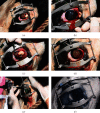
Objective: To document a case of limbal squamous cell carcinoma (SCC) in an adult Black Baldy cow treated with photodynamic therapy (PDT) as an adjunctive therapy following surgical excision. Animals Studied. One privately owned 8-year-old female, entire, Black Baldy cow. Procedures. A complete ophthalmic examination was performed on an adult Black Baldy cow for assessment of a mass affecting the left eye. Following a routine partial incision superficial lamellar keratectomy and conjunctivectomy under local analgesia using a Peterson retrobulbar block, photodynamic therapy was performed as an adjunctive treatment to lower the chance for recurrence and improve the prognosis for the globe.
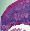
Results: Histopathologic analysis of the limbal mass was reported to be consistent with a squamous cell carcinoma, removed with clean margins. The patient was comfortable and visual with no signs of tumor recurrence 11 months after surgery.
Conclusion: Superficial lamellar keratectomy and conjunctivectomy with adjunctive photodynamic therapy is an effective treatment for limbal squamous cell carcinoma and may be performed as an alternative to enucleation, exenteration, euthanasia, or slaughtering in cattle.
Copyright © 2023 Alexandra T. J. Ng et al.
Conflict of interest statement
The authors declare that they have no conflicts of interest.
Figures


References
-
- Cordy D. R. Nervous system and eye. In: Moulton J. E., editor. Tumors in Domestic Animals . Los Angeles, CA, USA: University of California Press; 1990. pp. 654–660.
-
- Spadbrow P. B., Hoffman D. Bovine ocular squamous cell carcinoma. The Veterinary Bulletin . 1980;50:449–459.
-
- Monlux A. W., Anderson W. A., Davis C. L. The diagnosis of squamous cell carcinoma of the eye (cancer eye) in cattle. American Journal of Veterinary Research . 1957;18:5–34. - PubMed
Publication types
LinkOut - more resources
Full Text Sources
Research Materials
