

Vitamin D3 alleviates inflammation in ulcerative colitis by activating the VDR-NLRP6 signaling pathway
- PMID: 36845152
- PMCID: PMC9944717
- DOI: 10.3389/fimmu.2023.1135930
Vitamin D3 alleviates inflammation in ulcerative colitis by activating the VDR-NLRP6 signaling pathway
Abstract
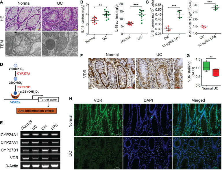
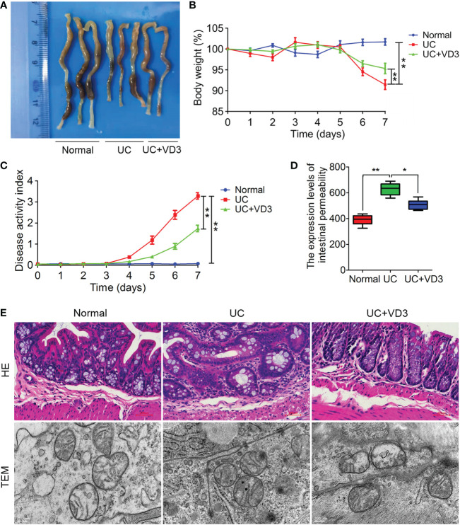
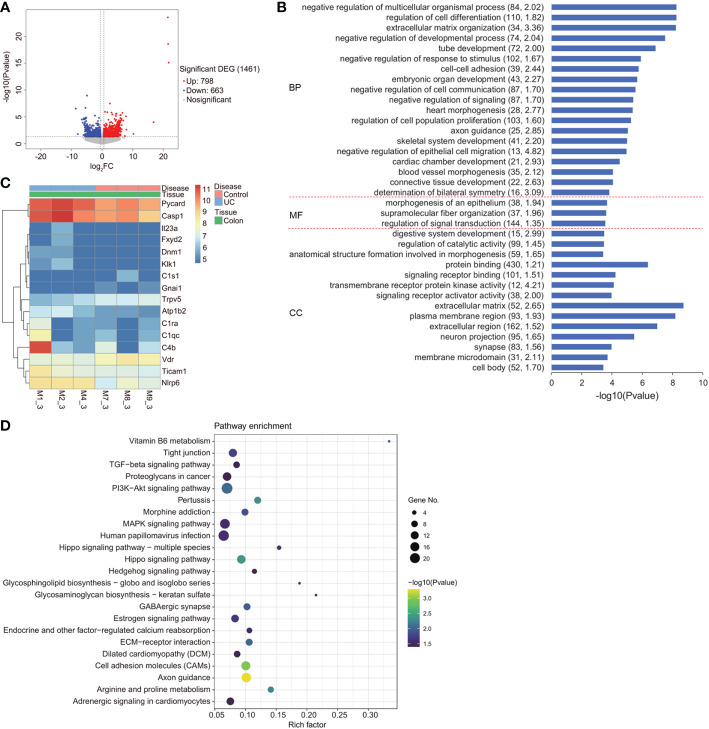
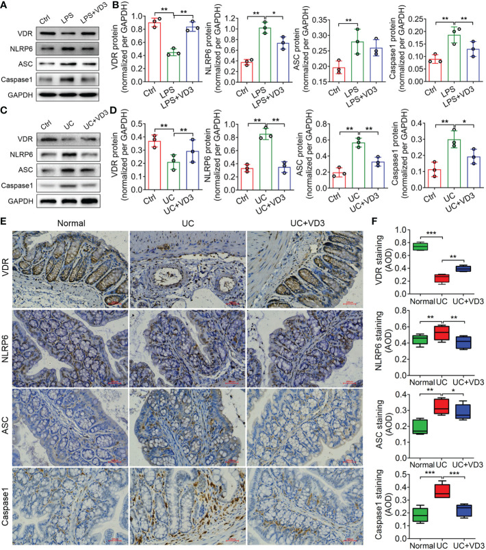
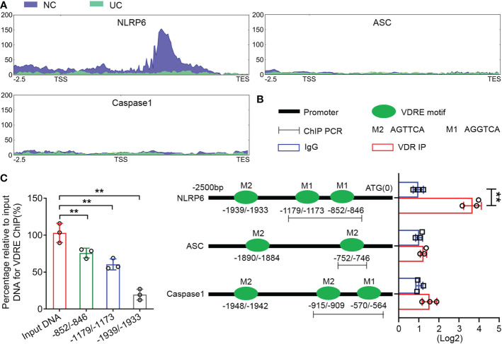
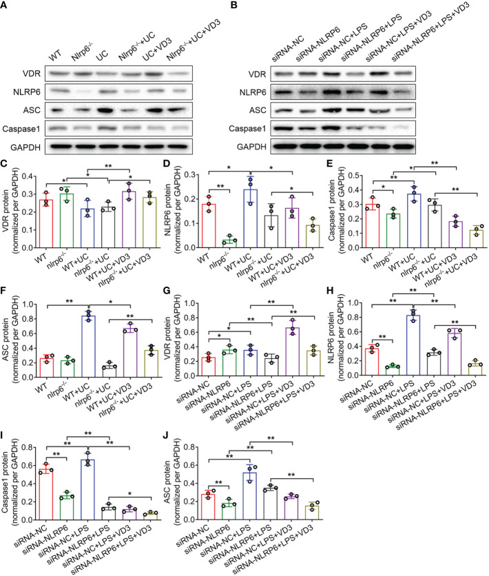
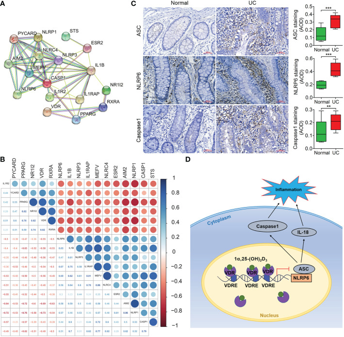
Inflammation is a key factor in the development of ulcerative colitis (UC). 1,25-dihydroxyvitamin D3 (1,25(OH)2D3, VD3), as the major active ingredient of vitamin D and an anti-inflammatory activator, is closely related to the initiation and development of UC, but its regulatory mechanism remains unclear. In this study, we carried out histological and physiological analyses in UC patients and UC mice. RNA sequencing (RNA-seq), assays for transposase-accessible chromatin with high-throughput sequencing (ATAC-seq), chromatin immunoprecipitation (ChIP) assays and protein and mRNA expression were performed to analyze and identify the potential molecular mechanism in UC mice and lipopolysaccharide (LPS)-induced mouse intestinal epithelial cells (MIECs). Moreover, we established nucleotide-binding oligomerization domain (NOD)-like receptor protein nlrp6 -/- mice and siRNA-NLRP6 MIECs to further characterize the role of NLRP6 in anti-inflammation of VD3. Our study revealed that VD3 abolished NOD-like receptor protein 6 (NLRP6) inflammasome activation, suppressing NLRP6, apoptosis-associated speck-like protein (ASC) and Caspase-1 levels via the vitamin D receptor (VDR). ChIP and ATAC-seq showed that VDR transcriptionally repressed NLRP6 by binding to vitamin D response elements (VDREs) in the promoter of NLRP6, impairing UC development. Importantly, VD3 had both preventive and therapeutic effects on the UC mouse model via inhibition of NLRP6 inflammasome activation. Our results demonstrated that VD3 substantially represses inflammation and the development of UC in vivo. These findings reveal a new mechanism by which VD3 affects inflammation in UC by regulating the expression of NLRP6 and show the potential clinical use of VD3 in autoimmune syndromes or other NLRP6 inflammasome-driven inflammatory diseases.
Keywords: ATAC-seq; NLRP6 inflammasome; VD3; VDR; ulcerative colitis.
Copyright © 2023 Gao, Zhou, Zhang, Gao, Li and Li.
Conflict of interest statement
The authors declare that the research was conducted in the absence of any commercial or financial relationships that could be construed as a potential conflict of interest.
Figures
Similar articles
-
1,25(OH)2 D3 alleviates DSS-induced ulcerative colitis via inhibiting NLRP3 inflammasome activation.J Leukoc Biol. 2020 Jul;108(1):283-295. doi: 10.1002/JLB.3MA0320-406RR. Epub 2020 Apr 1. J Leukoc Biol. 2020. PMID: 32237257
-
Qing-Chang-Hua-Shi granule ameliorates DSS-induced colitis by activating NLRP6 signaling and regulating Th17/Treg balance.Phytomedicine. 2022 Dec;107:154452. doi: 10.1016/j.phymed.2022.154452. Epub 2022 Sep 13. Phytomedicine. 2022. PMID: 36150347
-
Vitamin D3 deficiency induced intestinal inflammatory response of turbot through nuclear factor-κB/inflammasome pathway, accompanied by the mutually exclusive apoptosis and autophagy.Front Immunol. 2022 Sep 8;13:986593. doi: 10.3389/fimmu.2022.986593. eCollection 2022. Front Immunol. 2022. PMID: 36159807 Free PMC article.
-
The NLRP6 inflammasome.Immunology. 2021 Mar;162(3):281-289. doi: 10.1111/imm.13293. Epub 2020 Dec 27. Immunology. 2021. PMID: 33314083 Free PMC article. Review.
-
Progress on Regulation of NLRP3 Inflammasome by Chinese Medicine in Treatment of Ulcerative Colitis.Chin J Integr Med. 2023 Aug;29(8):750-760. doi: 10.1007/s11655-023-3551-1. Epub 2023 Apr 29. Chin J Integr Med. 2023. PMID: 37148482 Review.
Cited by
-
Tissue-Specific Metabolic Reprogramming in Innate Lymphoid Cells and Its Impact on Disease.Immune Netw. 2025 Feb 7;25(1):e3. doi: 10.4110/in.2025.25.e3. eCollection 2025 Feb. Immune Netw. 2025. PMID: 40078781 Free PMC article. Review.
-
Microbial sensing in the intestine.Protein Cell. 2023 Nov 8;14(11):824-860. doi: 10.1093/procel/pwad028. Protein Cell. 2023. PMID: 37191444 Free PMC article. Review.
-
The Role of Vitamin D in Patients with Inflammatory Bowel Disease Treated with Vedolizumab.Nutrients. 2023 Nov 20;15(22):4847. doi: 10.3390/nu15224847. Nutrients. 2023. PMID: 38004241 Free PMC article.
-
Influence of Vitamin D Receptor Signalling and Vitamin D on Colonic Epithelial Cell Fate Decisions in Ulcerative Colitis.J Crohns Colitis. 2024 Oct 15;18(10):1672-1689. doi: 10.1093/ecco-jcc/jjae074. J Crohns Colitis. 2024. PMID: 38747639 Free PMC article.
-
Confronting the global obesity epidemic: investigating the role and underlying mechanisms of vitamin D in metabolic syndrome management.Front Nutr. 2024 Aug 9;11:1416344. doi: 10.3389/fnut.2024.1416344. eCollection 2024. Front Nutr. 2024. PMID: 39183985 Free PMC article. Review.
References
Publication types
MeSH terms
Substances
LinkOut - more resources
Full Text Sources
Medical
Molecular Biology Databases
Miscellaneous