Review
. 2023 Feb 9:17:1087912.
doi: 10.3389/fnins.2023.1087912. eCollection 2023.

Functional imaging of the exposed brain

Affiliations
Review

Functional imaging of the exposed brain

Sadaf Soloukey et al. Front Neurosci. .

Abstract

When the brain is exposed, such as after a craniotomy in neurosurgical procedures, we are provided with the unique opportunity for real-time imaging of brain functionality. Real-time functional maps of the exposed brain are vital to ensuring safe and effective navigation during these neurosurgical procedures. However, current neurosurgical practice has yet to fully harness this potential as it pre-dominantly relies on inherently limited techniques such as electrical stimulation to provide functional feedback to guide surgical decision-making. A wealth of especially experimental imaging techniques show unique potential to improve intra-operative decision-making and neurosurgical safety, and as an added bonus, improve our fundamental neuroscientific understanding of human brain function. In this review we compare and contrast close to twenty candidate imaging techniques based on their underlying biological substrate, technical characteristics and ability to meet clinical constraints such as compatibility with surgical workflow. Our review gives insight into the interplay between technical parameters such sampling method, data rate and a technique's real-time imaging potential in the operating room. By the end of the review, the reader will understand why new, real-time volumetric imaging techniques such as functional Ultrasound (fUS) and functional Photoacoustic Computed Tomography (fPACT) hold great clinical potential for procedures in especially highly eloquent areas, despite the higher data rates involved. Finally, we will highlight the neuroscientific perspective on the exposed brain. While different neurosurgical procedures ask for different functional maps to navigate surgical territories, neuroscience potentially benefits from all these maps. In the surgical context we can uniquely combine healthy volunteer studies, lesion studies and even reversible lesion studies in in the same individual. Ultimately, individual cases will build a greater understanding of human brain function in general, which in turn will improve neurosurgeons' future navigational efforts.

Keywords: brain mapping; craniotomy; exposed brain; functional imaging; functional ultrasound; imaging techniques; neuronal activity; surgical decision-making.


Conflict of interest statement

The authors declare that the research was conducted in the absence of any commercial or financial relationships that could be construed as a potential conflict of interest.

Figures

FIGURE 1
FIGURE 1
Overview of the biological substrates underlying different functional imaging techniques. (A) Graphical overview of the relationship between blood dynamics, vascular dynamics and hemodynamics. Blood dynamics consists of changes in CBV (the volume of blood in a volume of brain) and changes in CBF (the speed with which a volume of blood perfuses a volume of brain). Vascular dynamics specifically encompasses vasoconstriction and vasodilatation, mediated by smooth muscle cells (SMCs) surrounding larger veins, arteries and smaller arterioles. “Hemodynamics” is used as an overarching term encompassing blood and vascular dynamics. (B) Electrical techniques (depicted in green) rely on detection of electrical signal (ECoG, EEG, MEG) or direct proxies thereof (Ca-I, V-i) to measure neuronal activity. Alternatively, techniques such as ESM or ECoG rely on introduction of electrical current into the brain to actively produce or interrupt neuronal activity. Through the principles of neurovascular coupling (NVC), electrical neuronal activity is linked to changes in hemodynamics, which underlies techniques such as dOCT, fUS, LDI, and LSCI (depicted in red). This neurovascular response is closely interwoven with the metabolic requirements for neuronal activity known as neurometabolic coupling (NMC). Techniques such as IFF-I, IT, OISI, PAI, fNIRS, fPET/SPECT, and fMRI rely primarily on measurements of metabolites as a proxy for functional activity (depicted in blue). White boxes indicate pre-operative techniques specifically. ECoG, electrocorticography; EEG, electroencephalography; MEG, magnetoencephalography; ESM, electrocortical stimulation; TMS, transcranial magnetic stimulation; Ca-i, calcium-imaging; V-i, voltage-imaging; NVC, neurovascular coupling; PAI, photo-acoustic imaging; fNIRS, functional near-infrared spectroscopy; dOCT, Doppler optical coherence tomography; fUS, functional ultrasound; LDI, laser Doppler imaging; LSCI, laser speckle contrast imaging; RBC, red blood cells; NMC, neurometabolic coupling; IFF-I, intrinsic functional fluorescence-imaging; IT, infrared thermography; OISI, optical intrinsic signal imaging; fMRI, functional magnetic resonance Imaging; fPET, functional position emission tomography; SPECT, single-photon emission computed tomography; FP, flavoprotein.
FIGURE 2
FIGURE 2
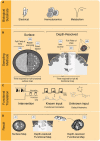
The process of functional image acquisition. (A) Functional imaging techniques rely on detection of one or more underlying biological substrates which serve as indicators of functional activity. (B) Roughly, techniques can be subdivided in surface and depth-resolved, penetrative techniques, which either build their full field of view at once (in 2D or 3D) or using subsamples, such as point-based, raster-based or line-by-line scanning. (C) To translate from acquired data to functional information, techniques rely on one or more of three methods. They can be intervention-based, probing the brain by interrupting or producing certain behavior. They can rely on known input patterns of pre-defined functional tasks to use for functional correlation or they can use functional connectivity analyses without known inputs (i.e., determining resting-state or default networks). (D) Once functional analyses have been performed, functional maps are displayed as, e.g., 2D surface maps or 2D/3D depth-resolved functional maps.
FIGURE 3
FIGURE 3
The landscape of intra-operative functional imaging techniques and their respective technical characteristics. Metabolism-, Electrical- and Hemodynamics-based techniques are placed within a grid spanning spatial resolution (y-axis) and penetrative depth (x-axis). The techniques field of view, current status of use, intra-operative applicability and temporal resolution are also taken into considerations. Many of the techniques illustrate a trade-off between penetrative depth and spatial resolution, with high penetration, lower resolution techniques such as SPECT and fPET on the one end and low penetration, high resolution techniques such as V-i and Ca-i on the other end of the spectrum. Unique positions within the domain are taken up by techniques such as MEG, which is able to combine full brain penetration with good spatiotemporal resolutions. A similar position is taken up by fUS, which combines sufficient penetration depths with exceptional spatiotemporal resolution, while showing actual intra-operative applicability. ECoG, electrocorticography; EEG, electroencephalography; MEG, magnetoencephalography; ESM, electrocortical stimulation; Ca-i, calcium-imaging; V-i, voltage-imaging; TMS, transcranial magnetic stimulation; NVC, neurovascular coupling; PAI, photo-acoustic imaging; fNIRS, functional near-infrared spectroscopy; dOCT, Doppler optical coherence tomography; fUS, functional ultrasound; LDI, laser Doppler imaging; LSCI, laser speckle contrast imaging; RBC, red blood cells; NMC, neurometabolic coupling; IFF-I, intrinsic functional fluorescence-imaging; IT, infrared thermography; OISI, optical intrinsic signal imaging; fMRI, functional magnetic resonance imaging; fPET, functional position emission tomography; SPECT, single-photon emission computed tomography; Pre-op, pre-operatively available only; Intra-op, intra-operatively available.
FIGURE 4
FIGURE 4
Requirements for the intra-operative context. Here we see an overview of the functional neuro-imaging landscape, drawn up using parameters with a clinical and intra-operative relevance. In the upper right corner, we see a group of three pre-operative techniques (MEG, fMRI, fPET/SPECT) with high costs and limited mobility, partially explaining their limited intra-operative applicability. Also the contact-level needed for the technique to function can dictate the technique’s intra-operative applicability. Optical techniques such as OISI and IT, make use of stand-alone cameras or integrated versions into conventional surgical microscopes, which does not require any substantial interruption of the surgical flow. Techniques such as fUS or PAI come in mobile, hand-held acquisition units, which do not take up much space in the operating room. A technique’s multimodal potential is also important and can be complicated by electrical or susceptibility artifacts. Finally, the level of invasiveness can severely determine its clinical success. As Ca-i and V-i require introduction of alien fluorescent markers, their clinical applications is more complicated than techniques such as OISI and IFF-i which rely on intrinsic fluorescence. ECoG, electrocorticography; EEG, electroencephalography; MEG, magnetoencephalography; ESM, electrocortical stimulation; Ca-i, calcium-imaging; V-i, voltage-imaging; TMS, transcranial magnetic stimulation; NVC, neurovascular coupling; PAI, photo-acoustic imaging; fNIRS, functional near-infrared spectroscopy; dOCT, Doppler optical coherence tomography; fUS, functional ultrasound; LDI, laser Doppler imaging; LSCI, laser speckle contrast imaging; RBC, red blood cells; NMC, neurometabolic coupling; IFF-I, intrinsic functional fluorescence-imaging; IT, infrared thermography; OISI, optical intrinsic signal imaging; fMRI, functional magnetic resonance imaging; fPET, functional position emission tomography; SPECT, single-photon emission computed tomography; Pre-op, pre-operatively available only; Intra-op, intra-operatively available.

References

    1. Abbott A. (2009). Neuroscience: Opening up brain surgery. Nature 461 866–868. 10.1038/461866a - DOI - PubMed
    1. Agarwal S., Sair H. I., Gujar S., Pillai J. J. (2019). Language mapping with fMRI: Current standards and reproducibility. Top. Magn. Reson. Imaging 28 225–233. 10.1097/RMR.0000000000000216 - DOI - PubMed
    1. Aksenov D. P., Li L., Miller M. J., Iordanescu G., Wyrwicz A. M. (2015). Effects of anesthesia on BOLD signal and neuronal activity in the somatosensory cortex. J. Cereb. Blood Flow Metab. 35 1819–26. 10.1038/jcbfm.2015.130 - DOI - PMC - PubMed
    1. Almasian M., Wilk L. S., Bloemen P. R., van Leeuwen T. G., ter Laan M., Aalders M. C. G. (2019). Pilot feasibility study of in vivo intraoperative quantitative optical coherence tomography of human brain tissue during glioma resection. J. Biophoton. 12:e201900037. 10.1002/jbio.201900037 - DOI - PMC - PubMed
    1. Ances B. M. (2004). Coupling of changes in cerebral blood flow with neural activity: What must initially dip must come back up. J. Cereb. Blood Flow Metab. 24 1–6. 10.1097/01.WCB.0000103920.96801.12 - DOI - PubMed

LinkOut - more resources