

A fast and intuitive method for calculating dynamic network reconfiguration and node flexibility
- PMID: 36845440
- PMCID: PMC9949291
- DOI: 10.3389/fnins.2023.1025428
A fast and intuitive method for calculating dynamic network reconfiguration and node flexibility
Abstract
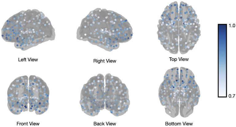
Dynamic interactions between brain regions, either during rest or performance of cognitive tasks, have been studied extensively using a wide variance of methods. Although some of these methods allow elegant mathematical interpretations of the data, they can easily become computationally expensive or difficult to interpret and compare between subjects or groups. Here, we propose an intuitive and computationally efficient method to measure dynamic reconfiguration of brain regions, also termed flexibility. Our flexibility measure is defined in relation to an a-priori set of biologically plausible brain modules (or networks) and does not rely on a stochastic data-driven module estimation, which, in turn, minimizes computational burden. The change of affiliation of brain regions over time with respect to these a-priori template modules is used as an indicator of brain network flexibility. We demonstrate that our proposed method yields highly similar patterns of whole-brain network reconfiguration (i.e., flexibility) during a working memory task as compared to a previous study that uses a data-driven, but computationally more expensive method. This result illustrates that the use of a fixed modular framework allows for valid, yet more efficient estimation of whole-brain flexibility, while the method additionally supports more fine-grained (e.g. node and group of nodes scale) flexibility analyses restricted to biologically plausible brain networks.
Keywords: community detection; dynamic functional connectivity; dynamical network analysis; modular structure; network neuroscience; task-based fMRI; template-based flexibility.
Copyright © 2023 Chinichian, Kruschwitz, Reinhardt, Palm, Wellan, Erk, Heinz, Walter and Veer.
Conflict of interest statement
The authors declare that the research was conducted in the absence of any commercial or financial relationships that could be construed as a potential conflict of interest.
Figures


Similar articles
-
Persistency and flexibility of complex brain networks underlie dual-task interference.Hum Brain Mapp. 2015 Sep;36(9):3542-62. doi: 10.1002/hbm.22861. Epub 2015 Jun 12. Hum Brain Mapp. 2015. PMID: 26095953 Free PMC article.
-
Reconfiguration of Brain Network Architectures between Resting-State and Complexity-Dependent Cognitive Reasoning.J Neurosci. 2017 Aug 30;37(35):8399-8411. doi: 10.1523/JNEUROSCI.0485-17.2017. Epub 2017 Jul 31. J Neurosci. 2017. PMID: 28760864 Free PMC article.
-
Dynamic reconfiguration of frontal brain networks during executive cognition in humans.Proc Natl Acad Sci U S A. 2015 Sep 15;112(37):11678-83. doi: 10.1073/pnas.1422487112. Epub 2015 Aug 31. Proc Natl Acad Sci U S A. 2015. PMID: 26324898 Free PMC article.
-
Demystifying cognitive flexibility: Implications for clinical and developmental neuroscience.Trends Neurosci. 2015 Sep;38(9):571-8. doi: 10.1016/j.tins.2015.07.003. Trends Neurosci. 2015. PMID: 26343956 Free PMC article. Review.
-
On the Importance of Being Flexible: Dynamic Brain Networks and Their Potential Functional Significances.Front Syst Neurosci. 2022 Jan 21;15:688424. doi: 10.3389/fnsys.2021.688424. eCollection 2021. Front Syst Neurosci. 2022. PMID: 35126062 Free PMC article. Review.
Cited by
-
Modeling brain network flexibility in networks of coupled oscillators: a feasibility study.Sci Rep. 2024 Mar 8;14(1):5713. doi: 10.1038/s41598-024-55753-8. Sci Rep. 2024. PMID: 38459077 Free PMC article.
-
Mindfulness Meditation and Network Neuroscience: Review, Synthesis, and Future Directions.Biol Psychiatry Cogn Neurosci Neuroimaging. 2025 Apr;10(4):350-358. doi: 10.1016/j.bpsc.2024.11.005. Epub 2024 Nov 17. Biol Psychiatry Cogn Neurosci Neuroimaging. 2025. PMID: 39561891 Review.
-
Global motion filtered nonlinear mutual information analysis: Enhancing dynamic portfolio strategies.PLoS One. 2024 Jul 11;19(7):e0303707. doi: 10.1371/journal.pone.0303707. eCollection 2024. PLoS One. 2024. PMID: 38990955 Free PMC article.
-
Unveiling the neuroplastic capacity of the bilingual brain: insights from healthy and pathological individuals.Brain Struct Funct. 2024 Dec;229(9):2187-2205. doi: 10.1007/s00429-024-02846-9. Epub 2024 Sep 18. Brain Struct Funct. 2024. PMID: 39289268 Free PMC article.
References
-
- Bazzi M., Porter M. A., Williams S., McDonald M., Fenn D. J., Howison S. D. (2016). Community detection in temporal multilayer networks, with an application to correlation networks. Multiscale. Model Simul. 14, 1–41. 10.1137/15M1009615 - DOI
-
- Blondel V. (2022). The Louvain Method for Community Detection in Large Networks. Available online at: https://www.nasa.gov/nh/pluto-the-other-red-planet (accessed December 12, 2022).
LinkOut - more resources
Full Text Sources