

Toxoplasma Neuroretinitis
- PMID: 36845446
- PMCID: PMC9944202
- DOI: 10.1159/000526682
Toxoplasma Neuroretinitis
Abstract
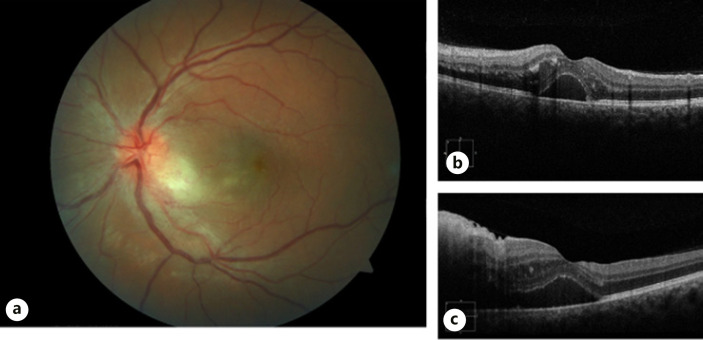
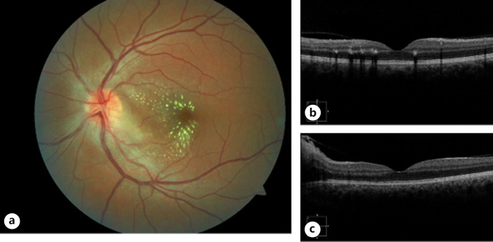
Neuroretinitis is an inflammatory condition with rapid unilateral vision loss, optic disc edema, and macular star formation. While neuroretinitis is commonly due to infectious causes such as Bartonella henselae, neuroretinitis due to toxoplasmosis is uncommon. A 29-year-old male presents to our neuro-ophthalmology clinic on December 7, 2021, at the University of Arkansas for Medical Sciences with symptoms of left eye pain and blurred vision. Subsequent workup led to the diagnosis and treatment of toxoplasma neuroretinitis. The fundus exam eventually demonstrated a notable macular star. Treatment was well tolerated, and the patient regained total visual acuity in the affected eye. Toxoplasma neuroretinitis is known for a characteristic appearance of optic disc edema prior to appearance of stellate maculopathy with vitreous inflammation and peripheral chorioretinal scars. Although loss of vision due to toxoplasmosis is rare, it should be included as part of the differential diagnosis with pertinent history.
Keywords: Neuro-ophthalmology; Ocular toxoplasmosis; Optic nerve/neurophthalmology; Toxoplasma gondii; Toxoplasma neuroretinitis.
Copyright © 2022 by The Author(s).Published by S. Karger AG, Basel.
Conflict of interest statement
The authors have no conflicts of interest to declare.
Figures
References
-
- Moreno RJ, Weisman J, Waller S. Neuroretinitis: an unusual presentation of ocular toxoplasmosis. Ann Ophthalmol. 1992;24((2)):68–70. - PubMed
-
- Fish RH, Hoskins JC, Kline LB. Toxoplasmosis neuroretinitis. Ophthalmol. 1993 Aug;100((8)):1177–1182. - PubMed
-
- Purvin V, Sundaram S, Kawasaki A. Neuroretinitis: review of the literature and new observations. J Neuroophthalmol. 2011 Mar;31((1)):58–68. - PubMed
-
- Ray S, Gragoudas E. Neuroretinitis. Int Ophthalmol Clin. 2001;41((1)):83–102. - PubMed
Publication types
LinkOut - more resources
Full Text Sources
