. 2023 Feb 20;13(4):e4613.
doi: 10.21769/BioProtoc.4613.

CRISPR/Cas9-based Engineering of Immunoglobulin Loci in Hybridoma Cells

Affiliations

CRISPR/Cas9-based Engineering of Immunoglobulin Loci in Hybridoma Cells

Camille Le M Gall et al. Bio Protoc. .

Abstract

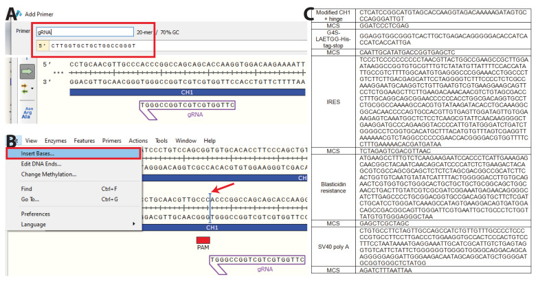
Development of the hybridoma technology by Köhler and Milstein (1975) has revolutionized the immunological field by enabling routine use of monoclonal antibodies (mAbs) in research and development efforts, resulting in their successful application in the clinic today. While recombinant good manufacturing practices production technologies are required to produce clinical grade mAbs, academic laboratories and biotechnology companies still rely on the original hybridoma lines to stably and effortlessly produce high antibody yields at a modest price. In our own work, we were confronted with a major issue when using hybridoma-derived mAbs: there was no control over the antibody format that was produced, a flexibility that recombinant production does allow. We set out to remove this hurdle by genetically engineering antibodies directly in the immunoglobulin (Ig) locus of hybridoma cells. We used clustered regularly interspaced short palindromic repeats (CRISPR)/CRISPR-associated protein 9 (Cas9) and homology-directed repair (HDR) to modify antibody's format [mAb or antigen-binding fragment (Fab')] and isotype. This protocol describes a straightforward approach, with little hands-on time, leading to stable cell lines secreting high levels of engineered antibodies. Parental hybridoma cells are maintained in culture, transfected with a guide RNA (gRNA) targeting the site of interest in the Ig locus and an HDR template to knock in the desired insert and an antibiotic resistance gene. By applying antibiotic pressure, resistant clones are expanded and characterized at the genetic and protein level for their ability to produce modified mAbs instead of the parental protein. Finally, the modified antibody is characterized in functional assays. To demonstrate the versatility of our strategy, we illustrate this protocol with examples where we have (i) exchanged the constant heavy region of the antibody, creating chimeric mAb of a novel isotype, (ii) truncated the antibody to create an antigenic peptide-fused Fab' fragment to produce a dendritic cell-targeted vaccine, and (iii) modified both the constant heavy (CH)1 domain of the heavy chain (HC) and the constant kappa (Cκ) light chain (LC) to introduce site-selective modification tags for further derivatization of the purified protein. Only standard laboratory equipment is required, which facilitates its application across various labs. We hope that this protocol will further disseminate our technology and help other researchers. Graphical abstract.

Keywords: Antibody engineering; CRISPR/Cas9; Hybridoma; Immunoglobulin; Immunology; Immunotherapy.


Conflict of interest statement

Competing interestsThe authors declare no competing interest.

Figures

Figure 1.
Figure 1.. Workflow overview.
Hybridoma are transfected on day 0 with a plasmid encoding the clustered regularly interspaced short palindromic repeats (CRISPR)/CRISPR-associated protein 9 (Cas9) and guide ribonucleic acid (gRNA), and a plasmid encoding a homology-directed repair (HDR) template containing an antibiotic resistance gene. At day 3, selection pressure is applied, and cells start to grow out. Resistant clones are expanded until reaching confluency, and single-cell clones (SCC) are prepared in 96-well plates. By day 30, positive clones are screened for production of the modified antibody and expanded for production.
Figure 2.
Figure 2.. Annotating the target sequence. A.
Access the genomic sequence of the locus of interest (e.g., on NCBI Gene). Using “sequence text” view, visualize exons and introns in the target locus. B. Remove annotations to visualize only the nucleotide sequence. For a given locus, exons are marked in red and introns in green. Here are highlighted constant heavy chain (CH)1, hinge, CH2, and CH3 in the Ighg1 gene (mIgG1 constant region). C. Copy the sequence in Snapgene and annotate exons to identify the target region. This typically depends on the intended modification. Here, we target the C-terminus of CH1 of mIgG1.
Figure 3.
Figure 3.. Identification of the target locus.
A. Identify the region to target for the knock in. Here, we selected the C-terminus of the CH1 domain to insert part of the hinge region, a short linker, a site-specific labeling site, and a polyhistidine tag. This effectively transforms a mAb into a Fab’ fragment bearing a site-specific labelling site at the C-terminus of its heavy chain (HC). B. Copy the selected sequence to CRISPOR. C. Choose the appropriate host (here, Mus musculus) and protospacer adjacent motif (PAM) (typically, -NGG) and submit.
Figure 4.
Figure 4.. Selection of gRNAs.
A. Protospacer adjacent motif (PAM) sequences and corresponding gRNA candidates are identified by CRISPOR. All possible PAM sequences (-NGG here) are identified below the input sequence. B. Sequences and characteristics of potential gRNAs are displayed as a table, ordered by predicted efficacy. The insertion site should be less than 10 bp away from the double strand break if possible. When targeting a new region, select at least two or three gRNAs based on their proximity to the intended site of insertion and precited score. Here, we selected gRNA_m1_HC (5′-CTTGGTGCTGCTGGCCGGGT-3′). By clicking on “cloning,” a new window opens with sequences for cloning into a variety of Cas9-containing vectors. C. We used a U6 expression system (U6 promoter, pX330 vectors, and derivatives) (Cong et al., 2013). Here, we used a 20 nt gRNA, but it was recently proposed to use a 19 nt gRNA for improved efficiency (Kim et al., 2020). Select the appropriate primers and follow the instructions to clone the gRNAs into the pX330 vector at the BbsI site.
Figure 5.
Figure 5.. Inserting the desired sequence at the knock-in site.
A. Copy the gRNA sequence(s) into Snapgene and B. visualize the protospacer adjacent motif (PAM) sequence(s). Insert the sequence to knock in as close as possible to the PAM sequence. C. Insert the predetermined nucleotides containing the desired insert. Here, we inserted part of the hinge region (VPRDC) allowing association with the light chain (LC) downstream of CH1, followed by a multiple cloning site (MCS), a flexible linker (GGGGS), a sortag motif (LAETGG), and a polyhistidine tag (HHHHHH), followed by a stop codon. The insert is followed by an internal ribosomal entry site (IRES) to allow for transcription of blasticidin resistance gene (BSD) and a simian vacuolating virus 40 (SV40) poly A tail to dissociate ribosomes. Each domain is surrounded by a MCS to allow easy exchange of any part of the plasmid. Here, we additionally optimized part of the CH1 to improve association with the LC (PACSTKVDKKI, S>C mutation).
Figure 6.
Figure 6.. Homology arms (HA) and final HDR template design.
The HAs span ca. 0.6 kb on each side of the insert. Their large size allows a few mismatches between the HAs and the target locus without affecting HA annealing to the genomic locus during HDR. Therefore, small adjustments can be done in the HA sequence compared to the germline sequence initially obtained from the genome browser without affecting HDR efficacy. A. It is necessary to mutate the “NGG” protospacer adjacent motif (PAM) sequence(s). To inactivate the PAM sequences, mutate “NGG” to “NGC,” “NGA,” or “NGT.” If the PAM sequence is located within the coding region, ensure that it is a silent mutation. Alternatively, remove the PAM sequences all together. Cells having undergone successful HDR will not contain PAM sequence(s) and the Cas9 will be inactivated. B. To define 5’HA and 3’HA, select ~0.6 kb on each side of the insert.
Figure 7.
Figure 7.. Antibiotic titration, transfection, and expansion of resistant cells.
A. Titration of puromycin on wild-type (WT) anti-human CD20 [NKI.B20/1]. Serial dilution of 0–12.5 μg/mL puromycin. After 48 h, cells start dying in the treated conditions. Healthy hybridoma cells are round and regular and grow in clusters. Dead or dying cells lose their round shape and become irregular. Dead cells can be spotted at 0.5 μg/mL, and some live cells remain up to 4 μg/mL. From 8 μg/mL on, cells are clearly dead. A trained eye can easily recognize how viable the cells are. B. To quantify the viability, Trypan blue staining allows to discern live (white) from dead (blue) cells. Here, we selected 8 μg/mL puromycin to conduct the procedure. C. AMAXA cuvette and program (Cell line SF, CQ-104). Carefully pipette the suspension containing cells and DNA between the electrodes (arrow head) without introducing air bubbles. D. After selection, small clusters start to grow out in HDR-transfected plates among a majority of dying or dead cells (day 6). These resistant cells have successfully undergone HDR. Passage the plates until larger clusters start to grow out (day 10) and grow towards confluency (day 13). In contrast, green fluorescent protein (GFP)-transfected hybridoma remain dead and no cluster can be seen (day 13, right). When the plate is full, perform a limiting dilution. E. After limiting dilution, monoclonal cell lines can be easily identified at the bottom of their respective well (arrow heads). Expand and characterize these cell lines.
Figure 8.
Figure 8.. Validation of three engineered cell lines at the genetic, protein, and functional level.
A. HDR template and gRNA for isotype switching of rat IgG2a hybridoma to mouse IgG2a. B. HDR template structure and gRNA for rat IgG2a hybridoma to Fab’ antigen (Fab’Ag). Ag presented here, OTI and OTII peptide. C. HDR template and gRNA for editing of the LC of mouse IgG1 hybridoma. In this strategy, HC and LC are both edited to form a Fab’ fragment bearing two site-selective modification sites (LC editing shown here). D. PCR amplification of gDNA isolated from a m2a and m2a(silent) resistant clone using a location specific (1) and HDR specific (2) primer set. E. PCR amplification of gDNA isolated from Fab’OTI and Fab’OTII resistant clones using a location-specific (1) and HDR-specific (2) primer set. F. PCR amplification of gDNA isolated from a mκ resistant clone using a location-specific (1) and HDR-specific (2) primer set. G. After selecting monoclonal hybridoma for each isotype, supernatant was incubated with PD-L1–expressing target cells (CT26) and cells were stained with anti r2a, anti m2a, or anti-His-tag. Displayed plots demonstrate that supernatants exclusively contain the engineered isotype variant with a C-terminal His-tag, while the original rIgG2a mAbs is absent. H. Western blot of Fab’-OTI and -OTII, stained for His-tag (green) and rat IgG2a (blue). Protein G–isolated NLDC-145 antibodies were used as negative control (WT). I. SDS-PAGE in-gel fluorescence (Sypro staining) visualization of WT mAb CD20, Fab’HC, and Fab’HC+LC proteins shows a change in molecular weight after editing of the HC and LC. J. Representative sensograms display interactions of MIH5 WT and MIH5-engineered mAbs (WT r2a, m2a, m2asilent) for immobilized murine FcγR (FcγRI, FcγRIIb, and FcγRIV) at increasing concentrations (0.49–1000 nM). Binding to FcγR is expressed in resonance units (RU). K. In vitro antibody-dependent cellular cytotoxicity (ADCC) assay to compare effector function of MIH5 isotype variants. MC38 cells were 51Cr-labeled, opsonized with m2a or m2a(silent) MIH5 isotype variants, and exposed to whole blood from C57BL/6 mice for 4 h. Specific lysis was quantified by measuring 51Cr release (n = 3, mean ± SEM, * p < 0.05) and shows that m2a induces specific lysis of PD-L1-expressing cells, with no activity of m2a(silent) in vitro. L. In vivo depletion assay. Splenic B cells labeled with Violet and Red tracer dye were used as target cells for in vivo depletion and opsonized with either m2a(silent) variant or m2a. Subsequently, B cells were mixed 1:1 (m2a:m2a(silent) or m2a(silent):m2a(silent)) and injected intravenously into C57BL/6 mice. Twenty-four hours later, spleens were isolated, and ratios between fluorescently labeled cell populations were determined via flow cytometry to quantify the isotype-specific depletion of target cells, showing that m2a and m2a(silent) are both functional in vivo (n = 3, mean ± SEM, * p < 0.05). M. DEC-205-expressing murine dendritic cells were generated from bone marrow using Flt3L and OP9-DL-1 feeder layer (Kirkling et al., 2018). At day 8, CD11c+ cells were isolated using CD11c microbeads and incubated with 1 μM Fab’OTI, 5 nM OVA257-263 (SIINFEKL, positive control) or 10 μM OVA. After 2 h, cells were washed and exposed to freshly isolated fluorescently labeled OTI CD8 T cells in media supplemented with 0.3 μg/mL lipopolysaccharide (LPS). After 72 h, cells were analyzed by flow cytometry for proliferation (cell dye dilution) and activation markers (not shown), and supernatant was analyzed by ELISA for production of pro-inflammatory IFNγ and IL-2 cytokines. Fab’OTI-treated dendritic cells induced CD8 T cell proliferation and activation, showing that it was taken up by DEC-205-expressing dendritic cells and processed, and that the CD8 T cell epitope was presented on class I major histocompatibility complex (MHC) (n = 3, mean ± SD, ns: not significant, **** p < 0.0001, one-way ANOVA with Tukey’s correction for multiple testing). N. Day 8 DEC-205-expressing murine dendritic cells were incubated with 1 μM Fab’OTII, 1 μM OVA323-339 (ISQAVHAAHAEINEAGR, positive control), or 10 μM OVA. After 2 h, cells were washed and exposed to freshly isolated fluorescently labeled OTII CD4 T cells in media supplemented with 0.3 μg/mL LPS. After 72 h, cells were analyzed by flow cytometry for proliferation and activation marker expression (not shown) and supernatant was analyzed by ELISA for production of pro-inflammatory IFNγ and IL-2 cytokines. Fab’OTII-treated dendritic cells induced CD4 T cell proliferation and activation, showing that it was taken up by DEC-205-expressing dendritic cells and processed, and that the CD4 T cell epitope was presented on class II MHC (n = 3, mean ± SD, ns: not significant, * p < 0.05, ** p < 0.01, one-way ANOVA with Tukey’s correction for multiple testing). O. SDS-PAGE analysis of Fab′CD20 sequentially site-specifically labeled with H-GGG-C-K(FITC)-NH2 on the HC and H-GGG-K(N3)-NH2 on the LC confirms introduction of two distinct cargos site-specifically onto the engineered Fab′ and functionality of the introduced motifs. P. Antigen binding competition assay of each engineered protein against mAbNKI.B20/1-AF647 reveals that proteins do not lose binding affinity to their target following CRISPR/Cas9 editing and sequential dual site-specific labeling. D, G, J, K, L. Adapted from van der Schoot et al. (2019) E, H, M, N. Adapted from Fennemann et al. (2021) F, I, O, P. Adapted from Le Gall et al. (2021).

References

    1. Adhikari P., Zacharias N., Ohri R. and Sadowsky J.(2020). Site-Specific Conjugation to Cys-Engineered THIOMABTM Antibodies. In: Tumey, L.(Ed.). Antibody-Drug Conjugates. Methods in Molecular Biology. pp. 51-69. - PubMed
    1. Beers S. A., Glennie M. J. and White A. L.(2016). Influence of immunoglobulin isotype on therapeutic antibody function. Blood 127(9): 1097-1101. - PMC - PubMed
    1. Cong L., Ran F. A., Cox D., Lin S., Barretto R., Habib N., Hsu P. D., Wu X., Jiang W., Marraffini L. A., et al. .(2013). Multiplex Genome Engineering Using CRISPR/Cas Systems. Science 339(6121): 819-823. - PMC - PubMed
    1. Evers M., Stip M., Keller K., Willemen H., Nederend M., Jansen M., Chan C., Budding K., Nierkens S., Valerius T., et al. .(2021). Anti-GD2 IgA kills tumors by neutrophils without antibody-associated pain in the preclinical treatment of high-risk neuroblastoma. J Immunother Cancer 9(10): e003163. - PMC - PubMed
    1. Fennemann F. L., Le Gall C. M., van der Schoot J. M. S., Ramos-Tomillero I., Figdor C. G., Scheeren F. A., and Verdoes M.(2021). Generation of dendritic cell targeted anti-cancer vaccines by CRISPR- Cas9-editing of the hybridoma immunoglobulin locus. In: Molecularly Defined Dendritic Cell-Targeted Vaccines for Cancer Immunotherapy. pp. 42-63.

LinkOut - more resources