Longitudinal Study on Seroreactivity of Goats Exposed to Colostrum and Milk of Small Ruminant Lentivirus-infected Dams
- PMID: 36846043
- PMCID: PMC9945002
- DOI: 10.2478/jvetres-2022-0071
Longitudinal Study on Seroreactivity of Goats Exposed to Colostrum and Milk of Small Ruminant Lentivirus-infected Dams
Abstract
Introduction: Small ruminant lentivirus (SRLV) causes caprine arthritis-encephalitis in goats and maedi-visna disease in sheep. Transmission is via ingestion of colostrum and milk from infected dams or long-term direct contact between animals. Lifelong seroconversion can occur several weeks after infection via ingestion. However, sub-yearling lambs that ingest contaminated colostrum may be able to clear the infection and become seronegative. Whether a similar phenomenon occurs in goats remains unknown. Therefore, the serological status of goats was studied longitudinally from the moment of natural exposure to colostrum and milk of SRLV-positive dams through the age of 24 months.
Material and methods: Between February 2014 and March 2017 a dairy goat herd was studied which had been infected with SRLV for more than 20 years and carried maedi-visna virus-like genotype A subtype A17. Thirty-one kids born to dams seropositive for SRLV for at least a year beforehand were followed. They ingested colostrum immediately after birth and then remained with their dams for three weeks. The goats were tested serologically every month using two commercial ELISAs. The clinical condition of the goats was also regularly assessed.
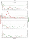
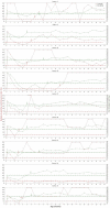
Results: Out of 31 goats, 13 (42%) seroconverted at the age ranging from 3 to 22 months with a median of 5 months. Two goats seroconverted in the second year of life. The other eleven did so before the age of one year; two of these reverted to seronegative status. Only 9 out of 31 goats (29%) seroconverted in the first year of life and remained seropositive. They were early and stable seroreactors to which SRLV was transmitted lactogenically. The age at which they seroconverted ranged from 3 to 10 months with a median of 5 months. In 8 of the 18 persistently seronegative goats, a single isolated positive result occurred. No goats showed any clinical signs of arthritis. The level of maternal antibodies at the age of one week did not differ significantly between the stable seroreactors and the remainder.
Conclusion: Seroconversion appears to occur in less than 50% of goats exposed to heterologous SRLV genotype A via ingestion of colostrum and milk from infected dams and is delayed by 3-10 months. The natural lactogenic route of transmission of SRLV genotype A in goats appears to be less effective than this route of genotype B transmission reported in earlier studies.
Keywords: caprine arthritis-encephalitis; colostral antibodies; humoral immunity; maternal antibodies; seroconversion.
© 2022 J. Kaba et al. published by Sciendo.
Conflict of interest statement
Conflict of Interest Conflict of Interests Statement: The authors declare that there is no conflict of interests regarding the publication of this article.
Figures


Similar articles
-
The herd-level prevalence of caprine arthritis-encephalitis and genetic characteristics of small ruminant lentivirus in the Lithuanian goat population.Prev Vet Med. 2024 Dec;233:106363. doi: 10.1016/j.prevetmed.2024.106363. Epub 2024 Oct 23. Prev Vet Med. 2024. PMID: 39486103
-
Interspecific transmission of small ruminant lentiviruses from goats to sheep.Braz J Microbiol. 2015 Jul 1;46(3):867-74. doi: 10.1590/S1517-838246320140402. eCollection 2015 Jul-Sep. Braz J Microbiol. 2015. PMID: 26413072 Free PMC article.
-
The effect of the subclinical small ruminant lentivirus infection of female goats on the growth of kids.PLoS One. 2020 Mar 24;15(3):e0230617. doi: 10.1371/journal.pone.0230617. eCollection 2020. PLoS One. 2020. PMID: 32208446 Free PMC article.
-
Retroviral infections in sheep and goats: small ruminant lentiviruses and host interaction.Viruses. 2013 Aug 19;5(8):2043-61. doi: 10.3390/v5082043. Viruses. 2013. PMID: 23965529 Free PMC article. Review.
-
Risk factors for transmission and methods for control of caprine arthritis-encephalitis virus infection.Vet Clin North Am Food Anim Pract. 1997 Mar;13(1):35-53. doi: 10.1016/s0749-0720(15)30363-7. Vet Clin North Am Food Anim Pract. 1997. PMID: 9071745 Review.
Cited by
-
A Look Under the Carpet of a Successful Eradication Campaign Against Small Ruminant Lentiviruses.Pathogens. 2025 Jul 20;14(7):719. doi: 10.3390/pathogens14070719. Pathogens. 2025. PMID: 40732765 Free PMC article.
-
Serological testing of an equal-volume milk sample - a new method to estimate the seroprevalence of small ruminant lentivirus infection?BMC Vet Res. 2023 Feb 10;19(1):43. doi: 10.1186/s12917-023-03599-z. BMC Vet Res. 2023. PMID: 36759821 Free PMC article.
-
The Gene Expression Profile of Milk Somatic Cells of Small Ruminant Lentivirus-Seropositive and -Seronegative Dairy Goats (Capra hircus) During Their First Lactation.Viruses. 2025 Jul 3;17(7):944. doi: 10.3390/v17070944. Viruses. 2025. PMID: 40733561 Free PMC article.
-
Milk Transmission of Mammalian Retroviruses.Microorganisms. 2023 Jul 8;11(7):1777. doi: 10.3390/microorganisms11071777. Microorganisms. 2023. PMID: 37512949 Free PMC article. Review.
-
In vivo evaluation of the antiretroviral activity of Melia azedarach against small ruminant lentiviruses in goat colostrum and milk.Braz J Microbiol. 2024 Mar;55(1):875-887. doi: 10.1007/s42770-023-01174-0. Epub 2023 Nov 27. Braz J Microbiol. 2024. PMID: 38010582 Free PMC article.
References
-
- Adams D.S., Klevjer-Anderson P., Carlson J.L., McGuire T.C., Gorham J.R.. Transmission and control of caprine arthritisencephalitis virus. Am J Vet Res. 1983;44:1670–1675. - PubMed
-
- Ahmad A., Morisset R., Thomas R., Menezes J.. Evidence for a defect of antibody-dependent cellular cytotoxic (ADCC) effector function and anti-HIV gp120/41-specific ADCC-mediating antibody titres in HIV-infected individuals. J Acquir Immune Defic Syndr. 1994;7:428–437. - PubMed
-
- Altman D.G., Machin D., Bryant T.N., Gardner M.J. Statistics with Confidence: Confidence Intervals and Statistical Guidelines, Second Edition. BMJ Books; London: 2000.
-
- Alvarez V., Arranz J., Daltabuit-Test M., Leginagoikoa I., Juste R.A., Amorena B., de Andrés D., Luján L.L., Badiola J.J., Berriatua E.. Relative contribution of colostrum from Maedi-Visna virus (MVV) infected ewes to MVV-seroprevalence in lambs. Res Vet Sci. 2005;78:237–243. doi: 10.1016/j.rvsc.2004.09.006.. - DOI - PubMed
LinkOut - more resources
Full Text Sources
