Review
. 2023 Feb 4:38:102122.
doi: 10.1016/j.jcot.2023.102122. eCollection 2023 Mar.

Pediatric spinal injuries- current concepts

Affiliations
Review

Pediatric spinal injuries- current concepts

Guna Pratheep Kalanjiyam et al. J Clin Orthop Trauma. .

Abstract

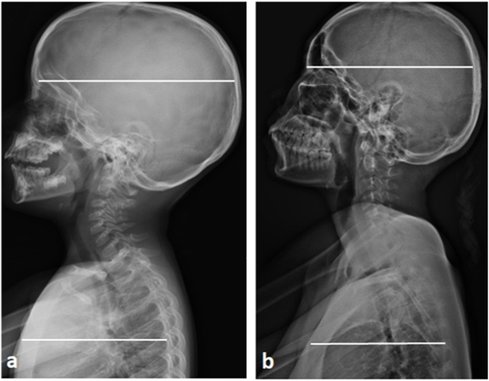
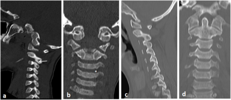
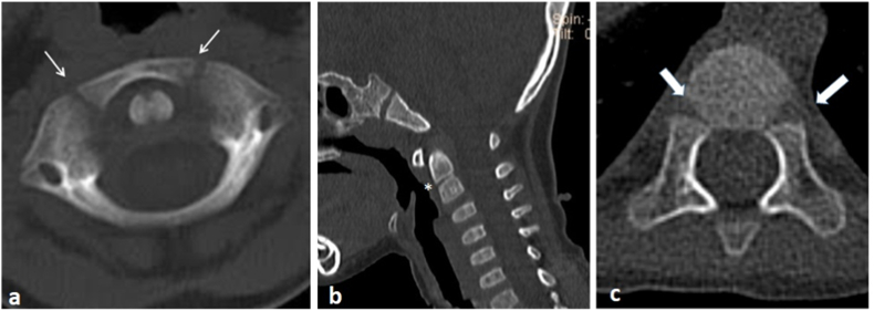
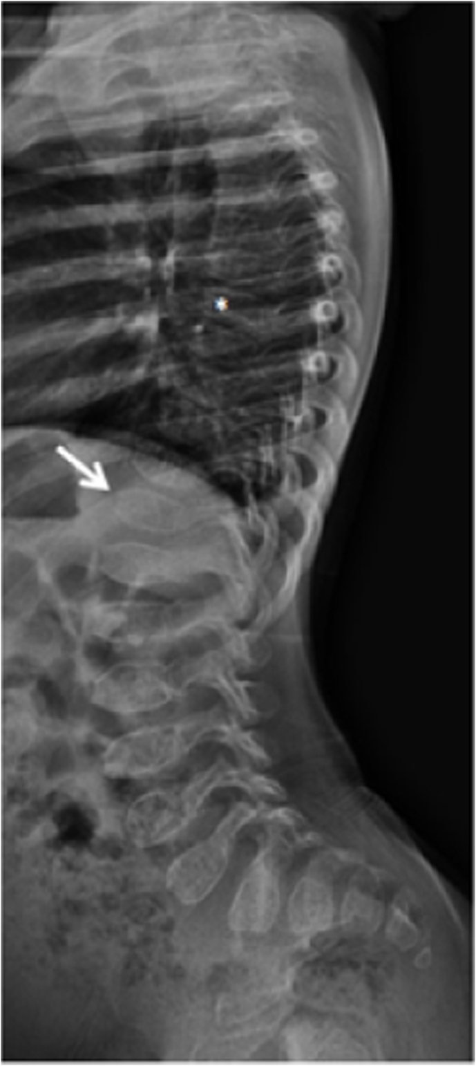
Spinal injuries in children contribute to the highest mortality and morbidity among all pediatric injuries. Fortunately, these injuries are a rare clinical entity but pose a difficulty in diagnosis due to challenges in neurological evaluation of a child and varied radiological presentation. Anatomical and biomechanical aspects of developing musculoskeletal system, relative plasticity of the pediatric spine make children vulnerable to spine injuries. Though motor vehicle collisions are common, children also suffer non-accidental trauma, falls and sports injuries. More chances of cervical spine involvement, higher susceptibility of spinal cord to tensile forces and associated multisystem injuries result in devastating consequences in children compared to adults. Injuries like SCIWORA, vertebral apophyseal injuries, birth-related spinal cord injuries are more specific injuries in pediatric age group. A vigilant clinical, neurological and radiological evaluation is mandatory in all children with suspected spinal injuries. Normal radiological features like ossification centers, pseudosubluxation and physiological vertebral wedging should be carefully noted as they could be misinterpreted as injuries. While CT scans help in better understanding of the fracture pattern, Magnetic Resonance Imaging in children is beneficial especially in detecting SCIWORA and other soft tissue injuries. Management principles of these pediatric spinal injuries are similar to adults. Literature evidences support conservative management in injuries like SCIWORA, unless there is an ongoing spinal cord compression. As in adults, the role of high dose methylprednisolone is still controversial in pediatric spinal cord injuries. Stable spinal injuries can be managed conservatively using orthosis or halo. Instrumentation by both anterior and posterior techniques has been described, but it is challenging due to smaller anatomy and poor implant purchase. In addition to pedicle screw instrumentation, wiring techniques are very beneficial especially in younger children.

Keywords: Limbus fractures; Pediatric spinal injury; Pseudosubluxation; SCIWORA; Surgical fixation.


Conflict of interest statement

All authors declare that there are no conflicts of interests.

Figures

Fig. 1
Fig. 1
Lateral radiograph of cranium with cervical spine and thorax of a 3 year old child (a) and a 17 year old adult (b) showing the difference in their head-trunk ratio between the pediatric and adult population.
Fig. 2
Fig. 2
(a)Mid-sagittal T2W Magnetic Resonance Image of Lumbar spine of a 4 years old child showing thick, well hydrated discs; (b)Lateral lumbo-sacral spine radiograph of the child showing well-maintained disc height.
Fig. 3
Fig. 3
(a-b)Parasagittal CT cervical spine showing shallow facets (white arrow)in a child; (b)Coronal CT cervical spine of the same patient showing under-developed uncus (∗); (c-d)Note the deeper, well articulated facet joints and uncovertebral joint in 38 years old male patient.
Fig. 4
Fig. 4
(a)Axial CT scan image of Atlas showing unfused apophysis of the anterior arch (white arrows); (b)Midsagittal CT scan image of a 5 years old child showing apophysis between dens and body of C2 (∗); (c)Thoracic Axial CT scan image of the same child showing unfused ossification centers between the neural arches and vertebral body (thick white arrow).
Fig. 5
Fig. 5
Lateral cervical radiograph of a 6 year old child showing mild subluxation of C2 over C3 (white arrow); Note the ‘Spinolaminar line’ (line connecting the junction of lamina and spinous process in the cervical spine) is intact (∗) indicating C2–C3 “pseudosubluxation”.
Fig. 6
Fig. 6
Lateral radiograph of dorsal with lumbosacral spine of a 3 year old child with mucopolysaccharidosis showing anterior beaking of vertebral bodies (white arrow). Also, note the flattening of the vertebral body-platyspondyly (∗).
Fig. 7
Fig. 7
(a)Lateral cervical spine radiograph of a 25 year old male showing C5 Burst fracture with increased Pre-vertebral soft tissue shadow (white line) following a trauma due to fall from height; (b) Lateral C- spine radiograph of a 2 year old child showing increase in the pre-vertebral shadow without any injury mechanism. The widening of soft tissue shadow is due to cry of the baby.
Fig. 8
Fig. 8
(a)Antero-posterior radiograph of a 6 years old child showing reduced disc space at T2-T3 level following a motor vehicle collision; (b)Mid-sagittal CT scan image showing dislocation at T2-T3 level with fracture of the spinous process; (c)T2W mid-sagittal MRI scan showing significant spinal cord compression at T2-T3 level.
Fig. 9
Fig. 9
(a–b) Mid-sagittal Computed Tomography and T2W Magnetic Resonance Imaging of a 12 years old child who sustained a road traffic accident. Note contiguous vertebral fractures at T3, T4, T5 and T6 levels.
Fig. 10
Fig. 10
(a–b) Lateral spine radiograph and midsagittal CT scan image of an adolescent male showing avulsion of the inferior endplate of L4 vertebra. Note the irregularities at the posterior aspect of L4 inferior end plate; (c)Midsagittal T2W MRI showing significant spinal canal narrowing and thecal sac compression; (d)Axial MRI image showing endplate defect (white arrow) at L4.
Fig. 11
Fig. 11
(a–b) Antero-posterior and lateral Whole Spine radiographs of a 3 years old child, who presented with history of fall while playing showing no significant abnormality; (c) Computed Tomography of the upper dorsal spine showing no obvious bony injury; (d) T2W MRI showing hyperintense signal changes in the spinal cord at T1-T2 level; (e) Hypointense signal is noted at the same level in T1W imaging indicating a contused spinal cord.
Fig. 12
Fig. 12
(a) Open mouth radiograph in a toddler could not give a clear visualization of the cervical spine; (b) Lateral view of cervical spine shows better details of the vertebrae.
Fig. 13
Fig. 13
(a) Lateral Cervical radiograph of a 14 years old boy showing C5 fracture; (b-d)Sagittal, Coronal and Axial CT scan showing involvement of the posterior vertebral wall indicating burst type fracture pattern; (e)His Axial CT scan image showing C5 lamina fracture (white arrow); (f)Coronal CT scan image showing fracture also involving the left lateral mass of C5.
Fig. 14
Fig. 14
(a)Midsagittal T2W MRI image showing Anterior Atlanto-Occipital membrane (#), Alar ligament (∗), Tectorial membrane (white arrow); (b)Axial T2W MRI showing Transverse Atlantal ligament (white arrow).
Fig. 15
Fig. 15
(a-b)Lateral cervical spine radiograph and mid-sagittal CT scan image of a 2 year old child showing displaced Odontoid fracture following a non-accidental trauma; (c)Coronal CT scan image showing fracture line passing below the level of C1–C2 facet joint; (d-e)Anteroposterior and Lateral Post-operative radiographs showing reduction of the fracture and fixation using Sub-laminar wires.
Fig. 16
Fig. 16
(a)Lateral radiograph of dorsolumbar spine of a 11 year old boy showing L1 Burst fracture; (b)CT scan and T2W MRI showing flexion distraction type injury with retropulsion of fractured vertebral body into the spinal canal; (d-e)Postoperative radiographs following D11-L3 pedicle screw instrumentation and L1 decompression.

Similar articles

Cited by

References

    1. Cirak B., Ziegfeld S., Knight V.M., Chang D., Avellino A.M., Paidas C.N. Spinal injuries in children. J Pediatr Surg. 2004 Apr;39(4):607–612. - PubMed
    1. Jones T.M., Anderson P.A., Noonan K.J. Pediatric cervical spine trauma. J Am Acad Orthop Surg. 2011 Oct;19(10):600–611. - PubMed
    1. Benmelouka A., ShamsEldin L.S., Nourelden A.Z., Negida A. A review on the etiology and management of pediatric traumatic spinal cord injuries. Adv J Emerg Med. 2019 Oct 10;4(2):e28. - PMC - PubMed
    1. Hasler R.M., Exadaktylos A.K., Bouamra O., et al. Epidemiology and predictors of spinal injury in adult major trauma patients: European cohort study. Eur Spine J. 2011 Dec;20(12):2174–2180. - PMC - PubMed
    1. Leonard J.R., Jaffe D.M., Kuppermann N., Olsen C.S., Leonard J.C. Cervical spine injury patterns in children. Pediatrics. 2014 May 1;133(5):e1179–e1188. - PMC - PubMed

LinkOut - more resources