. 2022 Dec 8;3(2):100261.
doi: 10.1016/j.xops.2022.100261. eCollection 2023 Jun.

Macular Telangiectasia Type 2: A Classification System Using MultiModal Imaging MacTel Project Report Number 10

Affiliations

Macular Telangiectasia Type 2: A Classification System Using MultiModal Imaging MacTel Project Report Number 10

Emily Y Chew et al. Ophthalmol Sci. .

Abstract

Purpose: To develop a severity classification for macular telangiectasia type 2 (MacTel) disease using multimodal imaging.

Design: An algorithm was used on data from a prospective natural history study of MacTel for classification development.

Subjects: A total of 1733 participants enrolled in an international natural history study of MacTel.

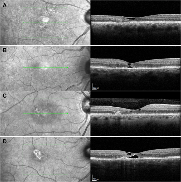
Methods: The Classification and Regression Trees (CART), a predictive nonparametric algorithm used in machine learning, analyzed the features of the multimodal imaging important for the development of a classification, including reading center gradings of the following digital images: stereoscopic color and red-free fundus photographs, fluorescein angiographic images, fundus autofluorescence images, and spectral-domain (SD)-OCT images. Regression models that used least square method created a decision tree using features of the ocular images into different categories of disease severity.

Main outcome measures: The primary target of interest for the algorithm development by CART was the change in best-corrected visual acuity (BCVA) at baseline for the right and left eyes. These analyses using the algorithm were repeated for the BCVA obtained at the last study visit of the natural history study for the right and left eyes.

Results: The CART analyses demonstrated 3 important features from the multimodal imaging for the classification: OCT hyper-reflectivity, pigment, and ellipsoid zone loss. By combining these 3 features (as absent, present, noncentral involvement, and central involvement of the macula), a 7-step scale was created, ranging from excellent to poor visual acuity. At grade 0, 3 features are not present. At the most severe grade, pigment and exudative neovascularization are present. To further validate the classification, using the Generalized Estimating Equation regression models, analyses for the annual relative risk of progression over a period of 5 years for vision loss and for progression along the scale were performed.

Conclusions: This analysis using the data from current imaging modalities in participants followed in the MacTel natural history study informed a classification for MacTel disease severity featuring variables from SD-OCT. This classification is designed to provide better communications to other clinicians, researchers, and patients.

Financial disclosures: Proprietary or commercial disclosure may be found after the references.

Keywords: BCVA, best-corrected visual acuity; BLR, blue light reflectance; CART, Classification and Regression Trees; CF, color fundus; Classification; Classification and Regression Trees (CART); EZ, ellipsoid zone; FAF, fundus autoflorescence; FLIO, fluorescence lifetime imaging ophthalmoscopy; MacTel, macular telangiectasia type 2; Machine learning; Macular telangiectasia type 2; NHOR, natural history observation registry; NHOS, natural history observation study; Neurovascular degeneration; OCTA, OCT angiography; SD-OCT, spectral domain-OCT; VA, visual acuity.


Figures

Figure 1
Figure 1
Color fundus (CF) image analysis (all left eyes), The presence and distribution of a loss of retinal transparency and of brown perivascular pigment were analyzed in CF images using an International Classification (IC) grid centered on the fovea. An IC grid consists of 3 concentric circles of 1000, 3000, and 6000 -μm diameter and 4 spokes dividing the outer circles into superior, nasal, inferior, and temporal subfields (A and B). A loss of retinal transparency (increased scatter) is also detectable in confocal scanning laser ophthalmoscopy images using short wavelength (blue) light (B). (C) demonstrates a loss of retinal transparency (white arrow) in the temporal inner subfield of the grid. (D) shows a loss of retinal transparency in all 4 quadrants of the middle ring of the IC grid, as well as superficial retinal crystals and microvascular anomalies in the temporal subfield. (E) shows a loss of retinal transparency, a few crystals and a small dark pigment associated with 2 converging venules on the boundary of the central and the temporal subfields. (F) demonstrates a bifocal pigmentation (white arrow) with an additional suspected pigmentation in the deeper layers of the retina on the nasal side of the foveal center (thin black arrow). (G) demonstrates a unifocal larger dense dark brown pigment plaque with tissue contraction demonstrated by straight radial and tortuous circumferential vessels. (H–J) demonstrate various stages of multifocal brown pigment that may also occur at the apparent foveal center.
Figure 2
Figure 2
Analysis of fundus fluorescein angiographic (FFA) images (all left eyes). As in color fundus images, FFA images were analyzed using an International Classification (IC) grid. (A) shows an early phase of the angiogram (with only partial filling of the veins). At this phase small vessel anomalies are well visible. (B) shows a late phase angiogram, with the IC grid superimposed over the image. At this phase, the presence and distribution of leakage of the dye on the level of the deep capillary plexus and the retinal pigment epithelium are measured by grid subfield and by type of leakage (focal/diffuse/mixed). The type of leakage was determined based on whether the source of the leakage could be identified just after the transit of the dye through the capillaries. All cases presented in this figure represent focal leakage, although at the late phase all appear diffuse. Leakage may be present without clear visible vascular morphological anomalies, as in (C, D), nasal versus the temporal part of the perifovea. (C, E, G, I) demonstrate increasing involvement of the perifovea in early phase FFA images, whereas (D, F, H, J) show the same structures in late phase angiograms. In (C), slightly dilated capillaries are apparent on the temporal side of the fovea. In (E), dilated capillaries and dilated and blunted venules are apparent. In (G) dilated capillaries and a right-angle vein on the temporal side are evident. In (I) a full circle of involvement of dilated capillaries and corresponding late FFA leakage are demonstrated.
Figure 3
Figure 3
Fundus autofluorescence (FAF) image analysis (right eyes, top row standard FAF images, bottom row magnified central features for better visibility of detail), (A) and (B) show a loss of the normally present central luteal pigment peak, and a faintly increased autofluorescence (AF) temporal to the foveal center. The shadow gram of a blunted, right-angle vein is apparent temporal to the fovea (green arrow in 3B). In (C) and (D), a clear temporal wedge of increased AF (suggestive of loss of luteal pigment) is apparent with a fine right-angle vein (green arrow in [D]). In (E) and (F) the few vessels converge towards foci of decreased AF with straight end branches, against a background of increased AF temporal to the foveal center (green arrow in [F]), perivascular pigment plaque, and loss of luteal pigment.
Figure 4
Figure 4
OCT features. (A) demonstrates a focal discontinuity in the ellipsoid zone (EZ) with a loss of the outer and a disorganization of the inner retinal layers, not involving the foveal center. A dark area just external to the retinal surface represents an inner retinal low-reflective space. (B) demonstrates a discontinuity of the EZ with smaller low reflective spaces in the outer and the inner retina and a slight hyper-reflectivity of the external limiting membrane. (C) shows discontinuity temporal to the fovea not involving the center of fovea, as well as a hyper-reflective lesion in the outer retina. (D) shows outer retinal hyper-reflectivity. OCT hyper-reflectivity is defined as mounds of hyper-reflective material extending internally from the retinal pigment epithelium. They are associated with linear vertical or oblique hyper-reflective streaks. They may or may not be associated with shadowing corresponding to the retinal pigment seen on color fundus or fundus autofluorescence images.
Figure 5
Figure 5
Representative images of grades 3 to 6 in color fundus (left column) and OCT B-scan images (right column). The middle column shows infrared images with green markers indicating the OCT grid placement and the position of the representative OCT B-scan within the scan volume. The top row (A) shows a small patch of noncentral pigment, a wide ellipsoid zone (EZ) break with a small island of photoreceptor at the center and no OCT hyper-reflectivity. The second row (B) demonstrates the presence of OCT hyper-reflectivity within an EZ break involving the foveal center, but no pigment. The third row (C) shows brown pigment also present in the central subfield of the International Classification grid, an extensive EZ break, with no exudative neovascularization. The bottom row (D) shows an active neovascular lesion with small patches of hemorrhage in the color image and retinal thickening, indistinct lesion boundaries, and intraretinal fluid in the OCT B-scan.

References

    1. Heeren T.F., Clemons T., Scholl H.P., et al. Progression of vision loss in macular telangiectasia type 2. Invest Ophthalmol Vis Sci. 2015;56:3905–3912. - PubMed
    1. Charbel Issa P., Gillies M.C., Chew E.Y., et al. Macular telangiectasia type 2. Prog Retin Eye Res. 2013;34:49–77. - PMC - PubMed
    1. Clemons T.E., Gillies M.C., Chew E.Y., et al. The National eye institute visual function questionnaire in the macular telangiectasia (MacTel) Project. Invest Ophthalmol Vis Sci. 2008;49:4340–4346. - PMC - PubMed
    1. Charbel Issa P., Helb H.M., Rohrschneider K., et al. Microperimetric assessment of patients with type 2 idiopathic macular telangiectasia. Invest Ophthalmol Vis Sci. 2007;48:3788–3795. - PubMed
    1. Heeren T.F.C., Chew E.Y., Clemons T., et al. Macular telangiectasia type 2: visual acuity, disease end grade, and the MacTel area: MacTel Project report number 8. Ophthalmology. 2020;127:1539–1548. - PMC - PubMed

LinkOut - more resources