. 2023 Feb 8:11:1130058.
doi: 10.3389/fcell.2023.1130058. eCollection 2023.

The Usher syndrome 1C protein harmonin regulates canonical Wnt signaling

Affiliations

The Usher syndrome 1C protein harmonin regulates canonical Wnt signaling

Jessica Schäfer et al. Front Cell Dev Biol. .

Abstract

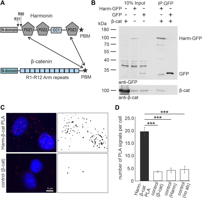
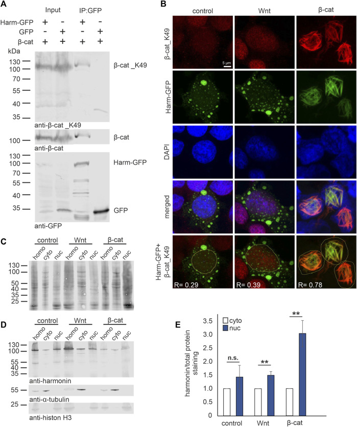
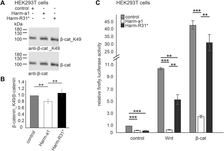
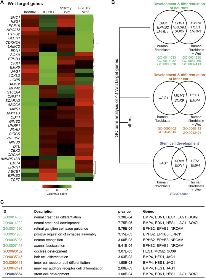
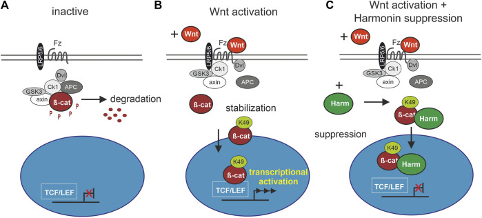
Human Usher syndrome (USH) is the most common form of hereditary combined deaf-blindness. USH is a complex genetic disorder, and the pathomechanisms underlying the disease are far from being understood, especially in the eye and retina. The USH1C gene encodes the scaffold protein harmonin which organizes protein networks due to binary interactions with other proteins, such as all USH proteins. Interestingly, only the retina and inner ear show a disease-related phenotype, although USH1C/harmonin is almost ubiquitously expressed in the human body and upregulated in colorectal cancer. We show that harmonin binds to β-catenin, the key effector of the canonical Wnt (cWnt) signaling pathway. We also demonstrate the interaction of the scaffold protein USH1C/harmonin with the stabilized acetylated β-catenin, especially in nuclei. In HEK293T cells, overexpression of USH1C/harmonin significantly reduced cWnt signaling, but a USH1C-R31* mutated form did not. Concordantly, we observed an increase in cWnt signaling in dermal fibroblasts derived from an USH1C R31*/R80Pfs*69 patient compared with healthy donor cells. RNAseq analysis reveals that both the expression of genes related to the cWnt signaling pathway and cWnt target genes were significantly altered in USH1C patient-derived fibroblasts compared to healthy donor cells. Finally, we show that the altered cWnt signaling was reverted in USH1C patient fibroblast cells by the application of Ataluren, a small molecule suitable to induce translational read-through of nonsense mutations, hereby restoring some USH1C expression. Our results demonstrate a cWnt signaling phenotype in USH establishing USH1C/harmonin as a suppressor of the cWnt/β-catenin pathway.

Keywords: USH1C; Usher syndrome; ataluren/PTC124; colorectal cancer; translational read-through; translational read-through inducing drugs; wnt signaling pathway; β-catenin.


Conflict of interest statement

The authors declare that the research was conducted in the absence of any commercial or financial relationships that could be construed as a potential conflict of interest.

Figures

FIGURE 1
FIGURE 1
In vitro and in situ interaction of harmonin with β-catenin. (A) Schematic representations of the domain structures of the scaffold protein USH1C/harmonin (isoform a) and β-catenin (β-cat). Pathogenic USH1C mutations c.91C>T; p.(R31*) and c.238dupC; p.(R80Pfs*69) are highlighted. Twoheaded arrows indicate that the C terminal PDZ (PDZ; PSD-95, DLG, and ZO-1) The pathogenic nonsense mutations c.91C>T; p.(R31*) and c.238dupC; p.(R80Pfs*69) in USH1C referred to in the Study are highlighted. Binding motif (PBM) of β-catenin is capable to bind to the PDZ1 and PDZ3 of harmonin. (B) Western blot analysis of a representative GFP-Trap® demonstrates interaction between harmonin a1-GFP (Harm-GFP) and β-catenin (β-cat) from lysates of HEK293T cells transfected with β-cat and Harm-GFP, or GFP, respectively. Harm-GFP but not GFP alone pulled down β-catenin; N = 3 experiments. (C) Representative fluorescence image of PLA signals (red, left) in HEK293T cells demonstrates interaction of intrinsic harmonin (Harm) and β-cat (upper panel). Signal is significantly decreased in the control using only β-cat antibody (lower panel). Cell nuclei are marked with DAPI (blue). (D) Statistical analysis reveals an increase in number of signals of 4-fold in the PLA compared to all three controls (Supplementary Figure S1). Two-tailed Student’s t-test, ***p ≤ 0.001; N = 3 experiments.
FIGURE 2
FIGURE 2
Harmonin interaction with acetylated β-catenin_K49 in the nucleus. (A) Western blot analysis of a representative GFP-Trap® pull down shows the interaction between harmonin a1-GFP (Harm-GFP) and acetylated β-catenin_K49 (β-cat_K49). HEK293T cells were transfected with β-cat and Harm-GFP or GFP alone, respectively. Harm-GFP but not GFP alone was able to precipitate β-catenin (β-cat) as well as β-cat_K49; 12% polyacrylamide gel; N = 3 experiments. (B) Immunofluorescence localization of Harm-GFP (green) and β-cat_K49 (red) under different stimulation. HEK293T cells were transfected with Harm-GFP and stimulated for Wnt signaling with Wnt medium (Wnt) or transfection of β-cat. DAPI (blue) marks the nucleus. In unstimulated (control) state (left panel) (Pearson correlation coefficient: Harm-GFP and β-cat_K49: R = 0.29; n = 136 cells; N = 3 experiments) and after Wnt stimulation (middle panel) (Pearson correlation coefficient: Harm-GFP and β-cat_K49: R = 0.39; n = 134 cells; N = 3 experiments), Harm-GFP is distributed in nucleus and cytosol of the cell. The β-cat_K49 staining is almost absent in the unstimulated state, however, there is an increase in anti-β-cat_K49 fluorescence in the nucleus after Wnt stimulation. Additional transfection with β-cat (left panel) results in β-cat_K49 bundles in the nucleus with which Harm-GFP colocalizes. Analysis of Pearson correlation coefficient (lowest panel) reveals strong interaction between Harm-GFP and β-cat_K49 with value of R = 0.78 (n = 169 cells; N = 3 experiments). (C–E) Western blot analysis of cell fractions (30 μg total protein loaded), separated from homogenate (homo) in cytosol (cyto) and nucleus (nuc). HEK293T cells were transfected with Harm-GFP and stimulated for Wnt signaling with Wnt medium or transfection of β-cat. Unstimulated HEK293 cells served as control. (C) Staining of total protein amount was compared to (D) Harm-GFP staining in the fractions; α-Tubulin marks the cytosol fraction and histon H3 the nucleus fraction (15% polyacrylamide gel). Statistical analysis (E) reveals a non-significant increase of 1.4-fold of harmonin in unstimulated cells (control) but a significant increase of 1.5-fold comparing nuclear to cytosolic harmonin (set to 1) in Wnt stimulated cells. Co-transfection with β-cat results in a significant increase of 3-fold of nuclear harmonin. Two-tailed Student’s t-test, **p ≤ 0.01; N = 3 experiments.
FIGURE 3
FIGURE 3
Harmonin overexpression leads to the decrease in Wnt signaling activity (A) Representative Western blot analysis of β-catenin (β-cat) expression in HEK293T cells transfected with harmonin a1-GFP (Harm-a1), truncated harmonin a1_R31*-GFP (Harm-R31*), or GFP (control) and β-catenin (β-cat). (B) Quantification reveals that the expression of full length harmonin a1 significantly decreases β-cat_K49/β-cat ratio (−0.2) while the expression of truncated harmonin a1-R31* did not alter the β-cat_K49/β-cat ratio when compared to control transfected HEK293T cells; 12% polyacrylamide gel. Two-tailed Student’s t-test, **p ≤ 0.01; N = 5 experiments. (C) Wnt response in transfected HEK293T cells measured by TCF/LEF luciferase activity assay. In unstimulated condition (control, left) luciferase activity is significantly decreased in Harm-a1 and Harm-R31* transfected cells compared to empty vector control. Stimulation with Wnt conditioned medium leads to an increase in luciferase activity for all, whereby control as well as Harm-R31* show significantly more cWnt signaling activity than Harm-a1 (Wnt, middle). Addition of β-cat results in the highest Wnt activity and reveals the largest difference of luciferase activity between Harm-a1 to control and Harm-R31* (β-cat, right). cWnt signaling is significantly decreased in Harm-a1 transfected cells. Two-tailed Student’s t-test, **p ≤ 0.01, **p ≤ 0.001; N = 3 experiments (three triplicates each condition).
FIGURE 4
FIGURE 4
RNAseq analysis uncovers differential expression of Wnt target genes in USH1C R31*/R80Pfs*69 patient-derived fibroblasts. (A) Heatmap shows transformed FPKM-values scaled by z-score identifying 40 differentially regulated Wnt target genes in unstimulated and Wnt stimulated healthy (column 1 and 3) and USH1C R31*/R80Pfs*69 patient-derived (USH1C) fibroblasts (column 2 and 4). Red areas correspond to a relative downregulation in gene expression, whereas green areas represent a relative upregulation. (B) GO enrichment analysis showing dysregulated Wnt target genes to be critical for distinct biological processes, such as “development and differentiation of neurons,” “development and differentiation of the inner ear,” as well as “stem cell development.” (C) Detailed description of GO terms summarized in (B).
FIGURE 5
FIGURE 5
RNAseq reveals differential expression of Wnt signaling pathway genes in USH1C R31*/R80Pfs*69 patient-derived fibroblasts. (A) Heatmap showing transformed FPKM-values scaled by z-score identifying 74 differentially regulated Wnt signaling pathway genes in unstimulated and Wnt stimulated healthy (column 1 and 3) and USH1C R31*/R80Pfs*69 patient-derived (USH1C) fibroblasts (column 2 and 4). Red areas correspond to a relative downregulation in gene expression, whereas green areas represent a relative upregulation. (B) Log2 transformed FPKM-values show the expression levels of Wnt pathway genes blotted due to their area of action within the Wnt signaling pathway for unstimulated and Wnt stimulated healthy and USH1CR31*/R80Pfs*69 (USH) fibroblasts, respectively.
FIGURE 6
FIGURE 6
Harmonin leads to a decrease in Wnt signaling activity in patient-derived USH1C fibroblasts baering the patogenic USH1C R31*/R80Pfs*69 mutations (A) Immunofluorescence analysis shows an increase of β-catenin_K49 (β-cat_K49) intensity (green) in Wnt stimulated human healthy fibroblasts. The nucleus is marked with DAPI (blue). Quantification reveals an increase of β-cat_K49 (1.74-fold) after stimulation with Wnt medium. Two-tailed Student’s t-test, **p ≤ 0.01; N = 12 experiments, cell number: Wnt- n = 1,219; Wnt+ n = 1,276. (B) Immunofluorescence analysis shows an increase of β-cat_K49 intensity (green) in Wnt stimulated USH1C R31*/R80Pfs*69 patient derived fibroblasts. The nucleus is marked with DAPI (blue). Quantification reveals an increase of β-cat_lys49 (1.34-fold) after stimulation with Wnt medium. Two-tailed Student’s t-test, *p ≤ 0.05; N = 12 experiments, cell number: Wnt-, n = 1,282; Wnt+, n = 1,140. (C) Unstimulated USH1C R31*/R80Pfs*69 fibroblasts show even higher levels of β-cat_K49 in stimulated healthy cells (unstimulated healthy = 1). Two-tailed Student’s t-test, *p ≤ 0.05, **p ≤ 0.01, ***p ≤ 0.001; N = 12 experiments.
FIGURE 7
FIGURE 7
Treatment with Ataluren rescues cWnt in USH1C/harmonin-deficient cells. (A) Scheme of translational read-through of premature termination codons. Ataluren (blue asterisk) enhances translational read-through of the premature stop codon UGA (red) resulting in expression of full-length harmonin. (B) cWnt response in harmonin a1_R31*-GFP (Harm-R31*) and β-catenin (β-cat) transfected HEK293T cells measured by TCF/LEF luciferase activity. Treatment with 5 μg/mL and 10 μg/mL Ataluren shows significant reduction of cWnt response in Harm-R31* transfected cells. DMSO in all cells at 0.2%. Two-tailed Student’s t-tests, ***p ≤ 0.001; N = 3 experiments (three triplicates each condition). (C) Immunofluorescence analysis reveals significant decrease of β-cat_K49 (green) in Wnt stimulated USH1C (USH1C R31*/R80Pfs*69) patient-derived fibroblasts after treatment with Ataluren (5, 10 μg/mL). The nucleus is marked with DAPI (blue). (D) Levels of β-cat_K49 were decreased by 0.3 (5 μg/mL) and 0.2 (10 μg/mL) in Ataluren treated cells. DMSO in all cells at 0.2%. Two-tailed Student’s t-tests, *p ≤ 0.05; N = 3 experiments, cell number: untreated, n = 246; Ataluren 5 μg/mL, n = 349 cells; 10 μg/mL, n = 277.
FIGURE 8
FIGURE 8
Schematic drawing of involvement of harmonin in cWnt signaling pathway. (A) In the inactive cWnt signaling pathway, absence of Wnt ligands leads to phosphorylation (P) of β-catenin (β-cat) by the destruction complex (grey and white circles) followed by protein degradation. (B) In presence of Wnt ligands, the pathway is activated by binding of Wnt to the receptor Frizzled (Fz), preventing the formation of the destruction complex. β-catenin can then be acetylated at K49 (β-cat_K49) resulting in its stabilization and nuclear translocation where it serves as transcriptional coactivator of TCF/LEF (T cell factor/lymphoid enhancing factor) Wnt target genes. (C) Harmonin (Harm) as negative regulator of cWnt signaling, suppresses the pathway by binding to β-cat_K49. The protein complex can still translocate to the nucleus but further transcription of cWnt target genes is blocked. Fz (Frizzled), LRP5/6 (lipoprotein receptor-related protein), Dvl (Dishevelled), APC (adenomatosis polyposis coli), axin, Ck1 (casein kinase 1α), GSK3 (glycogen synthase kinase 3).

Similar articles

Cited by

References

    1. Adzhubei I. A., Schmidt S., Peshkin L., Ramensky V. E., Gerasimova A., Bork P., et al. (2010). A method and server for predicting damaging missense mutations. Nat. Methods 7, 248–249. 10.1038/nmeth0410-248 - DOI - PMC - PubMed
    1. Angbohang A., Wu N., Charalambous T., Eastlake K., Lei Y., Kim Y. S., et al. (2016). Downregulation of the canonical WNT signaling pathway by TGFβ1 inhibits photoreceptor differentiation of adult human müller glia with stem cell characteristics. Stem Cells Dev. 25, 1–12. 10.1089/scd.2015.0262 - DOI - PMC - PubMed
    1. Archbold H. C., Yang Y. X., Chen L., Cadigan K. M. (2012). How do they do Wnt they do? Regulation of transcription by the wnt/beta-catenin pathway. Acta Physiol. (Oxf) 204, 74–109. 10.1111/j.1748-1716.2011.02293.x - DOI - PubMed
    1. Ashburner M., Ball C. A., Blake J. A., Botstein D., Butler H., Cherry J. M., et al. (2000). Gene ontology: Tool for the unification of biology. The gene ontology Consortium. Nat. Genet. 25, 25–29. 10.1038/75556 - DOI - PMC - PubMed
    1. Bilic J., Huang Y. L., Davidson G., Zimmermann T., Cruciat C. M., Bienz M., et al. (2007). Wnt induces LRP6 signalosomes and promotes dishevelled-dependent LRP6 phosphorylation. Science 316, 1619–1622. 10.1126/science.1137065 - DOI - PubMed

LinkOut - more resources