. 2023 Feb 7;18(3):22.
doi: 10.3892/br.2023.1604. eCollection 2023 Mar.

Potential prognostic and predictive value of UBE2N, IMPDH1, DYNC1LI1 and HRASLS2 in colorectal cancer stool specimens

Affiliations

Potential prognostic and predictive value of UBE2N, IMPDH1, DYNC1LI1 and HRASLS2 in colorectal cancer stool specimens

Yu-Nung Chen et al. Biomed Rep. .

Abstract

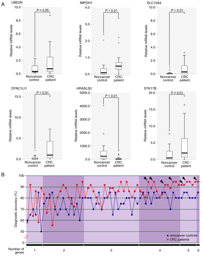
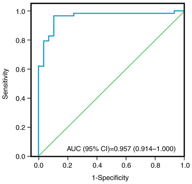
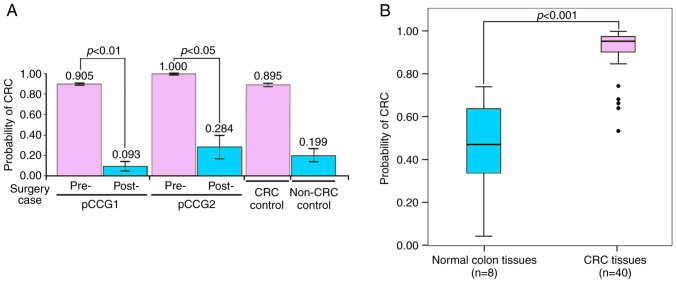
Colorectal cancer (CRC) is the most common gastrointestinal malignancy worldwide. The poor specificity and sensitivity of the fecal occult blood test has prompted the development of CRC-related genetic markers for CRC screening and treatment. Gene expression profiles in stool specimens are effective, sensitive and clinically applicable. Herein, a novel advantage of using cells shed from the colon is presented for cost-effective CRC screening. Molecular panels were generated through a series of leave-one-out cross-validation and discriminant analyses. A logistic regression model following reverse transcription-quantitative polymerase chain reaction (RT-qPCR) and immunohistochemistry was used to validate a specific panel for CRC prediction. The panel, consisting of ubiquitin-conjugating enzyme E2 N (UBE2N), inosine monophosphate dehydrogenase 1 (IMPDH1), dynein cytoplasmic 1 light intermediate chain 1 (DYNC1LI1) and phospholipase A and acyltransferase 2 (HRASLS2), accurately recognized patients with CRC and could thus be further investigated as a potential prognostic and predictive biomarker for CRC. UBE2N, IMPDH1 and DYNC1LI1 expression levels were upregulated and HRASLS2 expression was downregulated in CRC tissues. The predictive power of the panel was 96.6% [95% confidence interval (CI), 88.1-99.6%] sensitivity and 89.7% (95% CI, 72.6-97.8%) specificity at a predicted cut-off value at 0.540, suggesting that this four-gene panel testing of stool specimens can faithfully mirror the state of the colon. On the whole, the present study demonstrates that screening for CRC or cancer detection in stool specimens collected non-invasively does not require the inclusion of an excessive number of genes, and colonic defects can be identified via the detection of an aberrant protein in the mucosa or submucosa.

Keywords: colorectal cancer; dynein cytoplasmic 1 light intermediate chain 1; inosine monophosphate dehydrogenase 1; phospholipase A and acyltransferase 2; prognostic and predictive values; stool specimens; ubiquitin-conjugating enzyme E2 N.


Conflict of interest statement

The authors declare that they have no competing interests.

Figures

Figure 1
Figure 1
Development of gene panels in stool specimens for patients with CRC. (A) Statistical comparison of gene expressions between healthy donor control and CRC patients. Six genes were differentially expressed in healthy donor controls (n=20) and CRC patients (n=36). Differences in the relative levels of the target mRNAs among the samples were determined using the Mann-Whitney U test. P≤0.05 (B) Diagnostic accuracy of 63 different molecular panels. The 63 different molecular panels were comprised of one to six genes. Each panel was used to predict the healthy donor or CRC disease status using LOOCV analysis. The six genes used were UBE2N, IMPDH1, SLC15A4, DYNC1LI1, HRASLS2 and STK17B. Black arrowheads indicate the panels with higher sensitivity (≥90%) and specificity (≥85%). CRC, colorectal cancer; LOOCV, leave-one-out cross-validation; UBE2N, ubiquitin-conjugating enzyme E2 N; IMPDH1, inosine monophosphate dehydrogenase 1; SLC15A4, phospholipase A and acyltransferase 2 solute carrier family 15 member 4; DYNC1LI1, dynein cytoplasmic 1 light intermediate chain 1; HRASLS2, phospholipase A and acyltransferase 2; STK17B, serine/threonine kinase 17b.
Figure 2
Figure 2
COC1021 colon cancer tissue array. A total number of 102 available cores was enrolled (102 cores: x-axis, nos. 1 to 13; y-axis, a to h). N, non-CRC colonic tissue; CM, congenital megacolon; Ad, colon adenoma; I, AJCC stage I; II, AJCC stage II; III, AJCC stage III. Blue area, papillary adenocarcinoma; green area, mucinous adenocarcinoma; pink area, colon adenocarcinoma. CRC, colorectal cancer; AJCC, American Joint Committee on Cancer.
Figure 3
Figure 3
Immunohistochemical staining for the expression of HRASLS2 protein in the CRC tissue array. (A) Overview of a tissue array with the HRASLS2 immunoactivity. (B) Representative images of immunohistochemical staining for HRASLS2 protein. Protein signal densities of three CRC tissues (c12 and e7 at AJCC stage I; g9 at AJCC stage III) are shown as immunoreactive cells per mm2 and displayed in parentheses for the CRC fraction (T) and the adjacent non-CRC fraction (N). There were 102 cores (x-axis, nos. 1 to 13; y-axis, a to h). The scale bar corresponds to 500 µm. HRASLS2, phospholipase A and acyltransferase 2; CRC, colorectal cancer.
Figure 4
Figure 4
Immunohistochemical staining for the expression of UBE2N protein in the CRC tissue array. (A) Overview of a tissue array with the UBE2N immunoactivity. (B) Representative images of immunohistochemical staining for UBE2N protein. Protein signal densities of three CRC tissues (c12 and e7 at AJCC stage I; g9 at AJCC stage III) are shown as immunoreactive cells per mm2 and displayed in parentheses for the CRC fraction (T) and the adjacent non-CRC fraction (N). There were 102 cores (x-axis, nos. 1 to 13; y-axis, a to h). The scale bar corresponds to 500 µm. UBE2N, ubiquitin-conjugating enzyme E2 N; CRC, colorectal cancer; AJCC, American Joint Committee on Cancer.
Figure 5
Figure 5
Immunohistochemical staining for the expression of IMPDH1 protein in the CRC tissue array. (A) Overview of a tissue array with the IMPDH1 immunoactivity. (B) Representative images of immunohistochemical staining for IMPDH1 protein. Protein signal densities of three CRC tissues (c12 and e7 at AJCC stage I; g9 at AJCC stage III) are shown as immunoreactive cells per mm2 and displayed in parentheses for the CRC fraction (T) and the adjacent non-CRC fraction (N). There were 102 cores (x-axis, nos. 1 to 13; y-axis, a to h). The scale bar corresponds to 500 µm. IMPDH1, inosine monophosphate dehydrogenase 1; CRC, colorectal cancer; AJCC, American Joint Committee on Cancer.
Figure 6
Figure 6
Immunohistochemical staining for the expression of DYNC1LI1 protein in the CRC tissue array. (A) Overview of a tissue array with the DYNC1LI1 immunoactivity. (B) Representative images of immunohistochemical staining for DYNC1LI1 protein. Protein signal densities of three CRC tissues (c12 and e7 at AJCC stage I; g9 at AJCC stage III) are shown as immunoreactive cells per mm2 and displayed in parentheses for the CRC fraction (T) and the adjacent non-CRC fraction (N). There were 102 cores (x-axis, nos. 1 to 13; y-axis, a to h). The scale bar corresponds to 500 µm. DYNC1LI1, dynein cytoplasmic 1 light intermediate chain 1; CRC, colorectal cancer; AJCC, American Joint Committee on Cancer.
Figure 7
Figure 7
The analysis of the receiving operating characteristics curve demonstrating the ability of the predictive model (UBE2N, IMPDH1, DYNC1LI1 and HRASLS2) to discriminate the CRC group. UBE2N, ubiquitin-conjugating enzyme E2 N; IMPDH1, inosine monophosphate dehydrogenase 1; DYNC1LI1, dynein cytoplasmic 1 light intermediate chain 1; STK17B, serine/threonine kinase 17b; HRASLS2, phospholipase A and acyltransferase 2; CRC, colorectal cancer.
Figure 8
Figure 8
Model validity of patients with CRC and non-CRC controls. (A) Probability of CRC from pre- and postsurgical stool specimens. Data are presented as the mean ± standard error of the mean (n=2). (B) Boxplot showing the probability of CRC cDNA array. HCRT104 cDNA array (OriGene Technologies, Inc.) of 48 colon tissues was used. Data were presented as the mean ± standard error of the mean of non-CRC status (n=8) and four CRC stages (n=40). Black spot, outliers. CRC, colorectal cancer.

Similar articles

Cited by

References

    1. Taniguchi H, Moriya C, Igarashi H, Saitoh A, Yamamoto H, Adachi Y, Imai K. Cancer stem cells in human gastrointestinal cancer. Cancer Sci. 2016;107:1556–1562. doi: 10.1111/cas.13069. - DOI - PMC - PubMed
    1. Miyake M, Takemasa I, Matoba R, Tanino M, Niijima S, Ikeda M, Yamamoto H, Sekimoto M, Kuhara S, Okayama T, et al. Heterogeneity of colorectal cancers and extraction of discriminator gene signatures for personalized prediction of prognosis. Int J Oncol. 2011;39:781–789. doi: 10.3892/ijo.2011.1092. - DOI - PubMed
    1. Pitroda SP, Weichselbaum RR. Integrated molecular and clinical staging defines the spectrum of metastatic cancer. Nat Rev Clin Oncol. 2019;16:581–588. doi: 10.1038/s41571-019-0220-6. - DOI - PubMed
    1. Wang S, Guan X, Ma M, Zhuang M, Ma T, Liu Z, Chen H, Jiang Z, Chen Y, Wang G, Wang X. Reconsidering the prognostic significance of tumour deposit count in the TNM staging system for colorectal cancer. Sci Rep. 2020;10(89) doi: 10.1038/s41598-019-57041-2. - DOI - PMC - PubMed
    1. Das V, Kalita J, Pal M. Predictive and prognostic biomarkers in colorectal cancer: A systematic review of recent advances and challenges. Biomed Pharmacother. 2017;87:8–19. doi: 10.1016/j.biopha.2016.12.064. - DOI - PubMed