Review
. 2023 Feb 9:11:1036715.
doi: 10.3389/fchem.2023.1036715. eCollection 2023.

Recent advances in lanthanide-doped up-conversion probes for theranostics

Affiliations
Review

Recent advances in lanthanide-doped up-conversion probes for theranostics

Danyang Xu et al. Front Chem. .

Abstract

Up-conversion (or anti-Stokes) luminescence refers to the phenomenon whereby materials emit high energy, short-wavelength light upon excitation at longer wavelengths. Lanthanide-doped up-conversion nanoparticles (Ln-UCNPs) are widely used in biomedicine due to their excellent physical and chemical properties such as high penetration depth, low damage threshold and light conversion ability. Here, the latest developments in the synthesis and application of Ln-UCNPs are reviewed. First, methods used to synthesize Ln-UCNPs are introduced, and four strategies for enhancing up-conversion luminescence are analyzed, followed by an overview of the applications in phototherapy, bioimaging and biosensing. Finally, the challenges and future prospects of Ln-UCNPs are summarized.

Keywords: biomedicine; lanthanide-doped; probes for theranostics; up-conversion; up-conversion luminescence.


Conflict of interest statement

The authors declare that the research was conducted in the absence of any commercial or financial relationships that could be construed as a potential conflict of interest.

Figures

FIGURE 1
FIGURE 1
Mixing and high-temperature heating steps are important processes in the controlled thermal coprecipitation synthesis of sub-5-nm Na(Gd−Yb)F4:Tm. [Reprinted with permission from Ref. (Amouroux et al., 2019) Copyright 2019: American Chemical Society].
FIGURE 2
FIGURE 2
(A) Simulation of the electric field strength (|E|/|E0|) of Ln-UCNPs@mSiO2-Au NPs (15 nm–D–5 nm geometry) under irradiation at λexc = λSPR and λexc = 800 nm, with different silica spacers with thickness D of 5, 15, and 30 nm |E|/|E0| is the enhancement factor. The electric field is amplified as |E| > |E0|. [Reprinted with permission from Ref. (Lv et al., 2018a) Copyright 2018: Lv et al. (2018a)]. (B) DDSCAT simulation results. Extinction spectra of the corresponding electronic strength images of SPS@Au. [Reprinted with permission from Ref. (Lin et al., 2020) Copyright 2020: American Chemical Society].
FIGURE 3
FIGURE 3
Comparison of SA, HSA, PSO, and GA algorithms to the final average and maximum luminescence intensity. Variance of (A) all concentrations and (B) the Ce/Tb concentrations in different generations. [Reprinted with permission from Ref. (Lv et al., 2019) Copyright 2019: American Chemical Society].
FIGURE 4
FIGURE 4
(A) Schematic illustration of the alleviation of Aggregation-Caused Quenching (ACQ) and promotion of dye sensitization in aqueous phase by coating with DSPE-PEG. (B) Schematic illustration of improving dye-sensitization performance through eliminating ACQ of dye molecules. (C) Schematic illustration of improving dye sensitization performance through alleviating EBT from Nd3+ to dyes. [Reprinted with permission from Ref. (Liang et al., 2020a) Copyright 2020: John Wiley & Sons, Inc.].
FIGURE 5
FIGURE 5
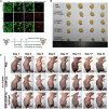
(A) In vitro PTT confocal images of ECA109 cells treated with PBS and BiNS@NaLnF4, with or without NIR irradiation. [Reprinted with permission from Ref. (Ma et al., 2021a) Copyright 2021: Royal Society of Chemistry]. (B) Images of tumor after treatment. [Reprinted with permission from Ref. (Wang et al., 2020) Copyright 2020: Royal Society of Chemistry]. (C) Photographs of female BALB/c nude mice bearing T24 tumors when treated with DNA–Ln-UCNPs-Au hydrogel, Ln-UCNPs-Au, and PBS samples over a period of 21 days after NIR irradiation in 3 min (1 W cm−2); s.c., subcutaneous injection. [Reprinted with permission from Ref. (Liu et al., 2020) Copyright 2020: John Wiley & Sons, Inc.].
FIGURE 6
FIGURE 6
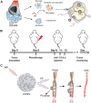
(A) Transmission electron microscope (TEM) images of Ln-UCNPs (a), Ln-UCNPs@PFNS (b), Ln-UCNPs@PFNS@MnCaP (c) and (Ln-UCNPs@PFNS/AQ4N) @MnCaP (d). (B) Schematic of the treatment of mice with intravenously injected (Ln-UCNPs@PFNS/AQ4N)@MnCaP and treatment by illumination. (C) The tumor growth curves for different treatments. Error bars indicate SD (n = 6). [Reprinted with permission from Ref. (Ji et al., 2019) Copyright 2019: Elsevier].
FIGURE 7
FIGURE 7
(A) Scheme of synergistic phototherapy for augmentation of antitumor immunity. Upon laser irradiation, nanoparticles ablate the primary tumor through phototherapy. (Reprinted with permission from Ref. (Yan et al., 2019) Copyright 2019: John Wiley & Sons, Inc.). (B) Schematic depiction of the experimental approach for the evaluation of the abscopal effects induced by Ln-UCNPs/ICG/RB-mal based phototherapy. (Reprinted with permission from Ref. (Wang et al., 2019a) Copyright 2019: John Wiley & Sons, Inc.). (C) Schematic of the design of a photoactivatable immunodevice through the integration of Ln-UCNPs with the UV light-responsive photoactivatable CpG (PCpG). Ln-UCNPs act as transducer to upconvert NIR light into UV light locally, thus liberate CpG oligonucleotides (ODN) from PCpG to achieve refined temporal control on its immunoactivity. [Reprinted with permission from Ref. (Chu et al., 2019) Copyright 2019: Nature Publishing Group].
FIGURE 8
FIGURE 8
Positive contrast enhancement evaluation in vivo. (A, B) T1-weighted MRI and corresponding pseudocolor images of tumor-bearing mice after intravenous injection of cell membrane coated-BSNPs (MSNPs) (A) and Magnevist (B) with the same dosage (2.5 μmol of Gd3+ for each mouse). Images were captured before and at different time points after the administration of contrast agents. The time points were collected at the midpoint of the time interval during each imaging acquisition. The dotted circles represent the regions of interest: 1) tumor, 2) muscle, 3) background, and 4) bladder. Scale bars are 5 mm for all images. The small spots on the corners are from the circulation apparatus in the MRI scanner. (C, D) Tumor-to-background (T/B) and tumor-to-muscle (T/M) contrast ratios based on the corresponding MRI images. Values represented as means ± s.d. (n = 3). [Reprinted with permission from Ref. (Yi et al., 2019) Copyright 2019: John Wiley & Sons, Inc.].
FIGURE 9
FIGURE 9
(A) Simple self-referenced luminescent pH sensors based on up-conversion nanocrystals and pH-sensitive fluorescent BODIPY dyes. (B) Time-dependent changes of the G/R ratio of the pH sensor layers treated with a suspension of E. coli with D-glucose and a suspension of E. coli without D-glucose or pure buffer. [Reprinted with permission from Ref. (Radunz et al., 2019) Copyright 2019: American Chemical Society].
FIGURE 10
FIGURE 10
A schematic diagram showing the CFC-Ln-UCNPs probe for Cu2+ ion sensing with electrochemical assistance. [Reprinted with permission from Ref. (Wong et al., 2019) Copyright 2019: Royal Society of Chemistry].
FIGURE 11
FIGURE 11
Illustration of the Ln-UCNPs@PDA@AP application for sensing intracellular Cyt c. The illustration is not drawn to scale. [Reprinted with permission from Ref. (Ma et al., 2017) Copyright 2017: Elsevier].

Similar articles

Cited by

References

    1. Abbaspour N., Hurrell R., Kelishadi R. (2014). Review on iron and its importance for human health. J. Res. Med. Sci. 19 (2), 164–174. - PMC - PubMed
    1. Abualrejal M. M. A., Eid K., Tian R., Liu L., Chen H., Abdullah A. M., et al. (2019). Rational synthesis of three-dimensional core-double shell upconversion nanodendrites with ultrabright luminescence for bioimaging application. Chem. Sci. 10 (32), 7591–7599. 10.1039/c9sc01586h - DOI - PMC - PubMed
    1. Achatz D. E., Meier R. J., Fischer L. H., Wolfbeis O. S. (2011). Luminescent sensing of oxygen using a quenchable probe and upconverting nanoparticles. Angew. Chemie-International Ed. 50 (1), 274–277. 10.1002/ange.201004902 - DOI - PubMed
    1. Adachi S. (2018). Photoluminescence properties of Mn4+-activated oxide phosphors for use in white-LED applications: A review. J. Luminescence 202, 263–281. 10.1016/j.jlumin.2018.05.053 - DOI
    1. Ali R., Saleh S. M., Meier R. J., Azab H. A., Abdelgawad I. I., Wolfbeis O. S. (2010). Upconverting nanoparticle based optical sensor for carbon dioxide. Sensors Actuators B-Chemical 150 (1), 126–131. 10.1016/j.snb.2010.07.031 - DOI

LinkOut - more resources