. 2023 May;165(4):563-586.
doi: 10.1111/jnc.15796. Epub 2023 Mar 21.

Identification of phosphorylated tau protein interactors in progressive supranuclear palsy (PSP) reveals networks involved in protein degradation, stress response, cytoskeletal dynamics, metabolic processes, and neurotransmission

Affiliations

Identification of phosphorylated tau protein interactors in progressive supranuclear palsy (PSP) reveals networks involved in protein degradation, stress response, cytoskeletal dynamics, metabolic processes, and neurotransmission

Rowan A W Radford et al. J Neurochem. 2023 May.

Abstract

Progressive supranuclear palsy (PSP) is a late-onset neurodegenerative disease defined pathologically by the presence of insoluble phosphorylated-Tau (p-Tau) in neurons and glia. Identifying co-aggregating proteins within p-Tau inclusions may reveal important insights into processes affected by the aggregation of Tau. We used a proteomic approach, which combines antibody-mediated biotinylation and mass spectrometry (MS) to identify proteins proximal to p-Tau in PSP. Using this proof-of-concept workflow for identifying interacting proteins of interest, we characterized proteins proximal to p-Tau in PSP cases, identifying >84% of previously identified interaction partners of Tau and known modifiers of Tau aggregation, while 19 novel proteins not previously found associated with Tau were identified. Furthermore, our data also identified confidently assigned phosphorylation sites that have been previously reported on p-Tau. Additionally, using ingenuity pathway analysis (IPA) and human RNA-seq datasets, we identified proteins previously associated with neurological disorders and pathways involved in protein degradation, stress responses, cytoskeletal dynamics, metabolism, and neurotransmission. Together, our study demonstrates the utility of biotinylation by antibody recognition (BAR) approach to answer a fundamental question to rapidly identify proteins in proximity to p-Tau from post-mortem tissue. The application of this workflow opens up the opportunity to identify novel protein targets to give us insight into the biological process at the onset and progression of tauopathies.

Keywords: Biotinylation; Tauopathy; mass spectrometry; neuropathology; progressive supranuclear palsy; tau.


Conflict of interest statement

The authors declare no conflict of interest.

Figures
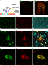
FIGURE 1
FIGURE 1
Targeted biotinylation of p‐Tau aggregates in post‐mortem tissue. (a) Process of biotinylation. Primary antibody recognizing p‐Tau was used to target the pathological aggregates of Tau in fixed PSP patient tissue. A secondary antibody conjugated to poly‐HRP was used to facilitate the biotinylation of proteins in proximity to the antibody complex. (b) Biotinylation is negligible when no primary antibody is used to target biotinylation. (c) Biotinylation occurs near Tau aggregates when a primary antibody is applied. Scale bar in (b) and (c) is 1000 μm. (d–f) Biotinylation profile is similar to the phospho‐Tau deposition in the post‐mortem tissue of patients. Scale bar = 100 μm. (g–i) The biotinylation profile is proximal to p‐Tau aggregates in glia with tufted astrocytes and coiled bodies in oligodendrocytes. The scale bar is 10 μm. (j–l) The close proximity of biotinylation in relation to p‐Tau neuronal pathology. Scale bar = 5 μm. This was conducted using tissue available from 4 patients with similar results in each case.
FIGURE 2
FIGURE 2
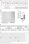
BAR leads to biotinylation of proteins in proximity to p‐Tau. (a) Workflow used to identify Tau interactome in patient tissue. (b) Biotinylation pattern of lysates derived from labeled tissue. N = 4 where “N” refers to the number of patients. (c) Densitometry of biotinylated profiles in Tau‐targeted samples and negative controls. Student's t‐test was conducted on paired samples. Actual p‐value = 0.0442, t‐value = 2.539, degrees of freedom = 6) (*p < 0.05). N = 4 where “N” refers to the number of patients. (d) Identification of peptides belonging to Tau. (e) Confidently identified phosphopeptides in PSP patients. Residues highlighted in blue represent the phosphorylation sites recognized by the p‐Tau antibody used. Residues in black boxes refer to phosphorylation sites that were unambiguously assigned. Residues in gray boxes (and with an * above) refer to phosphorylation sites that were assigned to one of two closely positioned amino acids. PSMs – peptide spectrum matches. A 0.01 or 100‐fold abundance respectively indicates the absence or presence of the proximal protein.
FIGURE 3
FIGURE 3
Proteins in proximity to Tau are enriched for known Tau interactions and proteins linked to neurological dysfunction. Groups of at least 10 proteins found in proximity to p‐Tau (ratio p‐Tau/control ≥2‐fold) that are known to be associated with neurological disorders – Frontotemporal degeneration spectrum disorder, Parkinson's disease, parkinsonism, Alzheimer disease, Huntington Disease, and disorder of the basal ganglia in red or neurological signs associated with PSP such as dyskinesia and cognitive impairment highlighted in gray. How Tau is related to these neurological dysfunctions and disorders is highlighted in green, while previously unreported proteins identified in close proximity to p‐Tau from Figure 4a are highlighted in blue. Results were obtained from proteomic studies using tissue from 4 different patients.
FIGURE 4
FIGURE 4
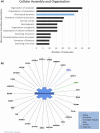
Grouping of proteins associated with Cellular Assembly and Organization (a) Groups of at least 10 proteins found in proximity to p‐Tau (ratio p‐Tau/control ≥2‐fold) associated with Cellular Assembly and Organization subfunctions, highlighted in blue is further broken down in (b). (b) Individual proteins in proximity to p‐Tau (ratio p‐Tau/control ≥2‐fold) were identified in a network associated with Microtubule dynamics. MAPT is highlighted in green, while a previously unreported protein identified proximal to p‐Tau from Figure 2a is highlighted in blue. Results were obtained from proteomic studies using tissue from 4 different patients.
FIGURE 5
FIGURE 5
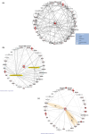
Protein networks associated with Tau. (a) Overlay of canonical pathways, “Protein Ubiquitination Pathway” and “Unfolded protein response.” (b) Proteins in proximity to p‐Tau in PSP brain tissue associated with the network: “Hereditary Disorder, Organismal Injury and Abnormalities, Skeletal and Muscular Disorders.” Increasing shades of red indicate a higher fold‐change ratio compared to the control. Double circles represent protein complexes. (c) Known protein–protein interactions within the network. Darker lines represent protein–protein interactions with Tau. Yellow highlights represent proteins with other relationships with Tau. BR, binding regulator; L, localization; U, ubiquitination, and I, inhibition. Results were obtained from proteomic studies using tissue from 4 different patients.
FIGURE 6
FIGURE 6
Brain enhanced expression and subcellular location of proximal proteins associated with p‐Tau in PSP. (a) of the 117 proteins associated with p‐Tau in PSP, 28.2% exhibited enhanced brain RNA expression shown in red. Proteins with tissue enhanced RNA expression in tissues other than the brain is green, while proteins with no enhanced RNA expression are shown in gray. (b) depicts subcellular locations of proteins associated with p‐Tau in PSP. The number of proteins found in each subcellular location is indicated with (=N). Results were obtained from proteomic studies using tissue from 4 different patients.
FIGURE 7
FIGURE 7
Predicted cell‐type enrichment of proteins found in proximity to p‐Tau (ratio p‐Tau/control ≥2‐fold) based on standardized human RNA‐seq datasets. (a) and (b) were constructed using an RNA‐seq dataset with cells sampled from the human temporal cortex (Zhang et al., 2016). (a) hierarchical cluster analysis and heatmap representation for transcriptional expression of proteins found proximal to p‐Tau in neurons, astrocytes, oligodendrocytes, oligodendrocyte precursor cells (OPCs), microglia, and endothelial cells. Z‐scores above 1.5 with a predicted enrichment are shown in green‐yellow. (b) principal component analysis (PCA) shows that the predicted expression variance of neurons and astrocytes is primarily explained in the first and second principal components. (c) and (d) were constructed using an RNA‐seq dataset with cells sampled from the human motor cortex (Bakken, Jorstad et al. 2020). (c) hierarchical cluster analysis and heatmap representation for transcriptional expression of proteins found proximal to p‐Tau in neurons, astrocytes, oligodendrocytes, oligodendrocyte precursor cells (OPCs), microglia, endothelial and vascular and leptomeningeal cells (VLMC). Z‐scores above 1.5 with a predicted enrichment are shown in green‐yellow. (d) principal component analysis (PCA) shows the predicted expression variance in the first and second dimensions are primarily explained by neurons and oligodendrocytes indicated by cos2. Results were obtained from proteomic studies using tissue from 4 different patients.
FIGURE 8
FIGURE 8
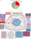
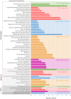
Selection of statistically significant canonical pathways associated with proteins found in proximity to p‐Tau (ratio p‐Tau/control ≥2‐fold). Pathways highlighted in green represent pathways associated with protein degradation systems, while those highlighted in red, blue, orange, and purple are associated with stress responses, cytoskeletal dynamics, metabolic processes, and neurotransmission. Particular pathways predicted to be enriched in neurons, astrocytes (Astro), and oligodendrocytes (o) are also displayed. Results were obtained from proteomic studies using tissue from 4 different patients.

Similar articles

Cited by

References

    1. Ahmed, Z. , Asi, Y. T. , Lees, A. J. , Revesz, T. , & Holton, J. L. (2013). Identification and quantification of oligodendrocyte precursor cells in multiple system atrophy, progressive supranuclear palsy and parkinson's disease. Brain Pathology, 23(3), 263–273. 10.1111/j.1750-3639.2012.00637.x - DOI - PMC - PubMed
    1. Allen, M. , Wang, X. , Serie, D. J. , Strickland, S. L. , Burgess, J. D. , Koga, S. , Younkin, C. S. , Nguyen, T. T. , Malphrus, K. G. , Lincoln, S. J. , Alamprese, M. , Zhu, K. , Chang, R. , Carrasquillo, M. M. , Kouri, N. , Murray, M. E. , Reddy, J. S. , Funk, C. , Price, N. D. , … Ertekin‐Taner, N. (2018). Divergent brain gene expression patterns associate with distinct cell‐specific tau neuropathology traits in progressive supranuclear palsy. Acta Neuropathologica, 136(5), 709–727. 10.1007/s00401-018-1900-5 - DOI - PMC - PubMed
    1. Alonso, A. D. C. , Grundke‐Iqbal, I. , Barra, H. S. , & Iqbal, K. (1997). Abnormal phosphorylation of tau and the mechanism of Alzheimer neurofibrillary degeneration: Sequestration of microtubule‐associated proteins 1 and 2 and the disassembly of microtubules by the abnormal tau. Proceedings of the National Academy of Sciences, 94(1), 298–303. 10.1073/pnas.94.1.298 - DOI - PMC - PubMed
    1. Ando, K. , Ndjim, M. , Turbant, S. , Fontaine, G. , Pregoni, G. , Dauphinot, L. , Yilmaz, Z. , Suain, V. , Mansour, S. , Authelet, M. , De Dekker, R. , Leroy, K. , Delatour, B. , Letournel, F. , Martin‐Négrier, M.‐L. , Chapon, F. , Godfraind, C. , Maurage, C.‐A. , Deramecourt, V. , … Brain Bank Neuro, C. E. B. N. N . (2020). The lipid phosphatase Synaptojanin 1 undergoes a significant alteration in expression and solubility and is associated with brain lesions in Alzheimer's disease. Acta Neuropathologica Communications, 8(1), 79. 10.1186/s40478-020-00954-1 - DOI - PMC - PubMed
    1. Arakhamia, T. , Lee, C. E. , Carlomagno, Y. , Duong, D. M. , Kundinger, S. R. , Wang, K. , Williams, D. , DeTure, M. , Dickson, D. W. , Cook, C. N. , Seyfried, N. T. , Petrucelli, L. , & Fitzpatrick, A. W. P. (2020). Posttranslational Modifications Mediate the Structural Diversity of Tauopathy Strains. Cell, 180(4), 633–644.e612. 10.1016/j.cell.2020.01.027 - DOI - PMC - PubMed

Publication types