Review
. 2023 Feb 24;53(1):44-57.
doi: 10.4274/tjo.galenos.2022.76436.

Avascular Peripheral Retina in Infants

Affiliations
Review

Avascular Peripheral Retina in Infants

Şengül Özdek et al. Turk J Ophthalmol. .

Abstract

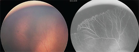
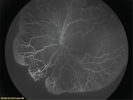
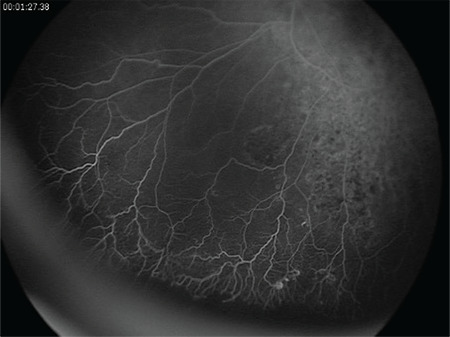
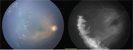
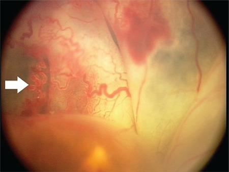
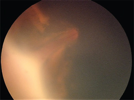
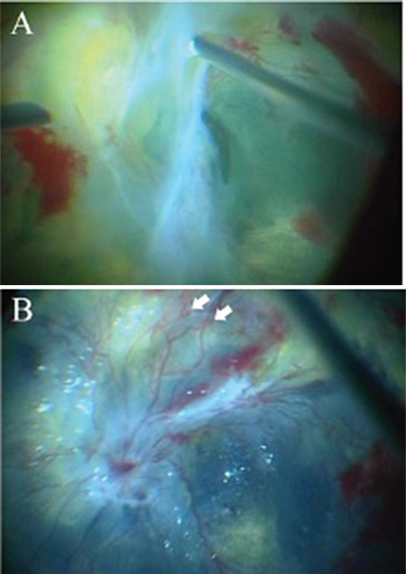
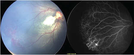
Avascular peripheral retina in an infant is a common characteristic of numerous pediatric retinal vascular disorders and often presents a diagnostic challenge to the clinician. In this review, key features of each disease in the differential diagnosis, from retinopathy of prematurity, familial exudative vitreoretinopathy, Coats disease, incontinentia pigmenti, Norrie disease, and persistent fetal vasculature, to other rare hematologic conditions and telomere disorders, will be discussed by expert ophthalmologists in the field.

Keywords: Avascular retina; Coats disease; Norrie disease; familial exudative vitreoretinopathy; incontinentia pigmenti; persistent fetal vasculature; retinopathy of prematurity.


Conflict of interest statement

Conflict of Interest: No conflict of interest was declared by the authors.

Figures

Figure 1
Figure 1
Color fundus image and corresponding fluorescein angiography of an avascular peripheral retina at 35 weeks of postmenstrual age in a preterm infant born at 27 weeks of gestational age with a birth weight of 890 grams (Courtesy Dr. Domenico Lepore)
Figure 2
Figure 2
Fluorescein angiography showing an avascular peripheral retina in a 2-year-old child born preterm (25 weeks of gestational age, birth weight 650 g) and treated with 0.2 mg/0.02 mL of ranibizumab at 34 weeks of postmenstrual age for type 1 retinopathy of prematurity (Courtesy of Dr. Domenico Lepore)
Figure 3
Figure 3
Fluorescein angiography of an avascular peripheral retina in a 2-year-old child who was born preterm (28 weeks of gestational age, birth weight 980 g) and had fully regressed stage 2 zone 2 retinopathy of prematurity diagnosed at 32 weeks (Courtesy of Dr. Domenico Lepore)
Figure 4
Figure 4
Avascular peripheral retina with neovascularization (NV) and typical posterior hyaloid yellow deposits in a term neonate with familial exudative vitreoretinopathy. Corresponding fluorescein angiography shows straightening of the vessels towards the periphery, avascular peripheral retina, and severe leakage from the NV at the vascular-avascular retinal junction (Courtesy of Dr. Şengül Özdek)
Figure 5
Figure 5
Fundus photo of a female patient aged 20 years. Peripheral avascular retina in familial exudative vitreoretinopathy never shows forward advancement during the lifetime, has fine but adherent vitreous condensation at the edge, and often the temporal raphe is involved more posteriorly, like a ‘notch’. Avascularity is present in all quadrants but is more prominent temporally. There are no ghost vessels (Courtesy of Dr. Subhadra Jalali)
Figure 6
Figure 6
Yellowish pupillary reflex in Coats’ disease. A bulbed-shaped retinal telangiectasia (white arrow) overlying the bullous retinal detachment suggests a diagnosis of Coats’ disease rather than retinoblastoma (Courtesy of Dr. Shunji Kusaka)
Figure 7
Figure 7
A case of retinopathy of prematurity (ROP) with exudative changes. A falciform retinal detachment with subretinal exudation in a patient with stage 5A ROP mimics Coats’ disease (Courtesy of Dr. Shunji Kusaka)
Figure 8
Figure 8
Intra-operative view of Coats’ disease with fibrovascular membrane. A fibrovascular membrane was localized posteriorly in a patient with Coats’ disease (A). After the removal of fibrovascular membrane, bulb-shaped telangiectasias (white arrows) and subretinal exudates became prominent under perfluorocarbon liquid (B) (Courtesy of Dr. Shunji Kusaka)
Figure 9
Figure 9
A case of Coats’ disease with a peripheral avascular retina, mimicking familial exudative vitreoretinopathy. Color fundus photograph of Coats’ disease shows peripheral vascular proliferations and branched vessels, and exudative changes in the posterior pole. Fluorescein angiography of the same case shows peripheral avascular retina with bulb-shaped telangiectasias (white arrow), suggesting the diagnosis of Coats’ disease (Courtesy of Dr. Şengül Özdek)
Figure 10
Figure 10
A 2-month-old girl with incontinentia pigmenti and hyperpigmented rash noted on the trunk and lower extremities (A, B). Fundus photograph of the right eye shows total tractional retinal detachment with peripheral avascular retina and fibrovascular membranes causing anteroposterior traction in the center (C). Fundus photograph of the left eye shows near-normal posterior pole and lasered peripheral avascular areas (D). Fluorescein angiography highlights the abnormal vasculature. The right eye shows severe leakage from the fibrovascular membranes and totally avascular periphery (E). The left eye shows leaking pathological vessels at the avascular-vascular junction, posterior to the laser scars (F, G). (Courtesy of Dr. Şengül Özdek)
Figure 11
Figure 11
Persistent fetal vasculature in a 6-month-old boy presenting with leukocoria and exotropia of the right eye.40 Color fundus photograph of the right eye revealed a fibrovascular stalk extending from the optic nerve to the posterior lens capsule (A, B, C). Fluorescein angiography of the right eye shows retinal vessels in posterior retinal folds pulled anteriorly into the stalk up to the posterior lens (a, c), as well as peripheral retinal capillary nonperfusion (b). Reprinted from Pictures & Perspectives, 124/4, Jeng-Miller KW, Joseph A, Baumal CR, Fluorescein Angiography in Persistent Fetal Vasculature, Page 455, Copyright (2016), with permission from Elsevier
Figure 12
Figure 12
A 40-day-old girl with Adams-Oliver syndrome presenting with tractional retinal detachment in both eyes (A, B). Fundoscopic examination of the right eye revealed extensive fibrosis involving the optic nerve and the posterior pole (A). The left eye showed a radial falciform retinal fold extending from the macula to temporal periphery (B). Fluorescein angiography of the right eye showed dilated leaking vessels at the center and abrupt termination of the vascularization with bulb-shaped ends in the midperiphery (C). The left eye showed tortuous vessels and capillary nonperfusion areas. There was an abrupt termination of vascularization at the temporal macula and straightening of vessels, as well as posterior leakage (D). The patient underwent lens-sparing vitrectomy with membrane peeling and endolaser in both eyes, which resulted in retinal attachment (E, F). Genetic testing revealed homozygous mutation in the DOCK6 gene. (Courtesy of Dr. Şengül Özdek)

References

    1. Spandau U, Kim SJ. Pediatric Retinal Vascular Diseases. 2019.
    1. Michaelson IC. The mode of development of the vascular system of the retina with some observations on its significance for certain retinal diseases. Trans Ophthalmol Soc UK. 1948;68:137–180.
    1. Flower RW, McLeod DS, Lutty GA, Goldberg B, Wajer SD. Postnatal retinal vascular development of the puppy. Invest Ophthalmol Vis Sci. 1985;26:957–968. - PubMed
    1. Smith LEH. Pathogenesis of retinopathy of prematurity. Semin Neonatol. 2003;8:469–473. - PubMed
    1. Chan-Ling T. Vasculogenesis and Angiogenesis in Formation of the Human Retinal Vasculature. In: Penn JS, ed Retinal and Choroidal Angiogenesis 1st ed. Springer. 2008:119–138.