. 2023 Mar 7;120(10):e2217199120.
doi: 10.1073/pnas.2217199120. Epub 2023 Feb 27.

Innate immune cell activation causes lung fibrosis in a humanized model of long COVID

Affiliations

Innate immune cell activation causes lung fibrosis in a humanized model of long COVID

Lu Cui et al. Proc Natl Acad Sci U S A. .

Abstract

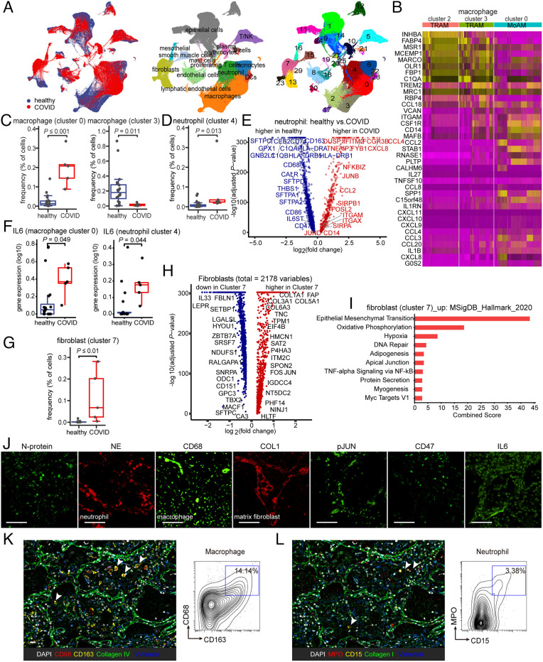
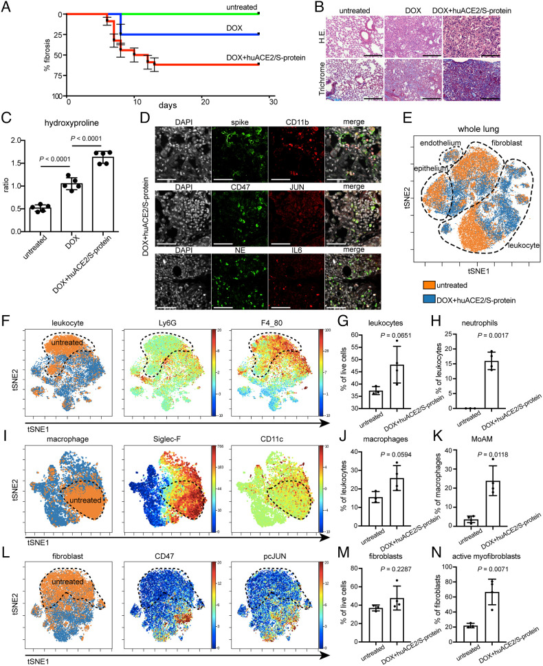
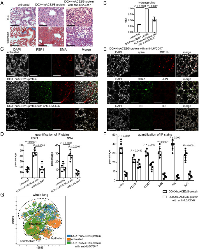
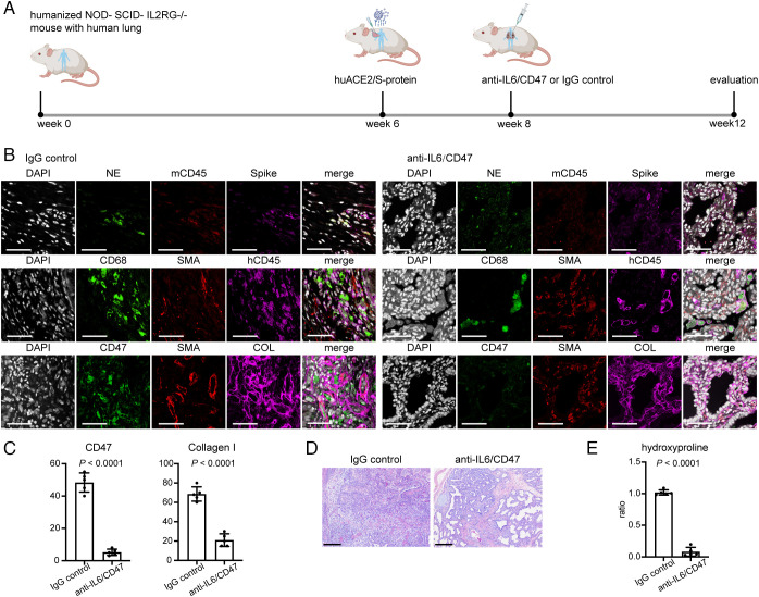
COVID-19 remains a global pandemic of an unprecedented magnitude with millions of people now developing "COVID lung fibrosis." Single-cell transcriptomics of lungs of patients with long COVID revealed a unique immune signature demonstrating the upregulation of key proinflammatory and innate immune effector genes CD47, IL-6, and JUN. We modeled the transition to lung fibrosis after COVID and profiled the immune response with single-cell mass cytometry in JUN mice. These studies revealed that COVID mediated chronic immune activation reminiscent to long COVID in humans. It was characterized by increased CD47, IL-6, and phospho-JUN (pJUN) expression which correlated with disease severity and pathogenic fibroblast populations. When we subsequently treated a humanized COVID lung fibrosis model by combined blockade of inflammation and fibrosis, we not only ameliorated fibrosis but also restored innate immune equilibrium indicating possible implications for clinical management of COVID lung fibrosis in patients.

Keywords: humanized mouse model; immune checkpoint therapy; innate immunity; long COVID pulmonary fibrosis.


Conflict of interest statement

The authors have patent filings to disclose: I.W. and G.W. have patents that have been licensed, and it is possible that they could receive royalties from them. No current consultation with companies in this field. I.W. is a founder and director of Bitterroot Bio, Inc, Pheast, Inc, and 48 Bio, Inc, none of which has programs in this field.

Figures

Fig. 1.
Fig. 1.
Immunological features in the lungs of COVID patients display fibrosis and innate immune activation. (A) UMAP plots demonstrating the major cell types and associated cluster in the lungs of five patients with COVID and 20 healthy lungs. Cells are colored according to donor origin, cell type classification, and clusters (for patient demographics, see Table 1). (B) Heat map comparing the expression of alveolar macrophage-related genes in two subsets of tissue-resident alveolar macrophages (TRAM, clusters 2 and 3) and one subset of monocyte-derived alveolar macrophages (MoAM, cluster 0). (C) Frequencies of macrophage subsets (clusters 0 and 3) which were presenting a significant difference between healthy and COVID cohorts. Each dot represents one individual patient. (D) The frequency of neutrophils (cluster 4) demonstrates a qualitative increase in COVID lung tissues. Each dot represents one individual patient. (E) Associations of genes with COVID status were determined using differential expression analysis for transcripts and linear regression in log-likelihood tests for neutrophil-related genes. The adjusted P values are plotted relative to the log2 fold change of the mean values between COVID and healthy lung cohorts. Blue indicates genes highly expressed in the healthy lung cohort and red genes highly expressed in the COVID lung cohort. (F) IL-6 is expressed in monocyte-derived alveolar macrophages (MoAM, cluster 0) and neutrophil (cluster 4) and in COVID lungs. Each dot represents one individual patient. (GI) Cluster 7 represents a cluster of fibroblasts uniquely enriched in COVID which demonstrates elevated expression of AP1 and fibroblast and epithelial-to-mesenchymal transition-related genes (fibroblasts are represented in clusters 7, 13, and 23). Each dot represents one individual patient. (J) Representative confocal images of lung tissue from COVID patients demonstrate positive staining of cells for SARS-CoV-2 N-protein, as well as indicating the presence of infiltrating neutrophils and macrophages by neutrophil elastase (NE) and CD68 staining, collagen accumulation with COL1, and activation of IL-6–pJUN–CD47 axis. (Scale bar, 100 µm.) (K and L) Representative CODEX images of COVID lung tissues; selected markers collagen IV, collagen I, and vimentin indicate fibrosis, CD68, and CD163 indicate macrophage (K), and myeloperoxidase (MPO) and CD15 indicate the presence of neutrophilic/myeloid cells (L). Contour plots display the double-positive populations. (Scale bar, 100 µm.) Data are expressed as mean ± SD of five COVID lung and 20 healthy lung cohorts. Data were analyzed by the two-tailed unpaired t test; P values were labeled.
Fig. 2.
Fig. 2.
Innate immune infiltrates and fibrosis in mice indicating the transition to COVID lung fibrosis. (A) Kaplan–Meier analysis depicting onset and % fibrosis in untreated (5 mice), doxycycline-treated (5 mice), and doxycycline plus human ACE2/S-protein–treated (10 mice) mice. (B) Histology demonstrating a drastic exacerbation of lung fibrosis with combined aerosol treatment of doxycycline for JUN induction and human ACE2/S-protein compared to doxycycline aerosol alone and untreated. (Scale bar, 100 µm.) (C) Hydroxyproline assay of mouse lung tissues confirmed that the addition of human ACE2/S-protein to doxycycline for JUN induction drastically increased the fibrosis also quantitatively (P < 0.0001). 5 mice per group were analyzed. (D) Representative immunofluorescence stains demonstrate positive cells for SARS-CoV-2 spike protein, macrophages, and monocytes (CD11b), neutrophils (neutrophil elastase, NE), JUN, CD47, and IL-6. (Scale bar, 100 µm.) (E) tSNE of single-cell CyTOF data of lung tissues of 4 mice in transition to lung exposed to Dox+huACE2/S-protein (blue) and three healthy lungs (orange). (F) tSNE plots represent leukocytes in the lungs of mice in transition to COVID and healthy controls. The healthy control is labeled with a dotted line. Ly6G and F4_80 expression is used to determine the abundance of neutrophils and macrophages. (G) The statistical analysis of the frequency of leukocytes in all live cells as indicated. (H) Quantification of neutrophil frequency in the lung as indicated. (I) tSNE alignment of macrophage clusters, Siglec-F, and CD11c abundance labeling heterogeneous macrophages. (J) The abundance of lung macrophages as indicated. (K) Dot plot shows the relative contributions of monocyte-derived alveolar macrophage (MoAM) per mouse. (L) tSNE of CyTOF profiling protein expression in fibroblasts as indicated. (M) Dot plots of percentages of fibroblast gated on live cells as indicated. (N) Activated myofibroblasts were drastically increased in the mouse lungs in transition to COVID. (Scale bar, 100 µm.) Data are expressed as mean ± SD of four mouse lungs in transition to COVID and three healthy control lungs. Data were analyzed by one-way ANOVA (C) and the two-tailed unpaired t test (G, H, J, K, M, and N); P values are indicated and deemed statistically significant if P < 0.05.
Fig. 3.
Fig. 3.
Mice in transition to COVID lung fibrosis and mice treated with anti-CD47/anti-IL-6 demonstrate improved fibrosis. (A) H&E and trichrome demonstrating lung histology after anti-CD47/anti-IL-6 treatment in mice in transition to COVID fibrosis. (Scale bar, 100 µm.) (B) Hydroxyproline assay calculated less extent of collagen accumulation. (C) Representative immunofluorescence images of FSP1 and SMA demonstrating that inhibiting CD47 and IL-6 improves lung fibrosis. (Scale bar, 100 µm.) (D) Quantification of expression of FSP1 and SMA in mice in transition to COVID with anti-CD47/anti-IL-6 treatment displaying decreased numbers of activated fibroblasts. (E) Confocal imaging of lung tissues showing attenuation of immune infiltration and fibrosis in mice in transition to COVID after anti-CD47/anti-IL-6 treatment. (Scale bar, 100 µm.) (F) Quantitation of marker expression in mouse lung tissues with or without anti-CD47/anti-IL-6 treatment. Data are shown as means ± SD from five different fields. (G) tSNE projection of CyTOF analysis probes global immune cellular changes after anti-CD47/anti-IL-6 treatment in the mouse lungs in transition to COVID. (Scale bar, 100 µm.) Data are shown as mean ± SD of five different mouse lungs per each group. Data were analyzed by one-way ANOVA (B and D) and multiple t tests (F); P values are indicated.
Fig. 4.
Fig. 4.
Fibrosis decreased in human lung tissues after treatment with blocking antibodies against CD47 and IL-6 in a humanized mouse model of COVID lung fibrosis. (A) Evaluation of therapeutic effects of CD47 and IL-6 inhibition on humanized mice grafted with human lung organoids infected with SARS-CoV-2 and treated with IgG or anti-IL-6/anti-CD47 blocking antibodies. (B) Representative immunofluorescence images of humanized COVID mouse treated with or without anti-CD47/anti-IL-6 antibodies highlighting the change of human immune cell infiltrates and extracellular matrix deposition. (Scale bar, 100 µm.). (C) Quantification of CD47 and collagen I expression in humanized mouse infected with SARS-CoV-2 without and with anti-CD47/anti-IL-6 treatment. (D) Representative images of histopathology demonstrating normalization of the lung structure in humanized COVID mouse model after anti-CD47/anti-IL-6 treatment. (Scale bar, 100 µm.). (E) Measurement of hydroxyproline content decreased with anti-CD47/anti-IL-6 treatment. (Scale bar, 100 µm.) Data are shown as mean ± SD of five different human lung tissue pieces per each group. Data were analyzed by the two-tailed unpaired t test; P values were labeled.

Comment in

References

    1. Huang C., et al. , Clinical features of patients infected with 2019 novel coronavirus in Wuhan, China. Lancet 395, 497–506 (2020). - PMC - PubMed
    1. Laing A. G., et al. , A dynamic COVID-19 immune signature includes associations with poor prognosis. Nat. Med. 26, 1623–1635 (2020). - PubMed
    1. Lucas C., et al. , Impact of circulating SARS-CoV-2 variants on mRNA vaccine-induced immunity. Nature 600, 523–529 (2021). - PMC - PubMed
    1. Wang D., et al. , Clinical characteristics of 138 hospitalized patients with 2019 novel coronavirus-infected pneumonia in Wuhan, China. JAMA 323, 1061–1069 (2020). - PMC - PubMed
    1. Tale S., et al. , Post-COVID-19 pneumonia pulmonary fibrosis. QJM 113, 837–838 (2020). - PMC - PubMed

Publication types

LinkOut - more resources