. 2023 Jan-Dec:37:3946320231161174.
doi: 10.1177/03946320231161174.

Immunopotentiation effects of apigenin on NK cell proliferation and killing pancreatic cancer cells

Affiliations

Immunopotentiation effects of apigenin on NK cell proliferation and killing pancreatic cancer cells

Yong-Bo Feng et al. Int J Immunopathol Pharmacol. 2023 Jan-Dec.

Abstract

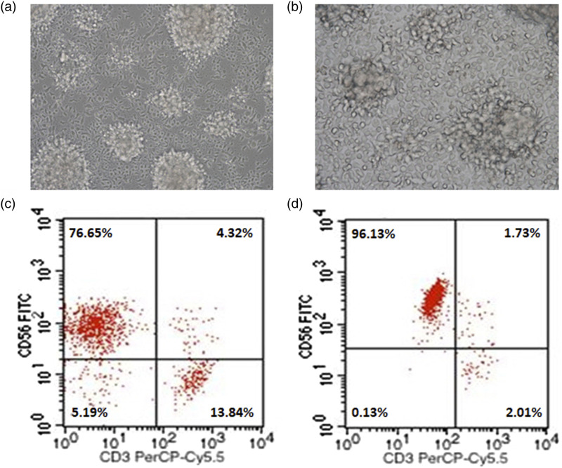
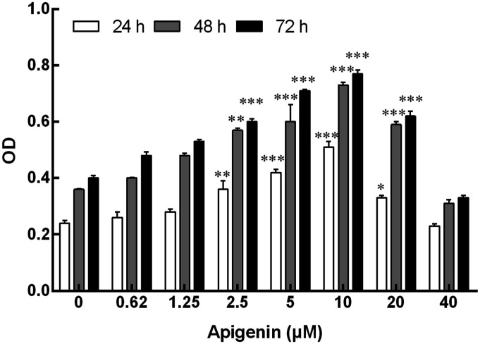
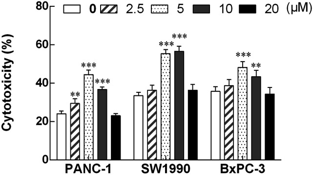
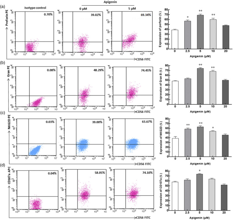
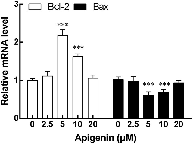
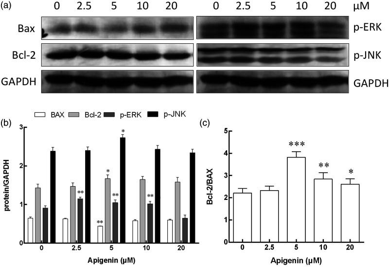
Apigenin is a kind of flavonoid with many beneficial biological effects. It not only has direct cytotoxicity to tumor cells, but also can boost the antitumor effect of immune cells by modulating immune system. The purpose of this study was to investigate the proliferation of NK cells treated with apigenin and its cytotoxicity to pancreatic cancer cells in vitro, and explore its potential molecular mechanism. In this study, the effect of apigenin on NK cell proliferation and killing pancreatic cancer cells were measured by CCK-8 assay. Perforin, granzyme B (Gran B), CD107a, and NKG2D expressions of NK cells induced with apigenin were detected by flow cytometry (FCM). The mRNA expression of Bcl-2, Bax and protein expression of Bcl-2, Bax, p-ERK, and p-JNK in NK cells were evaluated by qRT-PCR and western blotting analysis, respectively. The results showed that appropriate concentration of apigenin could significantly promote the proliferation of NK cells in vitro and enhance the killing activity of NK cells against pancreatic cancer cells. The expressions of surface antigen NKG2D and intracellular antigen perforin and Gran B of NK cells were upregulated after treating with apigenin. Bcl-2 mRNA expression was increased, while Bax mRNA expression was decreased. Similarly, the expression of Bcl-2, p-JNK, and p-ERK protein was upregulated, and the expression of Bax protein was downregulated. The molecular mechanism of the immunopotentiation effects of apigenin may be that it up-regulates Bcl-2 and down-regulates Bax expression at the gene and protein levels to facilitate NK cell proliferation, and up-regulates the expression of perforin, Gran B, and NKG2D through the activation of JNK and ERK pathways to enhance NK cell cytotoxicity.

Keywords: Apigenin; NK cell; immunopotentiation; killing activity; pancreatic cancer.


Conflict of interest statement

The author(s) declared no potential conflicts of interest with respect to the research, authorship, and/or publication of this article.

Figures

Figure 1.
Figure 1.
Morphological characteristics and phenotypic identification of NK cells after expansion. a: Cellular morphologies of NK cells in suspension growth state after 12 days of expansion (200×). b: Cellular morphologies of NK cells adhering to the wall of the culture flask (400×). c: Representative dot plot depicting the percentage of NK cells within the cultured PBMCs. d: Representative dot plot of NK cell percentage after magnetic cell sorting.
Figure 2.
Figure 2.
Effect of apigenin on proliferation of NK cells. NK cells were cultured with apigenin at concentrations of 0, 2.5, 5, 10, 20, 40 μM for 24 h, 48 h, and 72 h. Data are presented as mean ± standard deviation (SD). *p < 0.05, **p < 0.01, ***p < 0.001 compared with the respective control group (0 μM group). OD, optical density.
Figure 3.
Figure 3.
The cytotoxic activity of NK cells treated with apigenin for 72 h to three pancreatic cancer cell lines (PANC-1, SW1990 and BxPC-3). NK cells treated with various concentrations of apigenin were used as effector cells, and pancreatic cancer cells (PANC-1, SW1990, and BxPC-3) were used as target cells (E: T=10: 1). The cytotoxicity of NK cells against three pancreatic cancer cell lines was markedly enhanced after treatment with apigenin at the dose of 5 μM and 10 μM, and significantly higher than that of the corresponding control group.**p < 0.01, ***p < 0.001.
Figure 4.
Figure 4.
The expression of surface and intracellular antigen molecules on/in NK cells incubated with apigenin for 72h. a, b, c, and d represent the expression of perforin, Gran B, NKG2D, and CD107a in/on NK cells with apigenin treatment, respectively. Representative dot plot depicting the expression percentage of surface and intracellular antigen molecules on/in NK cells in the three columns on the left (from left to right, isotype control, 0 μM control group, and 5 μM treatment group). In the rightmost column, histograms describing the expression of perforin, Gran B, NKG2D, and CD107a on/in NK cells treated with various concentrations of apigenin are shown. Data are displayed in mean ± SD. *p < 0.05, **p < 0.01 compared with the 0 μM control group.
Figure 5.
Figure 5.
The mRNA expression of Bax and Bcl-2 in NK cells after treatment with apigenin for 72 h. The mRNA levels of Bcl-2 at 5 μM and 10 μM doses of apigenin were significantly higher than that of the control group. The change of Bax mRNA expression was contrary to that of Bcl-2, and the expression level decreased significantly at 5 μM and 10 μM doses. Results were expressed as mean ± SD, ***p < 0.001 VS compared with the 0 μM control group.
Figure 6.
Figure 6.
The protein expression of Bax, Bcl-2, p-ERK, p-JNK, and GAPDH in NK cells after treatment with apigenin for 72 h. a: Western blot analysis for Bax, Bcl-2, p-ERK, p-JNK, and GAPDH expression in NK cells treated with apigenin at concentrations of 0, 2.5, 5, 10, and 20 μM, respectively. b: Protein activation analysis of NK cells treated with apigenin at different concentrations, the means and standard deviation are depicted as diagrams. c: The ratio of Bcl-2 and Bax in NK cells. *p < 0.05, **p < 0.01, ***p < 0.001 versus the 0 μM control group.

References

    1. Siegel RL, Miller KD, Fuchs HE, et al. (2021) Cancer statistics, 2021. CA A Cancer J Clin 71(1): 7–33. - PubMed
    1. He Y, Zhou X, Fan X, et al. (2022) Disease Burden of Pancreatic Cancer - China, 1990-2019. China CDC Weekly 4(24): 527–531. - PMC - PubMed
    1. Lee JA, Ha SK, Cho E, et al. (2015) Resveratrol as a Bioenhancer to Improve Anti-Inflammatory Activities of Apigenin. Nutrients 7(11): 9650–9661. - PMC - PubMed
    1. Sánchez-Marzo N, Pérez-Sánchez A, Ruiz-Torres V, et al. (2019) Antioxidant and Photoprotective Activity of Apigenin and its Potassium Salt Derivative in Human Keratinocytes and Absorption in Caco-2 Cell Monolayers. Int J Mol Sci 20(9): 2148. - PMC - PubMed
    1. Meng S, Zhu Y, Li JF, et al. (2017) Apigenin inhibits renal cell carcinoma cell proliferation. Oncotarget 8(12): 19834–19842. - PMC - PubMed