. 2023 Apr;616(7956):326-331.
doi: 10.1038/s41586-023-05862-7. Epub 2023 Feb 27.

Ubiquitin-like conjugation by bacterial cGAS enhances anti-phage defence

Affiliations

Ubiquitin-like conjugation by bacterial cGAS enhances anti-phage defence

Justin M Jenson et al. Nature. 2023 Apr.

Abstract

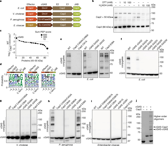
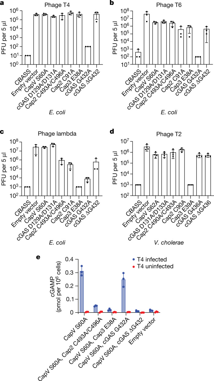
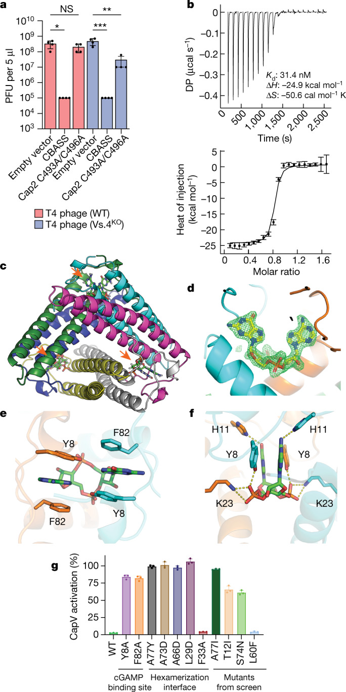
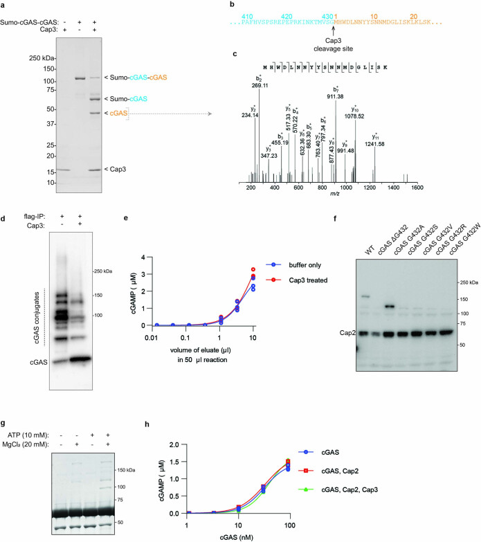
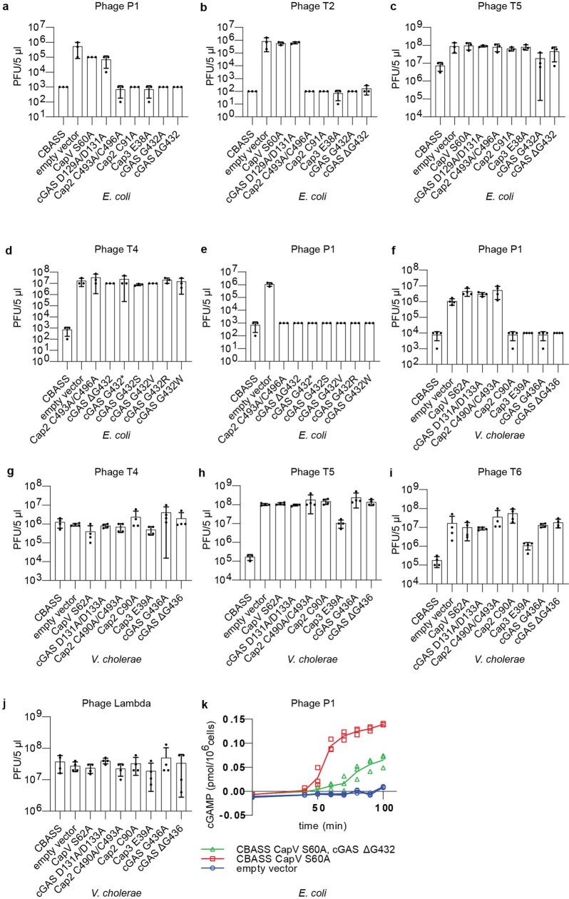
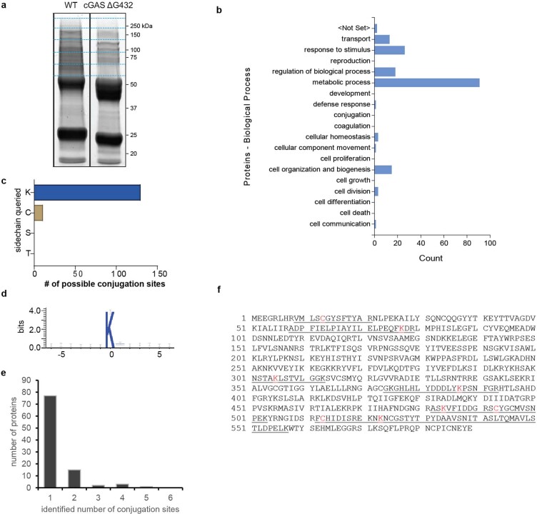
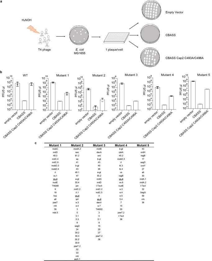
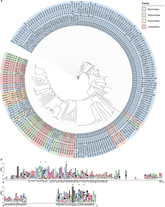
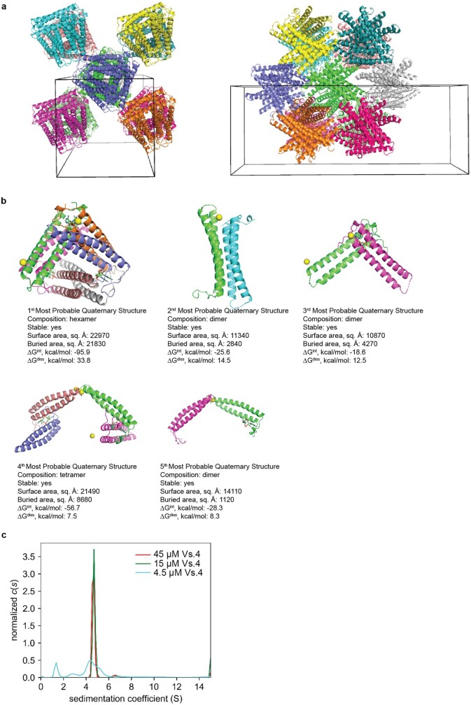
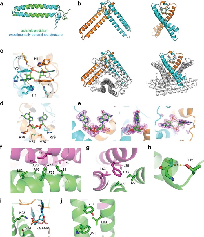
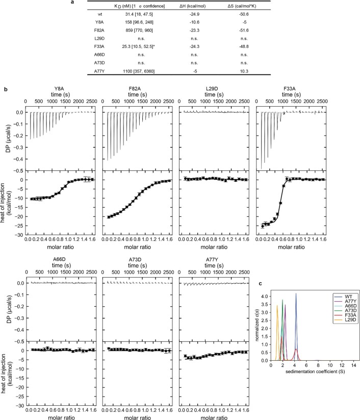
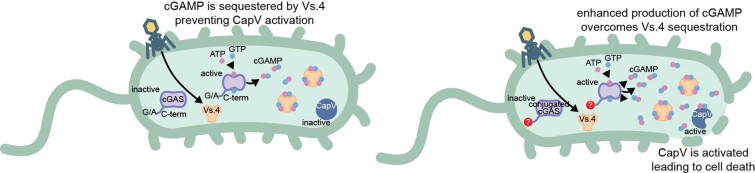
cGAS is an evolutionarily conserved enzyme that has a pivotal role in immune defence against infection1-3. In vertebrate animals, cGAS is activated by DNA to produce cyclic GMP-AMP (cGAMP)4,5, which leads to the expression of antimicrobial genes6,7. In bacteria, cyclic dinucleotide (CDN)-based anti-phage signalling systems (CBASS) have been discovered8-11. These systems are composed of cGAS-like enzymes and various effector proteins that kill bacteria on phage infection, thereby stopping phage spread. Of the CBASS systems reported, approximately 39% contain Cap2 and Cap3, which encode proteins with homology to ubiquitin conjugating (E1/E2) and deconjugating enzymes, respectively8,12. Although these proteins are required to prevent infection of some bacteriophages8, the mechanism by which the enzymatic activities exert an anti-phage effect is unknown. Here we show that Cap2 forms a thioester bond with the C-terminal glycine of cGAS and promotes conjugation of cGAS to target proteins in a process that resembles ubiquitin conjugation. The covalent conjugation of cGAS increases the production of cGAMP. Using a genetic screen, we found that the phage protein Vs.4 antagonized cGAS signalling by binding tightly to cGAMP (dissociation constant of approximately 30 nM) and sequestering it. A crystal structure of Vs.4 bound to cGAMP showed that Vs.4 formed a hexamer that was bound to three molecules of cGAMP. These results reveal a ubiquitin-like conjugation mechanism that regulates cGAS activity in bacteria and illustrates an arms race between bacteria and viruses through controlling CDN levels.


Conflict of interest statement

The authors declare no competing interests.

Figures

Fig. 1
Fig. 1. Cap2 catalyses covalent conjugation of cGAS.
a, Domain organization of the CBASS operons from E. coli, Vibrio cholerae, Pseudomonas aeruginosa and Enterobacter cloacae. b, E. coli MG1655, which lacks CBASS, was transformed with the four-gene operon (from a) including an N-terminally Flag-tagged Cap2. Bacterial cell lysates were treated for 30 min with the indicated amount of DTT or hydroxylamine (H2NOH), subjected to non-reducing SDS–PAGE and immunoblotted with an anti-Flag antibody. c, Mass spectrometry analysis of 45–55 kDa proteins conjugated to Cap2 that migrated at about 110 kDa on an SDS–PAGE gel. The y axis indicates enrichment of the proteins in bacteria with wild-type CBASS compared to those harbouring the C493A/C496A mutations of Cap2. The sum PEP score is the sum of negative logarithms of the posterior error probabilities (PEP) and represents the credibility of the spectral matches. d, Sequence logo of alignment of the C-terminal (C term.) sequences of cGAS from previously identified CBASS operons with the indicated gene architecture: operons containing full-length Cap2 and Cap3 (left), minimal operons with no Cap2 or Cap3 (centre) and operons lacking the E2 domain in Cap2 (right). ei, Immunoblotting of cGAS conjugates in bacterial lysates from cells harbouring the wild-tye (WT) four-gene CBASS operon with N-terminally Flag-tagged cGAS, or those with indicated mutations in the CBASS (e) and cGAS (f) genes from E. coli, V. cholerae (g), P. aeruginosa (h) and Enterobacter cloacae (i). Bacteria cell lysates were subjected to SDS–PAGE in the presence of β-mercaptoethanol, followed by immunoblotting with an anti-Flag antibody. j, Purified recombinant cGAS and Cap2 proteins were incubated in the presence of ATP for 30 min at room temperature, as indicated. Following the reaction, proteins were separated by SDS–PAGE and stained with Coomassie blue. All data are representative of at least two independent experiments. For gel source data, see Supplementary Fig. 1. Source data
Fig. 2
Fig. 2. cGAS conjugation enhances cGAMP production and anti-phage immunity in vivo.
ac, Viral titre of phage T4 (a), T6 (b) and lambda (c) after infection of bacterial strains with the wild-type operon from E. coli (CBASS), no operon (empty vector) or the indicated point mutations. Data are mean ± s.d. of n = 3 independent experiments with individual points overlaid. d, Viral titre of phage T2 after infection in bacterial strains harbouring the wild-type V. cholerae CBASS operon, no operon (empty vector) or the indicated point mutations. Data are mean ± s.d. of n = 4 independent experiments with individual points overlaid. e, E. coli harbouring the indicated CBASS operon was infected by the phage T4 at a multiplicity of infection of approximately 10. CapV in the CBASS operon was inactivated by a mutation in the active site (S60A) to avoid cell death from CBASS signalling. Samples from each bacterial culture were collected 60 min after infection, snap frozen and lysed by heating. Clarified lysates were incubated with THP1 Lucia ISG cells, which express a luciferase (Lucia) reporter gene under the control of an IRF-inducible promoter. Luminescence signal was measured and converted to cGAMP concentrations using a cGAMP standard curve. Data are mean ± s.d. of n = 3 technical replicates and is representative of two independent experiments. Source data
Fig. 3
Fig. 3. Vs.4 tightly binds and sequesters cGAMP.
a, Viral titre of WT and Vs.4 knockout T4 phage in strains of E. coli MG1655 containing the WT or Cap2 C493A/C496A CBASS or empty vector. Bar graph represents average values ±s.e.m. of n = 4 independent experiments with individual points overlaid. NS, not significant, *P < 0.05 (P = 0.0178), **P < 0.005 (P = 0.0010) and ***P < 0.0005 (P = 0.0005) by one-way analysis of variance test. b, Representative example of a raw titration trace of differential power (DP) versus time (top) and integrated data (bottom, data points depict integrated heat of injection and error bars depict the weighted root mean squared deviation of the difference between predicted and measured values) of an ITC experiment in which cGAMP was titrated into a solution of Vs.4. The global best fit of three titrations indicated a dissociation constant (Kd) of 31.4 nM with a 68% confidence interval of 18–47.5 nM. c, X-ray crystal structure of hexameric Vs.4 (cartoon representation, one colour per monomer) bound to three molecules of cGAMP (green stick representation indicated by orange arrows). d, Composite omit map calculated with the cGAMP ligand omitted. Simulated annealing was used to remove any memory of the ligand. The difference density map (green) is contoured at 3.5σ with cGAMP (yellow) superimposed in the binding pocket that is formed between two monomers (cyan and orange). e, Detailed view of the residues forming π–π interactions with cGAMP. f, Detailed view of hydrogen bonds and salt bridges formed with cGAMP in the Vs.4 binding pocket. g, CapV was incubated with the indicated Vs.4 mutant (10 µM), cGAMP (1 µM) and resorufin butyrate (a fluorogenic phospholipase substrate) to measure the enzymatic activity of CapV. Bar graph represents average values ±s.d. of n = 3 technical replicates with individual points overlaid and is representative of two independent experiments. Source data
Extended Data Fig. 1
Extended Data Fig. 1. Conjugation of cGAS and deconjugation by Cap3.
a, Cap3 cleaves at c-terminus of cGAS. Recombinant Cap3 was incubated with Sumo-cGAS-cGAS for 120 min at 37 °C, separated with SDS-PAGE, and stained with Coomassie blue. Data is representative of 3 independent experiments. The indicated bands were analyzed by LC-MS/MS. b, The Cap3 cut site identified by MS. c, A tandem mass spectrum of a peptide fragment from the Cap3 cleavage product showing the N-terminus of cGAS after cleavage from Sumo-cGAS. d, Flag-tagged cGAS proteins were immunopurified from cells harboring CBASS with inactivated Cap3. The isolated proteins were treated with Cap3 for 120 min at 37 °C followed by SDS-PAGE and immunostaining with an anti-Flag antibody. Data is representative of 2 independent experiments. e, cGAMP production of Cap3 treated and untreated isolates from d was measured using THP1-Lucia ISG cells (methods). Scatter plot represents mean ± SD of n = 3 technical replicates with individual points overlaid and is representative of two independent experiments. f, Immunoblot of cGAS thioester in bacterial lysates from cells harboring a CBASS operon with N-terminally Flag-tagged Cap2 and the indicated mutations. Cell lysates were subjected to non-reducing SDS-PAGE and immunoblotted with an anti-Flag antibody. The data are representative of two independent experiments. g, Recombinant cGAS and Cap2 proteins were incubated for 30 min at room temperature, separated by SDS-PAGE, and stained with Coomassie blue. Data are representative of two independent experiments. h, Enzymatic activity of recombinant cGAS in the presence of Cap2 and Cap3 was measured by incubating with ATP to allow conjugate formation before supplementing with ATP, GTP, and MgCl2 then incubating for 30 min at 37 °C. cGAMP generated in the reaction was measured in THP1-Lucia ISG cells. Scatter plot represents mean ± SD of n = 3 technical replicates with individual points overlaid and is representative of two independent experiments. For gel source data, see Supplementary Fig. 1. Source data
Extended Data Fig. 2
Extended Data Fig. 2. Viral titers of bacteriophages in bacteria harboring CBASS operons with point mutations in the indicated genes.
ac, E. coli MG1655 was transformed with the wild-type operon from E. coli TW11681 (CBASS) and the operon containing the indicated mutations. Empty vector indicates no CBASS operon. The strains were infected with Phage P1 (a), Phage T2 (b), or Phage T5 (c) and the plague forming units (PFU) were measured. Bar graph represents mean ± SD of n = 3 independent experiments for each bacterial strain. de, Viral titers of phage T4 (d) and P1 (e) after infection in bacterial strains harboring the wild-type CBASS operon from E. coli TW11681, no operon (empty vector), or the indicated point mutations. Bar graph represents average values ± SD of n = 3 independent experiments with individual points overlaid. fj, E. coli MG1655 was transformed with the wild-type operon from V. cholerae (CBASS) with Flag-tagged cGAS and the indicated mutations. Empty vector indicates no CBASS operon. The strains were infected with  Phage P1 (f), Phage T4 (g), Phage T5 (h), Phage T6 (i), or Phage Lambda (j) and the plague forming units (PFU) were measured. Bar graph represents mean ± SD of n = 4 independent experiments for each bacterial strain. k, E. coli harboring the indicated CBASS operon was infected by the phage P1 at a multiplicity of infection of 2. CapV in the CBASS operon was inactivated by a mutation in the active site (S60A) to inhibit cell death from CBASS signaling. Samples from each bacterial culture were collected at 10-min intervals starting at 40 min after infection, snap frozen, and lysed by heating and sonication. Clarified lysates were incubated with purified CapV in the presence of resorufin butyrate, a fluorogenic phospholipase substrate, to measure the bioactivity of cGAMP produced by the bacteria. Scatter plot represents mean ± SD of n = 3 technical replicates with individual points overlaid and is representative of two independent experiments. Source data
Extended Data Fig. 3
Extended Data Fig. 3. Identification of cGAS conjugation sites.
a, Flag-tagged cGAS and cGAS ΔG432 proteins were immunopurified from E. coli cells harboring the CBASS operons encoding these proteins. Isolated proteins were separated on SDS-PAGE and stained by Coomassie blue. Indicated protein bands were cut, alkylated, digested with trypsin, and analyzed by shot-gun LS-MS/MS analysis. Data is representative of three independent experiments: two with uninfected cells and one with cells infected with phage T4. b, Biological processes associated with proteins found to be conjugated by cGAS. c, The numbers of conjugation sites identified for lysine, cysteine, serine, and threonine is shown. d, Sequence alignment of the 130 cGAS conjugation sites surrounding the target lysine. e, Histogram of the number of conjugation sites per cGAS conjugated target. f, The protein sequence of Cap2. The cGAS conjugation sites are indicated in red and the peptides in which conjugation sites were identified by mass spectrometry are underlined. For gel source data, see Supplementary Fig. 1. Source data
Extended Data Fig. 4
Extended Data Fig. 4. Screen for loss of function mutants of T4 phage.
a, T4 phage was mutagenized with hydroxylamine (NH2OH) to produce a library, mixed with wild-type E. coli MG1655 and plated (Methods). Individual plaques (mutants) from this library were picked and transferred to wells of 96-well plates. These phage-containing solutions were pipetted onto the surface of top agar that had been inoculated with E. coli MG1655 harboring no CBASS (empty vector), wild-type CBASS (CBASS), or CBASS with inactivating Cap2 mutations (C493A/C496A). Mutants that formed plaques only on the empty vector plates were selected for further validation. b, efficiency of plating of wild-type T4 phage (WT) and the five mutant T4 phages (Mutants1–5) with more than 50-fold reduction in infectivity in bacteria containing the Cap2 C493A/C496A mutations. Bar plots represent mean ± SD of n = 3 independent experiments. c, table of genes found with non-synonymous mutations found in T4 phage mutants with increased sensitivity to CBASS defective in cGAS conjugation. Source data
Extended Data Fig. 5
Extended Data Fig. 5. Proteins with homology to Vs.4 are found in diverse phages.
a, Phylogenetic tree of 198 proteins with predicted homology to Vs.4 identified in Prokaryotic Virus Remote Homologous Groups database (see Methods). b, Vs.4 homologs were aligned with clustal omega and the multiple sequence alignment is shown. The sequence of Vs.4 from T4 phage is displayed on the x-axis. Insertions in the alignment are denoted with dashes. Residues in the cGAMP binding site are indicated with an *.
Extended Data Fig. 6
Extended Data Fig. 6. Quaternary structure assignment of Vs.4.
a, schematic representation of crystal packing and unit cell of Vs.4. Monomers of Vs.4 cluster in hexamers in the lattice. To illustrate hexamer clusters, each hexameric complex of Vs.4 is highlighted in a unique color. b, PISA was used to analyze the Vs.4 structure for possible higher-order assemblies of Vs.4. In total, 5 assemblies were predicted. The top prediction (top left) is the only hexameric assembly that was predicted to be stable (see Methods). c, analytical ultracentricugation of Vs.4 bound to cGAMP. Vs.4 forms a hexaxamer that is stable on the time scale of the sedimentation experiment at 45 μM and 15 μM with a sedimentation coefficient of 4.64 and a molecular weight >50.3 kDa (61.8 kDa expected for hexamer). Source data
Extended Data Fig. 7
Extended Data Fig. 7. Structural analysis of the Vs.4:cGAMP complex.
a, Alignment of the alphafold predicted structure of the Vs.4 monomer that was used for molecular replacement and the experimentally determined structure of Vs.4 bound to cGAMP. b, A cartoon representation of a single cGAMP binding pocket formed by Vs.4 dimers with one chain colored orange and the other cyan. cGAMP is bound between these monomers (green sticks). Other monomers are colored white to display the dimer in the context of the larger hexameric structure. c, Detail of the hydrogen-bonds formed with cGAMP in the binding pocket of Vs.4. d, Additional interactions between Vs.4 and cGAMP in the binding pocket. e, Polder map for the cGAMP ligand, which was calculated with cGAMP with 5Å radius omitted. The difference density map is contoured 3.0σ with cGAMP superimposed. f, Hexamer interface with key residues highlighted. gj, Residues found mutated in forward genetic screen shown in Extended Data Fig. 4a: A77I sterically clashes with the heximerization interface (g); T12I removes a stabilizing helix cap (h); S74N interferes with interactions with cGAMP (i); and the L60F may contribute to steric clashes (j).
Extended Data Fig. 8
Extended Data Fig. 8. Mutational analysis of the Vs.4:cGAMP complex.
a, Isothermal calorimetry (ITC) data from titrating cGAMP into a solution of the indicated Vs.4 mutant. Each experiment was performed at least twice and global analysis was used to determine KD, ΔH, and ΔS. Analysis of the F33A mutant indicated high incompetent fraction (0.496), as indicated with an *. Cases where no binding was observed are denoted as having no signal (n.s). b, Representative examples of raw titration traces (top) and integrated data (bottom, data points depict integrated heat of injection and error bars depict the weighted rmsd of the difference between predicted and measured values) of two independent ITC experiments reported in a. Titration data for WT Vs.4 is shown in Fig. 3b. c, Analytical ultracentrifugation of Vs.4 mutants. Vs.4 (WT) forms a hexamer that is stable on the time scale of the sedimentation experiment at 15 μM with a sedimentation coefficient of 4.3 and an estimated MW of 59 kDa (61.8 kDa expected). Mutations to the hexamerization interface causes dramatically reduced sedimentation coefficients, consistent with disruption of the quaternary structure. Source data
Extended Data Fig. 9
Extended Data Fig. 9. Model of the molecular antagonism between Vs.4 and cGAS conjugation.
In response to phage infection, bacterial cGAS (purple) is activated and produces the second messenger cGAMP from ATP (red) and GTP (blue). cGAMP is bound by the phage encoded protein Vs.4 (orange), which acts as a cGAMP ‘sponge’ that sequesters cGAMP and prevents activation of the bacterial ‘suicide’ effector protein CapV. This allows the phage to propagate within the bacterial cell, as shown in the left panel. In contrast, when cGAS is conjugated to its biological target, enhanced cGAS activity produces abundant cGAMP that overpowers Vs.4 inhibition and activates the phospholipase activity of CapV. CapV activation disrupts the bacterial membrane and kills the bacterial cell before the phage replication cycle is complete, thereby preventing phage propagation.

References

    1. Margolis SR, Wilson SC, Vance RE. Evolutionary origins of cGAS-STING signaling. Trends Immunol. 2017;38:733–743. doi: 10.1016/j.it.2017.03.004. - DOI - PubMed
    1. Duncan-Lowey B, Kranzusch PJ. CBASS phage defense and evolution of antiviral nucleotide signaling. Curr. Opin. Immunol. 2022;74:156–163. doi: 10.1016/j.coi.2022.01.002. - DOI - PubMed
    1. Wein T, Sorek R. Bacterial origins of human cell-autonomous innate immune mechanisms. Nat. Rev. Immunol. 2022;22:629–638. doi: 10.1038/s41577-022-00705-4. - DOI - PubMed
    1. Sun L, Wu J, Du F, Chen X, Chen ZJ. Cyclic GMP-AMP synthase is a cytosolic DNA sensor that activates the type I interferon pathway. Science. 2013;339:786–791. doi: 10.1126/science.1232458. - DOI - PMC - PubMed
    1. Wu J, et al. Cyclic GMP-AMP is an endogenous second messenger in innate immune signaling by cytosolic DNA. Science. 2013;339:826–830. doi: 10.1126/science.1229963. - DOI - PMC - PubMed

Publication types

MeSH terms

LinkOut - more resources