. 2023 Apr;299(4):103072.
doi: 10.1016/j.jbc.2023.103072. Epub 2023 Feb 26.

Constitutive and induced forms of membrane-bound proteinase 3 interact with antineutrophil cytoplasmic antibodies and promote immune activation of neutrophils

Affiliations

Constitutive and induced forms of membrane-bound proteinase 3 interact with antineutrophil cytoplasmic antibodies and promote immune activation of neutrophils

Carla Guarino et al. J Biol Chem. 2023 Apr.

Abstract

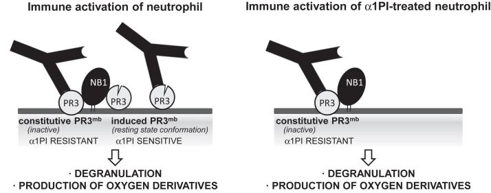
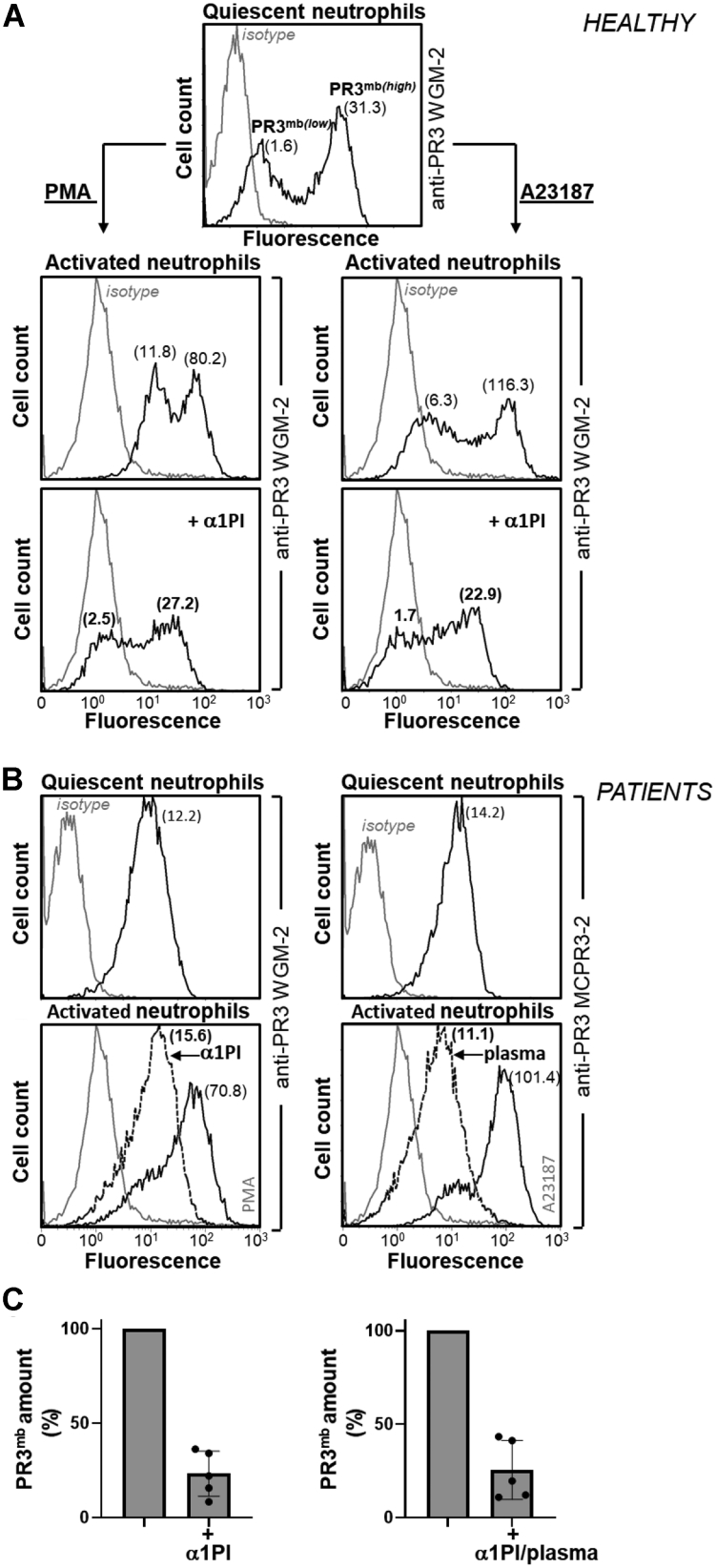
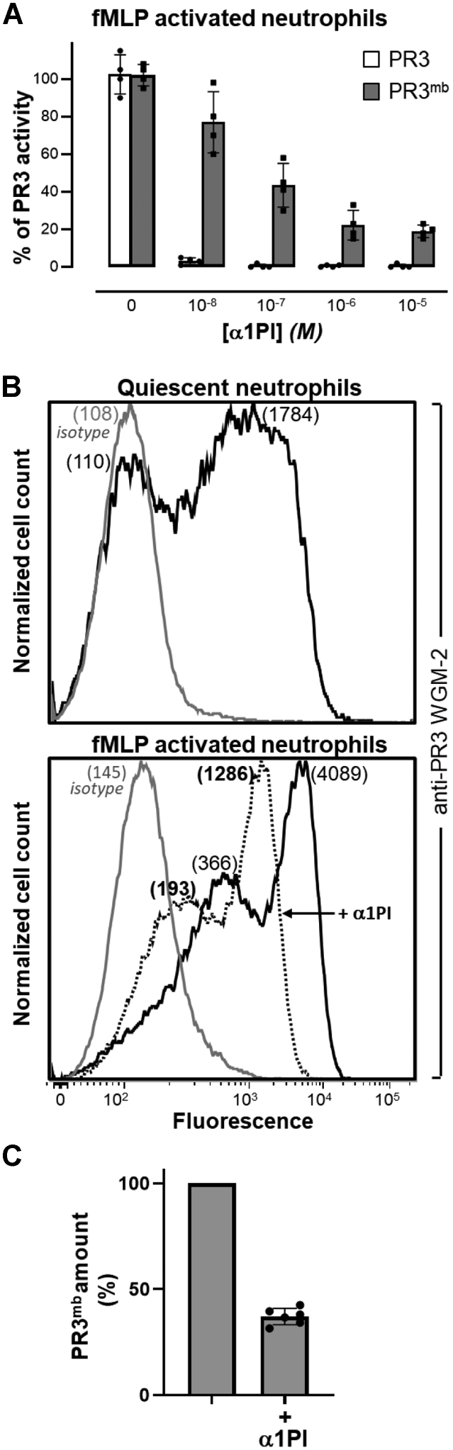
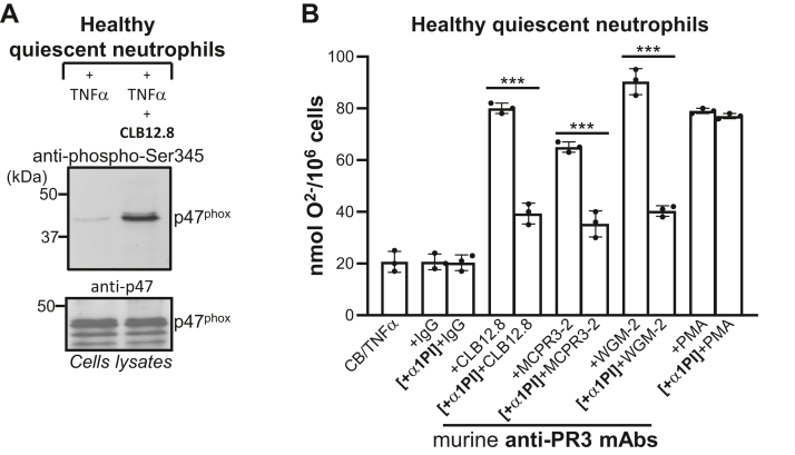
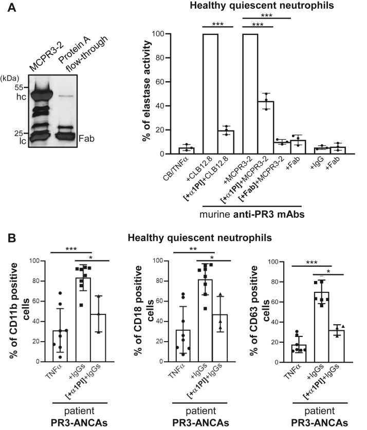
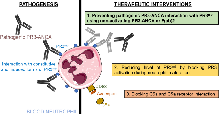
Proteinase 3 (PR3) is the main target antigen of antineutrophil cytoplasmic antibodies (ANCAs) in PR3-ANCA-associated vasculitis. A small fraction of PR3 is constitutively exposed on the surface of quiescent blood neutrophils in a proteolytically inactive form. When activated, neutrophils expose an induced form of membrane-bound PR3 (PR3mb) on their surface as well, which is enzymatically less active than unbound PR3 in solution due to its altered conformation. In this work, our objective was to understand the respective role of constitutive and induced PR3mb in the immune activation of neutrophils triggered by murine anti-PR3 mAbs and human PR3-ANCA. We quantified immune activation of neutrophils by the measurement of the production of superoxide anions and secreted protease activity in the cell supernatant before and after treatment of the cells by alpha-1 protease inhibitor that clears induced PR3mb from the cell surface. Incubation of TNFα-primed neutrophils with anti-PR3 antibodies resulted in a significant increase in superoxide anion production, membrane activation marker exposition, and secreted protease activity. When primed neutrophils were first treated with alpha-1 protease inhibitor, we observed a partial reduction in antibody-induced neutrophil activation, suggesting that constitutive PR3mb is sufficient to activate neutrophils. The pretreatment of primed neutrophils with purified antigen-binding fragments used as competitor significantly reduced cell activation by whole antibodies. This led us to the conclusion that PR3mb promoted immune activation of neutrophils. We propose that blocking and/or elimination of PR3mb offers a new therapeutic strategy to attenuate neutrophil activation in patients with PR3-ANCA-associated vasculitis.

Keywords: PR3-ANCA; alpha-1 protease inhibitor; anti-PR3 mAbs; autoantibody; immune activation; inflammation; neutrophil; proteinase 3; therapeutic strategy; vasculitis.


Conflict of interest statement

Conflict of interest The authors declare that they have no conflicts of interest with the contents of this article.

Figures

Figure 1
Figure 1
Exploration of α1PI for investigation of the respective role of constitutive and inducible PR3mbin neutrophils activation by anti-PR3 antibodies. The diagram illustrates two distinct pool of PR3mb colocalized with NB1 (CD177) (34) on activated neutrophils: one catalytically inactive, which is α1PI-resistant (constitutive PR3mb), and the other in an activatable conformation that is α1PI-sensitive (induced PR3mb) (10, 11). The property of α1PI to discriminate between constitutive and induced PR3mb was explored to assess their relative role in immune activation of neutrophils by anti-PR3 mAbs and PR3-ANCA. α1PI, alpha-1 protease inhibitor; ANCA, antineutrophil cytoplasmic antibody; PR3, proteinase 3.
Figure 2
Figure 2
Surface exposition of PR3 on activated healthy and patient neutrophils before and after treatment with α1PI.A, PR3mb on purified quiescent blood neutrophils from a healthy volunteer was stained using murine anti-PR3 WGM-2 (25 μg/ml). Flow cytometry analysis showed two populations of neutrophils with low and high amounts of constitutive PR3mb. Quiescent cells were then stimulated using PMA (100 ng/μl) or calcium ionophore A23187 (1 μM) for 15 min at 37 °C and the load of PR3mb before and after treatment with α1PI (50 μM) was quantified by flow cytometry using anti-PR3 WGM-2 (25 μg/ml). Flow cytometry studies showed that after neutrophil stimulation, PR3mb load was increased and as expected, the proportion of low and high PR3-exposing cells remained stable (14, 27). The displacement of fluorescence of the histogram plots to the left shows that induced but not constitutive PR3mb was cleared from the surface of triggered neutrophils by α1PI. Two populations of neutrophils with low and high amounts of constitutive PR3mb remained unchanged. Similar results were obtained in n ≥ 10 independents experiments using 10 or 50 μM of α1PI. B, purified blood neutrophils from two patients were stimulated using PMA or calcium ionophore A23187. The load of PR3mb before and after treatment with α1PI (50 μM) or plasma (dilution 1/3) was quantified by flow cytometry using murine anti-PR3 WGM-2 (25 μg/ml, epitope 3) or MCPR3-2 (40 μg/ml, epitope 4). A monomodal exposition of PR3mb on GPA patient neutrophils was observed as documented in (13, 14, 33). The gray lines represent nonspecific binding of mouse isotype-specific IgG control antibody used at the same concentration. C, PR3mb amount on PMA- or A23187-activated neutrophils after α1PI treatment by flow cytometry (PMA-activated cells, n = 5 independent experiments; A23187-activated cells, n = 5 independent experiments; See Fig. S1). Individuals results and the means ± SD are given. The results showed that the induced PR3mb was removed from the cell surface after α1PI treatment of activated healthy and patient neutrophils. α1PI, alpha-1 protease inhibitor; GPA, granulomatosis with polyangiitis; PMA, phorbol myristate acetate; PR3, proteinase 3.
Figure 3
Figure 3
Inhibition and surface exposition of PR3 on fMLP-activated neutrophils treated with α1PI.A, residual activity of soluble PR3 (gray bars) and PR3mb (black bars) after incubation with increasing amounts of α1PI (0.01–10 μM). Neutrophils were primed with CB (10 μg/ml) for 10 min and stimulated with chemotactic N-formyl-methionyl-leucyl-phenylalanine (fMLP, 1 μM) for 20 min at 37 °C and the activity of PR3mb was measured using FRET substrate ABZ-VAD(nor)VADYQ-EDDnp (20 μM) (43, 57) after incubation with increasing amounts of α1PI in comparison with that of soluble purified PR3. The starting rates of hydrolysis were adjusted on that of a 1 nM PR3 solution. Individuals results and the means ± SD are given (n = 4 independents experiments). B, representative flow cytometry analysis of fMLP-activated neutrophils as analyzed using anti-PR3 WGM-2 (25 μg/ml) showing a partial decrease of the load in PR3mb after incubation with α1PI (50 μM). The proportion of PR3mb over the total neutrophil population remained unchanged. The gray line represents nonspecific binding of mouse isotype-specific IgG control antibody used at the same concentration. C, PR3mb amount on fMLP-activated neutrophils after α1PI treatment by flow cytometry (n = 5 independents experiments, See Fig. S2). Individuals results and the means ± SD are given. α1PI, alpha-1 protease inhibitor; fMLP, formyl-methionyl-leucyl-phenylalanine; PR3, proteinase 3.
Figure 4
Figure 4
Inhibition of induced PR3mbby purified PR3-ANCA from patients.A, representative flow cytometry pattern of PR3mb on fMLP-activated neutrophils using purified PR3-ANCA. Neutrophils were labeled with purified PR3-ANCA (5 μg/ml) from two patients (sample (2, 4)) and revealed by FITC-conjugated antihuman IgG to visualize cell surface PR3 (black lines). The gray line represents nonspecific binding of human isotype-specific control IgG (5 μg/ml). B, residual activity of soluble (gray bars) and PR3mb (black bars) in the presence of increased concentrations of purified PR3-ANCA (1–20 μg/ml). The activities of soluble PR3 and PR3mb on the surface of fMLP-stimulated neutrophils were measured using the FRET substrate ABZ Tyr-Tyr-Abu-Asn-Glu-Pro-Tyr(3-NO2)-NH2 (58) (7 μM final) after a 30 min incubation time at 37 °C. The starting rates of hydrolysis were adjusted on that of a 1 nM PR3 solution. The rates of hydrolysis are expressed as the relative fluorescence unit per second (FU/s). Individuals results and the means ± SD are given (n = 4 independents experiments). Similar results were obtained using three other purified PR3-ANCA from patients (samples [(1), (3), (5)]). ANCA, antineutrophil cytoplasmic antibody; fMLP, formyl-methionyl-leucyl-phenylalanine; PR3, proteinase 3.
Figure 5
Figure 5
Effects of α1PI on superoxide anion production by anti-PR3–stimulated neutrophils.A, anti-PR3 CLB12.8–induced phosphorylation of p47phox from freshly prepared neutrophils from healthy volunteers pretreated by cytochalasin B (CB) and then primed by TNF-α. Phosphorylated p47phox was revealed using an anti-phospho-Ser345 antibody (p-Ser345). The intracellular level of p47phox was checked using an anti p47phox Ab. B, production of superoxide anion as quantified by the measurement at 550 nm of the SOD-inhibitable reduction of cytochrome c. Neutrophils were treated as in (A) and then incubated or not with α1PI (10 μM) before adding the anti-PR3 CLB12.8 antibody (40 μg/ml), an IgG mouse isotype (negative control, 40 μg/ml), or PMA (positive control). The same experiment was repeated using the MCPR3-2 antibody (40 μg/ml) and the WGM-2 antibody (40 μg/ml). The 30 to 40% decrease in superoxide anions generated by α1PI-treated cells indicates that activation occurs even when induced PR3 has been cleared from the membrane. The histogram shows the mean ± SD of three independent experiments, each done in duplicates. Statistical analysis of the data was performed using two-way ANOVA (Bonferroni’s multiple comparison tests), ∗∗∗p < 0.001. α1PI, alpha-1 protease inhibitor; PMA, phorbol myristate acetate; PR3, proteinase 3.
Figure 6
Figure 6
Effects of α1PI and purified Fab fragments on degranulation by anti-PR3–stimulated neutrophils.A, effects of α1PI and purified Fab fragments on secretion of elastase activity from neutrophils stimulated by anti-PR3 antibodies. (Left) SDS-PAGE (reducing conditions) of Fab fragments prepared from anti-PR3 MCPR3-2 mAbs after digestion with papain. Contaminating intact immunoglobulins and intact Fc parts were removed by the protein A affinity column. IgG heavy and light chains migrate respectively with an apparent molecular weight near 50 and 25 kDa, while Fab fragments display as a single band of 25 kDa. (Right) Purified neutrophils from healthy volunteers were primed, then incubated with anti-PR3 CLB12.8 (40 μg/ml), anti-PR3 WGM-2 (40 μg/ml), or anti-PR3 MCPR3-2 (40 μg/ml) as reported in Figure 5. The residual proteolytic activity of elastase in cell-free neutrophil supernatants was measured as an indicator of cell degranulation induced by anti-PR3 mAbs. Preincubation of the primed-cells with α1PI (50 μM) caused only a partial (50–60%) reduction in anti-PR3 mAbs-induced degranulation. Purified Fab fragments of anti-PR3 CLB12.8 or anti-PR3 MCPR3-2 mAbs were incubated with primed-neutrophils before the cells were treated with the entire antibody in a molar ratio of 4 and the cell degranulation measured as reported above. Results show that Fab fragments compete with the whole antibody for binding to both constitutive and inducible PR3mb, which results in a significant decrease in cell activation. The histogram shows the mean ± SD of three independent experiments, each done in duplicates. Statistical analysis of the data was performed using two-way ANOVA (Bonferroni’s multiple comparison tests), ∗∗∗p < 0.001. B, surface markers were analyzed using TNFα-primed neutrophils stimulated with IgGs from patients (200 μg/ml) before (eight independent experiments for CD11b and CD11b; seven independent experiments for CD63) and after incubation with α1PI (three independent experiments) as in A. After stimulation, cells were washed and stained with CD63-FITC or double stained with CD11b VioBlue/CD18 FITC before flow cytometry analysis. Results show that preincubation of the primed cells with α1PI caused only a partial reduction in anti-PR3–induced degranulation. Histogram shows the mean ± SD of independent experiments. Statistical analysis of the data was performed using a nonparametric test (Mann-Whitney test), ∗p < 0.05, ∗∗p < 0.01, ∗∗∗p < 0.001. α1PI, alpha-1 protease inhibitor; PR3, proteinase 3.
Figure 7
Figure 7
Targeting neutrophil activation in PR3-ANCA vasculitis. Our study focusing on the neutrophil-activating effects of pathogenic PR3-ANCA shows that both enzymatically inactive constitutive PR3mb as well as enzymatically active PR3mb contribute to the activation of neutrophils when interacting with anti-PR3 mAbs and pathogenic PR3-ANCA; this can be prevented completely by targeting both constitutive and induced PR3mb using nonactivating PR3-ANCA or F(ab)2, but not α1PI. This novel information is crucial for the design of a novel therapeutic approach (shown in green). Other therapeutic approaches are also depicted: the prevention of intracellular maturation of pro-PR3 to the active enzyme using CatC inhibition (shown in beige) is under clinical investigation, and inhibition of further neutrophil activation by blocking the interaction of C5a with its receptor (shown in salmon) has become an approved therapy for ANCA-associated vasculitis. ANCA, antineutrophil cytoplasmic antibody; CatC, cathepsin C; PR3, proteinase 3.

References

    1. Kitching A.R., Anders H.J., Basu N., Brouwer E., Gordon J., Jayne D.R., et al. ANCA-associated vasculitis. Nat. Rev. Dis. Primers. 2020;6:71. - PubMed
    1. Granel J., Korkmaz B., Nouar D., Weiss S.A.I., Jenne D.E., Lemoine R., et al. Pathogenicity of proteinase 3-anti-neutrophil cytoplasmic antibody in granulomatosis with polyangiitis: implications as Biomarker and future therapies. Front. Immunol. 2021;12 - PMC - PubMed
    1. Kettritz R. Vasculitis: a clear argument for targeting complement in ANCA vasculitis. Nat. Rev. Nephrol. 2017;13:448–450. - PubMed
    1. Specks U., Merkel P.A., Seo P., Spiera R., Langford C.A., Hoffman G.S., et al. Efficacy of remission-induction regimens for ANCA-associated vasculitis. N. Engl. J. Med. 2013;369:417–427. - PMC - PubMed
    1. Korkmaz B., Lesner A., Guarino C., Wysocka M., Kellenberger C., Watier H., et al. Inhibitors and antibody fragments as potential anti-inflammatory therapeutics targeting neutrophil proteinase 3 in human disease. Pharmacol. Rev. 2016;68:603–630. - PubMed

Publication types

MeSH terms