. 2023 Feb;11(2):e006130.
doi: 10.1136/jitc-2022-006130.

Synergistic therapeutic combination with a CAF inhibitor enhances CAR-NK-mediated cytotoxicity via reduction of CAF-released IL-6

Affiliations

Synergistic therapeutic combination with a CAF inhibitor enhances CAR-NK-mediated cytotoxicity via reduction of CAF-released IL-6

Young Eun Lee et al. J Immunother Cancer. 2023 Feb.

Abstract

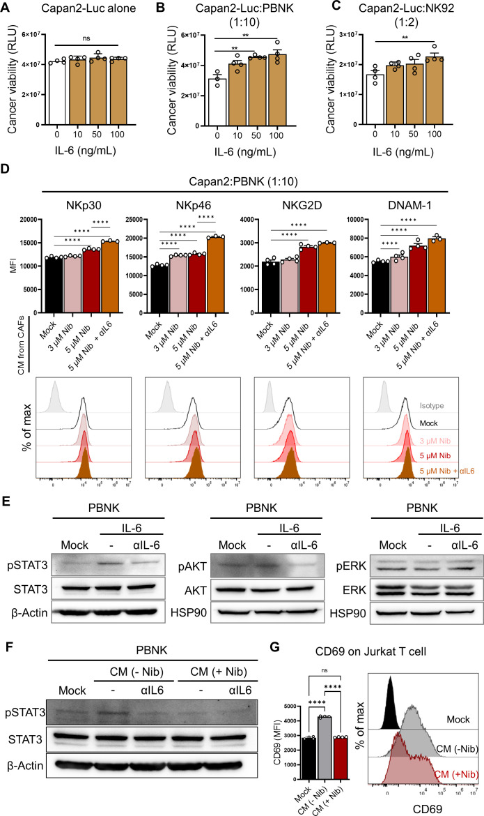
Background: Cancer-associated fibroblasts (CAFs) in the tumor microenvironment (TME) contribute to an impaired functionality of natural killer (NK) cells that have emerged as a promising therapeutic modality. The interaction between CAFs and NK cells within the TME exerts major inhibitory effects on immune responses, indicating CAF-targeted therapies as potential targets for effective NK-mediated cancer killing.

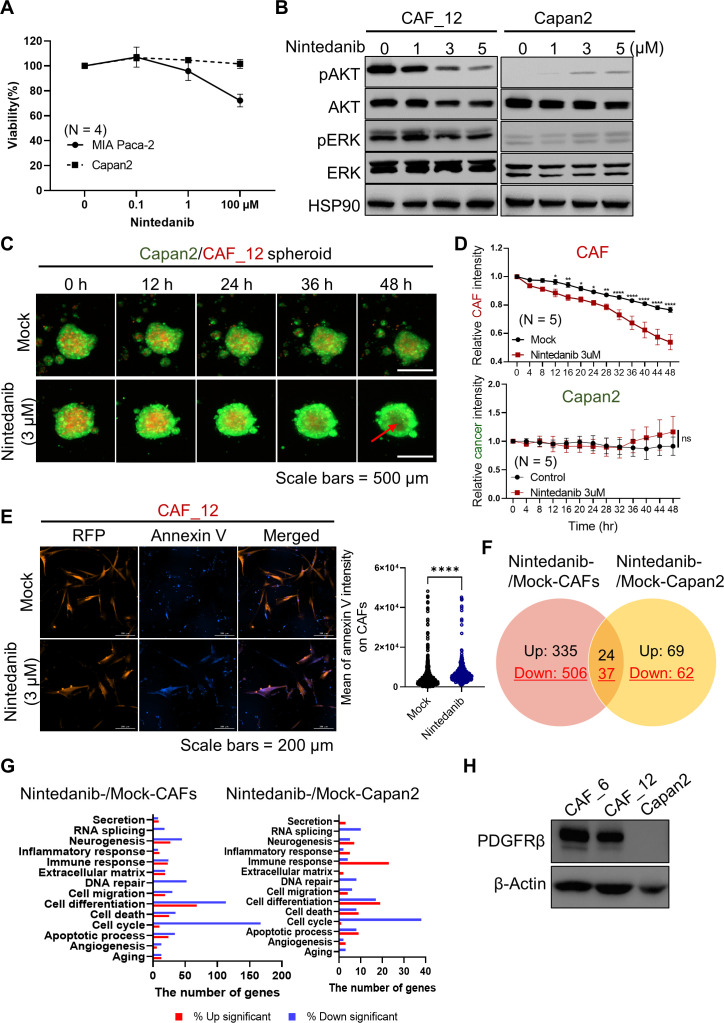
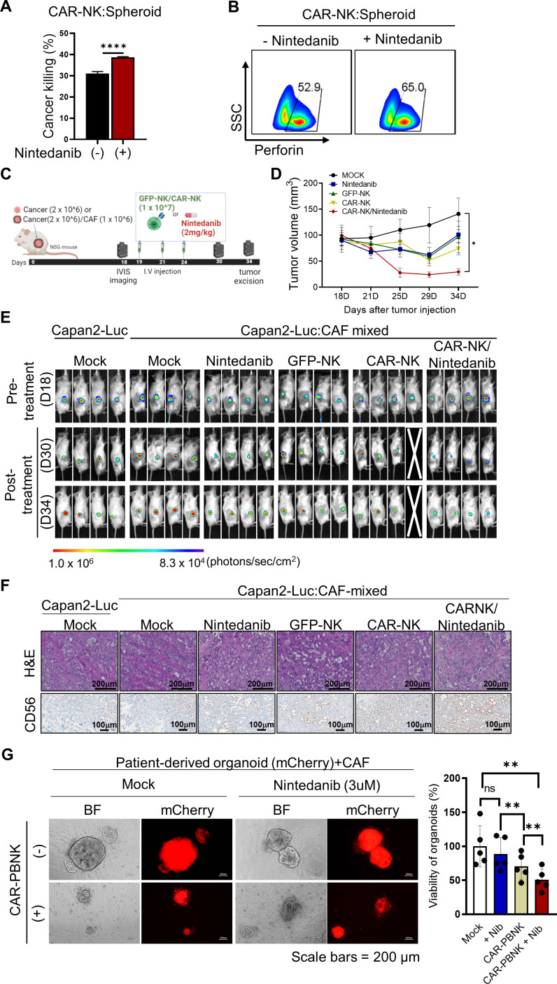
Methods: To overcome CAF-induced NK dysfunction, we selected an antifibrotic drug, nintedanib, for synergistic therapeutic combination. To evaluate synergistic therapeutic efficacy, we established an in vitro 3D Capan2/patient-derived CAF spheroid model or in vivo mixed Capan2/CAF tumor xenograft model. The molecular mechanism of NK-mediated synergistic therapeutic combination with nintedanib was revealed through in vitro experiments. In vivo therapeutic combination efficacy was subsequently evaluated. Additionally, the expression score of target proteins was measured in patient-derived tumor sections by the immunohistochemical method.

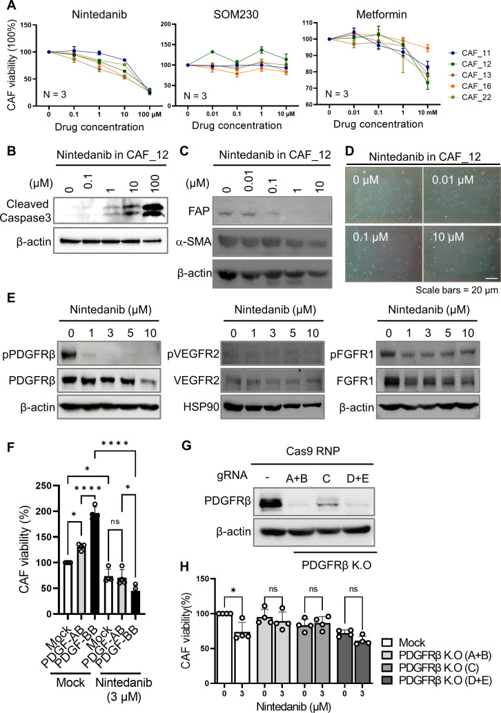
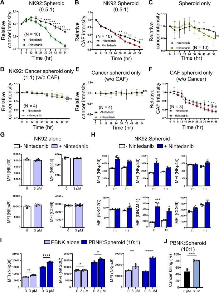
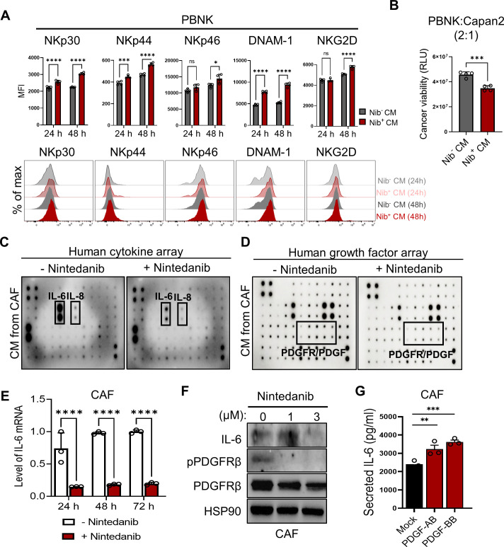
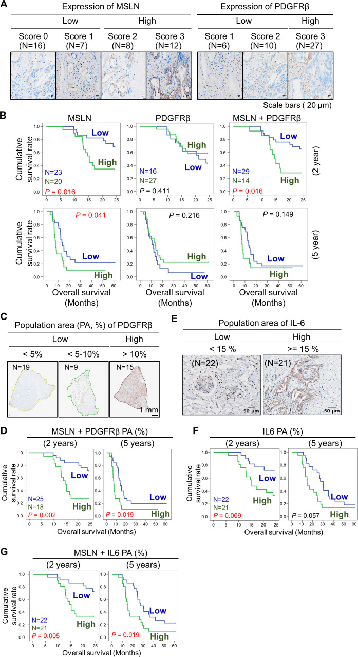
Results: Nintedanib blocked the platelet-derived growth factor receptor β (PDGFRβ) signaling pathway and diminished the activation and growth of CAFs, markedly reducing CAF-secreted IL-6. Moreover, coadministration of nintedanib improved the mesothelin (MSLN) targeting chimeric antigen receptor-NK-mediated tumor killing abilities in CAF/tumor spheroids or a xenograft model. The synergistic combination resulted in intense NK infiltration in vivo. Nintedanib alone exerted no effects, whereas blockade of IL-6 trans-signaling ameliorated the function of NK cells. The combination of the expression of MSLN and the PDGFRβ+-CAF population area, a potential prognostic/therapeutic marker, was associated with inferior clinical outcomes.

Conclusion: Our strategy against PDGFRβ+-CAF-containing pancreatic cancer allows improvements in the therapy of pancreatic ductal adenocarcinoma.

Keywords: Killer Cells, Natural; Receptors, Chimeric Antigen; Tumor Microenvironment.


Conflict of interest statement

Competing interests: None declared.

Figures

Figure 1
Figure 1
The selection of an antifibrotic drug for the depletion of patient-derived CAFs. (A) Cell viability was evaluated in five CAFs derived from patients with PDAC under treatment with nintedanib, SOM230, and metformin, respectively, as potential CAF-inhibiting drugs. Data were obtained from N=3 per each group. (B) Nintedanib-induced CAF cell death was validated by the level of cleaved caspase-3 using western blot experiments. (C) Nintedanib in CAF_12 lowered the expression of classical CAF markers in a dose-dependent manner. (D) Nintedanib-induced CAF cell death was observed using an optical microscope. (E) Putative molecular targets in response to nintedanib were accessed using western blot analysis. (F) The viability of CAF_12 in response to treatment with PDGF ligands was tested in the presence or absence of nintedanib. (*p<0.05, ****p<0.0001) (G, H) Nintedanib-induced CAF cell death was not affected in PDGFRβ K.O CAF cells achieved using CRISPR/Cas9.(*p<0.05) CAF, cancer-associated fibroblast; PDAC, pancreatic ductal adenocarcinoma; PDGFRβ, platelet-derived growth factor receptor β.
Figure 2
Figure 2
Prominent inhibitory effect of nintedanib in PDAC CAFs compared with cancer cells. (A) Cell viability was evaluated in cancer cells under treatment with nintedanib. (B) Nintedanib-treated CAFs exhibited decreased phosphorylation of AKT and ERK, members of the two major PDGF-mediated signaling pathways. Representative images were shown from one of three independent experiments. (C, D) Using a Capan2-CAF coculture spheroid system, live cell imaging was conducted in the presence or absence of nintedanib. Prior to coculture, GFP and RFP were introduced into Capan2 and CAF_12, respectively, for visualization. Quantitative data were obtained from N=5 per each group. P values were determined using one-way ANOVA followed by multiple comparison test. (*p<0.05, **p<0.01, ***p<0.001, ****p<0.0001) (E) Annexin V influx was measured on RFP expressing CAF cells to determine apoptotic death. Images from mock (N=861) and nintedanib (N=487) groups were visualized 24 hours post-treatment. P values were analyzed statistically using the unfired t-test. (****p<0.0001) (F, G) 3 µM nintedanib was added in CAF and Capan2, and then transcriptomic analysis was conducted in each sample (N=2). The number of differentially expressed genes (DEGs) with a fold change (FC) ≥2 and p<0.05 is shown in Venn diagram (F). The number of DEGs according to gene ontology enrichment (G). (H) Total expression of PDGFRβ on CAFs and Capan2 was accessed by a western blot experiment. ANOVA, analysis of variance; CAF, cancer-associated fibroblast; PDGFRβ, platelet-derived growth factor receptor β.
Figure 3
Figure 3
Nintedanib improved the NK-mediated cancer killing through the upregulated activation of markers. (A, B) To test the effect of nintedanib on NK-driven cytolysis, NK92 cells were incubated in the Capan2-CAF spheroid system with or without 3 µM nintedanib, and the relative intensities were measured in a time-dependent course based on fluorescence live cell imaging. (C) Cancer-CAF spheroids only, in the presence or absence of nintedanib. (D–F) Each single cell-type spheroid with or without NK92 was used as a control. (G–J) The direct or indirect effect of nintedanb on NK cytotoxic function was evaluated according to coculturing condition. Incubation with NK92 (G, H) and primary NK cells (I, J), respectively. Nintedanib ameliorated the function of NK cells only in Capan2-CAF spheroid condition. P values were determined using one-way ANOVA followed by multiple comparison test. *p<0.05, **p<0.01, ***p<0.001, ****p<0.0001. ANOVA, analysis of variance; CAF, cancer-associated fibroblast; NK, natural killer.
Figure 4
Figure 4
Nintedanib-treated CAFs exhibited a significant reduction in CAF-derived IL-6, consequently rescuing NK inhibition. (A, B) To reveal a NK-promoting role on treatment with nintedanib (Nib), the impact of nintedanib-treated condition media (CM) was evaluated through the expression of NK activating markers (A) or NK-mediated killing efficacy (B), respectively. p values were determined using one-way ANOVA followed by multiple comparison test. *p<0.05, **p<0.01, ***p<0.001, ****p<0.0001. Quantitative data were obtained from N=4 per each group. (C, D) Human cytokine array (C) and growth factor array (D), respectively, were conducted in the presence or absence of nintedanib to analyze the CAF-released secretome. (E, F) The level of IL-6 mRNA (****p<0.0001.) (E) or protein (F) is shown under treatment with nintedanib in CAFs. (G) Stimulation of PDGF ligands, such as PDGF-AB and PDGF-BB in CAFs induced the secretion of IL-6. Quantitative data were obtained from N=3 per each group. (**p<0.01, ***p<0.001) ANOVA, analysis of variance; CAF, cancer-associated fibroblast; NK, natural killer; PBNK, peripheral blood mononuclear cell-derived NK; PDGF, platelet-derived growth factor.
Figure 5
Figure 5
CAF-secreted IL-6 impeded NK cytotoxic function. (A–C) The effect of exogenous treatment with IL-6 in Capan2-Luc cells alone (A) and in coculture of Capan2-Luc with PBNK (B) or NK92 (C). For the luminescence-based tumor killing assay, nanoLuc luciferase-expressing Capan2 (Capan2-Luc) cells were generated. (**p<0.01) (B, C) PBNK or NK92 cells were cocultured with Capan2-Luc cells at a 10:1 or 2:1 ratio for 24 hours. (****p<0.0001) (D) The effect of the suppression of IL-6 on the activation of NK cells was investigated in the coculture of Capan2 with PBNK. Prior to coculture, PBNK cells were pretreated with CM from mock-, nintedanib-, and nintedanib plus αIL-6-treated CAFs for 24 hours. CAF-derived IL-6 attenuated the activation of NK cells. (E) IL-6-mediated signal transduction pathways in PBNK were validated under the treatment of IL-6 (100 ng/mL) with or without anti-IL-6 antibody (100 ng/mL) for 2 hours. (F) PBNK cells in the presence or absence of αIL-6 were incubated with CM from mock-, and nintedanib-treated CAFs for 2 h, and then the activation of STAT3 was detected by western blotting analysis. (G) Expression of CD69 on Jurkat T cells on treatment of CAF-derived CM in the presence or absence of nintedanib. Quantitative data were obtained from N=4 per group. (****p<0.0001) CAF, cancer-associated fibroblast; CM, conditioned media; NK, natural killer; PBNK, peripheral blood mononuclear cell-derived NK.
Figure 6
Figure 6
Optimal combination of MSLN-CAR-NK and nintedanib exhibited significant tumor killing in vitro and in vivo. (A, B) The CAR-NK-mediated cytotoxic function was significantly enhanced against MSLN-positive Capan2 under treatment with nintedanib (N=3). (****p<0.0001) (C) The scheme of the in vivo experiment for the combination of CAR-NK and nintedanib. For therapeutic studies, Capan2-Luc alone or a mixture of Capan2-Luc and CAF was subcutaneously injected into mice. (D, E) For therapeutic evaluation, the growth of each tumor (N=4) was monitored through size measurement using a caliper (*p<0.05) (D) or through visualization of the in vivo luminescence signal using the IVIS spectrum imaging system (E). (F) Histological analysis and IHC staining were conducted in excised tumor sections. (**p<0.01) (G) Synergistic cancer effect of CAR-PBNK with nintedanib, based on a patient-derived organoid/CAF co-culture system. CAF, cancer-associated fibroblast; CAR, chimeric antigen receptor; MSLN, mesothelin; NK, natural killer; IHC, immunohistochemical.
Figure 7
Figure 7
The combination of expression of MSLN and population of PDGFRβ+ CAFs was associated with inferior clinical outcomes and poor overall survival (OS). (A) Based on IHC staining intensity, the expression score of MSLN and PDGFRβ was measured in patient-derived tumor sections. The degree of staining intensity was scored and sorted into a low or high group. (B) Analysis of OS according to the score of target expression. Survival rates were determined using the Kaplan-Meier method according to MSLN, PDGFRβ, and the combination of MSLN and PDGFRβ; MSLN-low (N=23), high (N=20); PDGFRβ-low (N=16), high (N=27); MSLN+PDGFRβ-low (N=29), high (N=14). (C) Using the vs200 research slide scanner, the PDGFRβ+ population area (PA, %) was quantified and grouped into a low or high group; PDGFRβ PA-low (N=28), high (N=15). (D) Survival rates were analyzed according to the combination of MSLN and PDGFRβ PA; MSLN+PDGFRβ PA-low (N=25), high (N=18). (E) The IL-6 PA was quantified and grouped into a low and high group; IL-6 PA-low (N=22), high (N=21). (F) Analysis of OS according to the score of the IL-6 PA. (G) Survival rates were analyzed according to the combination of MSLN and IL-6 PA; MSLN+IL-6 PA-low (N=22), high (N=21). N indicates the number of clinical specimens. CAF, cancer-associated fibroblast; IHC, immunohistochemical; MSLN, mesothelin; PDGFRβ, platelet-derived growth factor receptor β.

References

    1. Kleeff J, Korc M, Apte M, et al. . Pancreatic cancer. Nat Rev Dis Primers 2016;2:16022. 10.1038/nrdp.2016.22 - DOI - PubMed
    1. Ligorio M, Sil S, Malagon-Lopez J, et al. . Stromal microenvironment shapes the intratumoral architecture of pancreatic cancer. Cell 2019;178:160–75. 10.1016/j.cell.2019.05.012 - DOI - PMC - PubMed
    1. Chu GC, Kimmelman AC, Hezel AF, et al. . Stromal biology of pancreatic cancer. J Cell Biochem 2007;101:887–907. 10.1002/jcb.21209 - DOI - PubMed
    1. Jacobetz MA, Chan DS, Neesse A, et al. . Hyaluronan impairs vascular function and drug delivery in a mouse model of pancreatic cancer. Gut 2013;62:112–20. 10.1136/gutjnl-2012-302529 - DOI - PMC - PubMed
    1. Olivares O, Mayers JR, Gouirand V, et al. . Collagen-derived proline promotes pancreatic ductal adenocarcinoma cell survival under nutrient limited conditions. Nat Commun 2017;8:16031. 10.1038/ncomms16031 - DOI - PMC - PubMed

Publication types