. 2023 Feb 27;13(1):3365.
doi: 10.1038/s41598-023-30528-9.

Structural basis for the toxic activity of MafB2 from maf genomic island 2 (MGI-2) in N. meningitidis B16B6

Affiliations

Structural basis for the toxic activity of MafB2 from maf genomic island 2 (MGI-2) in N. meningitidis B16B6

So Hyeon Park et al. Sci Rep. .

Abstract

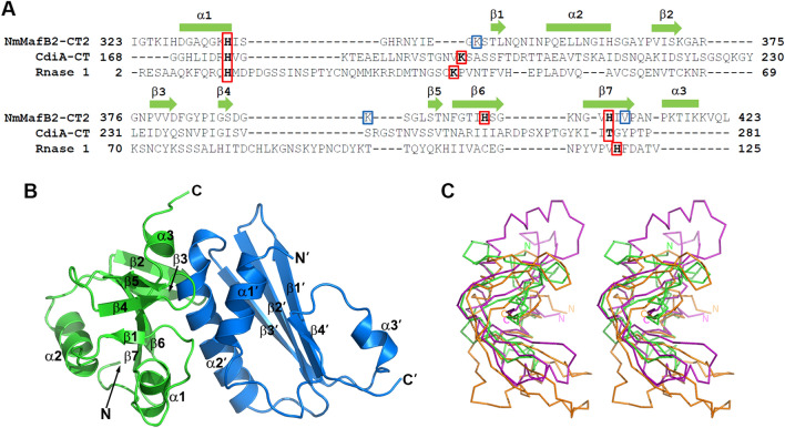
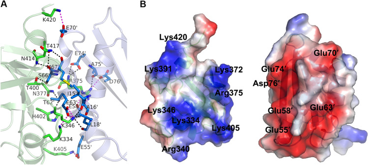
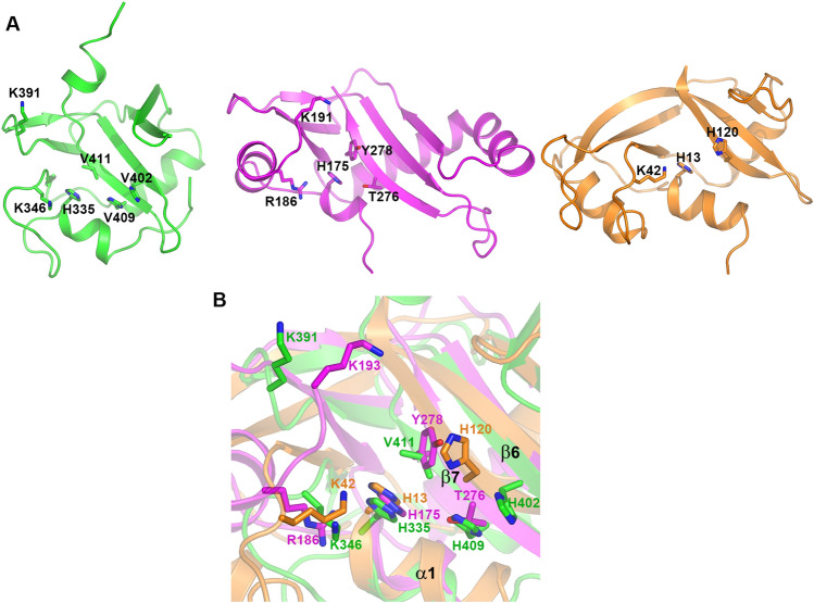
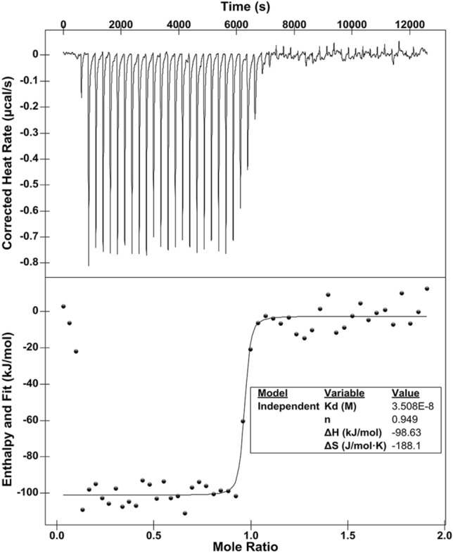
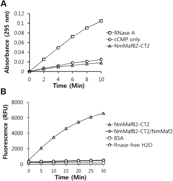
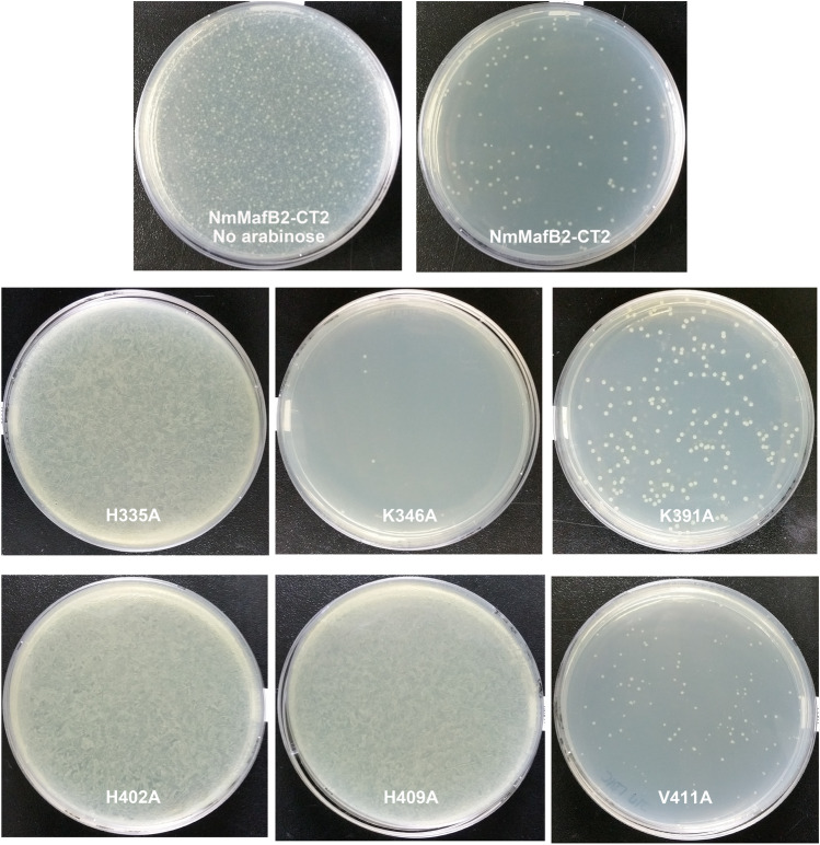
The Maf polymorphic toxin system is involved in conflict between strains found in pathogenic Neisseria species such as Neisseria meningitidis and Neisseria gonorrhoeae. The genes encoding the Maf polymorphic toxin system are found in specific genomic islands called maf genomic islands (MGIs). In the MGIs, the MafB and MafI encode toxin and immunity proteins, respectively. Although the C-terminal region of MafB (MafB-CT) is specific for toxic activity, the underlying enzymatic activity that renders MafB-CT toxic is unknown in many MafB proteins due to lack of homology with domain of known function. Here we present the crystal structure of the MafB2-CTMGI-2B16B6/MafI2MGI-2B16B6 complex from N. meningitidis B16B6. MafB2-CTMGI-2B16B6 displays an RNase A fold similar to mouse RNase 1, although the sequence identity is only ~ 14.0%. MafB2-CTMGI-2B16B6 forms a 1:1 complex with MafI2MGI-2B16B6 with a Kd value of ~ 40 nM. The complementary charge interaction of MafI2MGI-2B16B6 with the substrate binding surface of MafB2-CTMGI-2B16B6 suggests that MafI2MGI-2B16B6 inhibits MafB2-CTMGI-2B16B6 by blocking access of RNA to the catalytic site. An in vitro enzymatic assay showed that MafB2-CTMGI-2B16B6 has ribonuclease activity. Mutagenesis and cell toxicity assays demonstrated that His335, His402 and His409 are important for the toxic activity of MafB2-CTMGI-2B16B6, suggesting that these residues are critical for its ribonuclease activity. These data provide structural and biochemical evidence that the origin of the toxic activity of MafB2MGI-2B16B6 is the enzymatic activity degrading ribonucleotides.


Conflict of interest statement

The authors declare no competing interests.

Figures

Figure 1
Figure 1
Overall structure of the NmMafB2-CT2/NmMafI2 complex. (A) Structure-guided sequence alignment of NmMafB2-CT2 with CdiA-CTYkris and RNase 1. The rectangles (α-helixes) and arrows (β-strands) in the above alignment indicate the secondary structures of NmMafB2-CT2. Putative catalytic residues are shown in red boxes, and the residues (K346, K391 and V411) near the catalytic residues of CdiA-CTYkris or RNase 1 but not critical for toxicity activity are in blue boxes. (B) Ribbon diagram of the NmMafB2-CT2/NmMafI2 complex. NmMafB2-CT2 and NmMafI2 are shown in green and sky blue, respectively. N- and C-termini, and the secondary structure elements are labeled. A prime symbol is added for NmMafI2. (C) Stereoview of the structural overlay of NmMafB2-CT2, CdiA-CTYkris (PDB code: 5E3E) and RNase 1 (PDB code: 3TSR). The ribbon diagrams of NmMafB2-CT2, CdiA-CTYkris and RNase 1 are displayed in green, magenta and orange, respectively. The view was rotated by ~ 90° horizontally from that of (B) with the upper part toward the viewer.
Figure 2
Figure 2
Interface between NmMafB2-CT2 and NmMafI2. (A) Detailed view of the interface between NmMafB2-CT2 and NmMafI2. The residues involved in hydrogen bond and salt bridge interactions are shown in the stick model in green (NmMafB2-CT2) and sky blue (NmMafI2). The hydrogen bonds and salt bridges are shown as dash lines in black and red, respectively. The residues are labeled, and those of NmMafI2 are indicated with a prime symbol (′). (B) Open book view of the NmMafB2-CT2/NmMafI2 complex in a surface model with the electrostatic potentials. The potentials are shown in the range of − 75 kT/e (red, negative potential) to + 75 kT/e (blue, positive potential). The positively charged surface of NmMafI2 counteracts with the negatively charged surface of NmMafB2-CT2 in the interface of the complex. The positions of positively charged residues on the surface of NmMafB2-CT2 and negatively charged residues on the surface of NmMafI2 are indicated.
Figure 3
Figure 3
RNase fold of NmMafB2-CT2. (A) Ribbon diagram of NmMafB2-CT2 (left, green), CdiA-CTYkris (middle, magenta) and RNase 1 (right, orange). Catalytic residues are shown in a stick model. The view was rotated by ~ 90° vertically and clockwise from that of Fig. 1B. (B) Zoomed-in view of active sites. The stick model depicts residues of NmMafB2-CT2 superimposed on residues functioning as a base (H13 of RNase 1) or an acid (H120 of RNase 1 and T276 and Y278 of CdiA-CTYkris), those providing stabilizing environment during catalysis.
Figure 4
Figure 4
Interaction between NmMafB2-CT2 and NmMafI2. Isothermal titration calorimetry (ITC) for measuring the binding affinity of NmMafI2 to NmMafB2-CT2. The titration of NmMafB2-CT2 (0.2 mM) was performed by 50 injections of 1.3 mM NmMafI2.
Figure 5
Figure 5
Enzymatic assay of NmMafB2-CT2 in vitro. (A) Phosphodiester hydrolysis activity assay. The hydrolysis of cCMP was analyzed by measuring the increase in absorbance at 296 nm for 10 min. RNase A and cCMP were also included as the positive and negative controls, respectively. (B) RNase assay. The RNA cleavage activities were measured with a fluorophotometer using RNaseAlert fluorescent substrate at Ex/Em = 485/530 nm at 37 °C for 30 min. NmMafB2-CT2/NmMafI2 was the purified protein used to obtain NmMafB2-CT2. BSA: bovine serum albumin; RFU: relative fluorescence unit.
Figure 6
Figure 6
Toxicity of NmMafB2-CT2 in E. coli. The toxic activity of NmMafB2-CT2 and its mutants was assessed by counting the number of colonies. Overnight cultures of BL21 Star (DE3) transformed with the NmMafB2-CT2 or variants were diluted to an OD600 of 0.1 in fresh LB medium, and 0.2% L-arabinose was added to initiate toxin expression. After a further 6 h of incubation at 37 °C, the cultures were appropriately diluted and spread on agar plate and the plates were incubated at 37 °C overnight.

Similar articles

References

    1. Alouf JE. Bacterial protein toxins. An overview. Methods Mol. Biol. 2000;145:1–26. - PubMed
    1. Burga A, Ben-David E, Kruglyak L. Toxin-antidote elements across the tree of life. Annu. Rev. Genet. 2000;54:387–415. doi: 10.1146/annurev-genet-112618-043659. - DOI - PubMed
    1. Alkhatib Z, Abts A, Mavaro A, Schmitt L, Smits SH. Lantibiotics: How do producers become self-protected? J. Biotechnol. 2012;159:145–154. doi: 10.1016/j.jbiotec.2012.01.032. - DOI - PubMed
    1. Iyer LM, Zhang D, Rogozin IB, Aravind L. Evolution of the deaminase fold and multiple origins of eukaryotic editing and mutagenic nucleic acid deaminases from bacterial toxin systems. Nucl. Acids Res. 2011;39:9473–9497. doi: 10.1093/nar/gkr691. - DOI - PMC - PubMed
    1. Zhang D, Iyer LM, Aravind L. A novel immunity system for bacterial nucleic acid degrading toxins and its recruitment in various eukaryotic and DNA viral systems. Nucl. Acids Res. 2011;39:4532–4552. doi: 10.1093/nar/gkr036. - DOI - PMC - PubMed

Publication types

LinkOut - more resources