Review
. 2023 Jul;24(7):495-516.
doi: 10.1038/s41580-023-00583-1. Epub 2023 Feb 27.

Cell-extracellular matrix mechanotransduction in 3D

Affiliations
Review

Cell-extracellular matrix mechanotransduction in 3D

Aashrith Saraswathibhatla et al. Nat Rev Mol Cell Biol. 2023 Jul.

Abstract

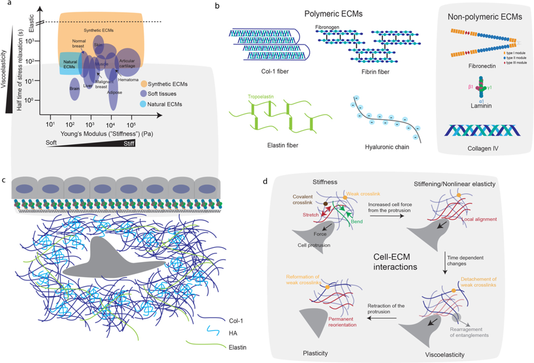
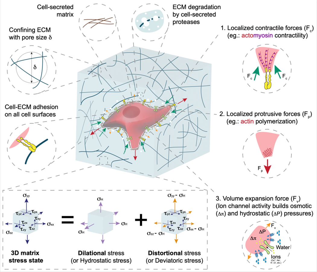
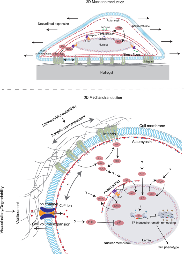
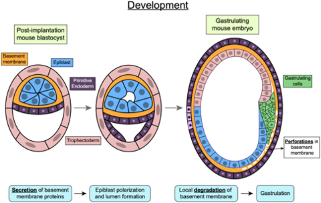
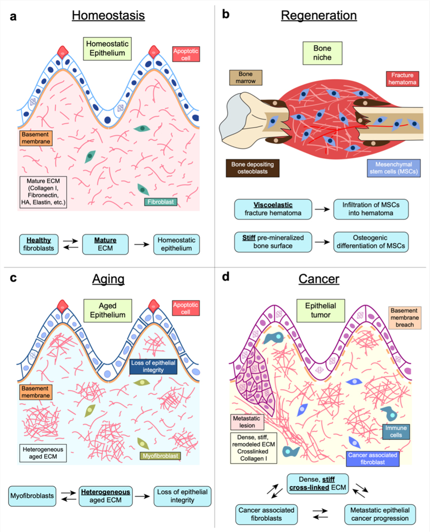
Mechanical properties of extracellular matrices (ECMs) regulate essential cell behaviours, including differentiation, migration and proliferation, through mechanotransduction. Studies of cell-ECM mechanotransduction have largely focused on cells cultured in 2D, on top of elastic substrates with a range of stiffnesses. However, cells often interact with ECMs in vivo in a 3D context, and cell-ECM interactions and mechanisms of mechanotransduction in 3D can differ from those in 2D. The ECM exhibits various structural features as well as complex mechanical properties. In 3D, mechanical confinement by the surrounding ECM restricts changes in cell volume and cell shape but allows cells to generate force on the matrix by extending protrusions and regulating cell volume as well as through actomyosin-based contractility. Furthermore, cell-matrix interactions are dynamic owing to matrix remodelling. Accordingly, ECM stiffness, viscoelasticity and degradability often play a critical role in regulating cell behaviours in 3D. Mechanisms of 3D mechanotransduction include traditional integrin-mediated pathways that sense mechanical properties and more recently described mechanosensitive ion channel-mediated pathways that sense 3D confinement, with both converging on the nucleus for downstream control of transcription and phenotype. Mechanotransduction is involved in tissues from development to cancer and is being increasingly harnessed towards mechanotherapy. Here we discuss recent progress in our understanding of cell-ECM mechanotransduction in 3D.


Conflict of interest statement

Competing interests

The authors declare no competing interests.

Figures

Figure 1.
Figure 1.. Tissue mechanics, ECM components, and cell-ECM mechanical interactions.
(a) Stiffness and half time of stress relaxation for various soft tissues, and the range of those properties accessible with reconstituted ECMs or synthetic ECMs (i.e. hydrogels). Data were taken from refs. ,,,,,,,. Stress relaxation half times, which provide a measure of viscoelasticity, are defined as the time for the stress to relax to half its original value in response to a constant deformation. Synthetic ECMs, including alginate hydrogels and PEG hydrogels, can be modified to mimic the physiological mechanics of soft tissues (check Box 1). (b) Schematics of structural components of major polymeric (left) and non-polymeric (right) ECMs in tissues. (c) Schematic of an epithelial monolayer with basement membrane underneath, and a stromal cell surrounded by fibrous ECMs such as col-1 and elastin in the underlying connective tissue. (d) As cells interact with ECM, these interactions are mediated by mechanical properties of the ECM including stiffness, nonlinear elasticity, viscoelasticity, and plasticity. As the cell push/pull on the ECM, the ECM may resist the cellular force through bending and stretching of the ECM fibers (left top). With increased forces from the cell, the ECM may stiffen (i.e. exhibit greater resistance) due to local alignment in fibers (right top). Over the time of force application, the ECM may undergo creep and stresses may relax due to detachment of weak crosslinks and fiber rearrangements (bottom right). Once the cell detaches from the ECM, the ECM may retain permanent deformations resulting from reformation of weak crosslinks that lock in changes in fibre position and alignment (bottom left).
Figure 2.
Figure 2.. Cell–matrix interactions in 3D.
Cells in 3D are confined in all directions due to restrictions imposed by the matrix and can form cell–matrix adhesions on all surfaces contacting ECM. Cells exert contractile, protrusive, and volumetric forces on the matrix, generating dilational and distortional stresses in the ECM, which in turn regulate various cell behaviours (Table 1). Hydrostatic stress is volumetric or dilational stress that acts to increase or decrease the volume of an object on which it acts, without changing its shape. Deviatoric stress is distortional stress that acts to change the shape of an object on which it acts, without changing its volume. Hydrostatic and deviatoric stresses combine to produce net 3D stress fields which cells perceive. These stress fields directly deform cells and influence their behaviour including proliferation, migration and differentiation. In addition to this, such stresses change over time depending on ECM properties such as viscoelasticity, degradability and plasticity, and form a positive feedback loop with the cell-generated forces.
Figure 3.
Figure 3.. 2D and 3D Mechanotransduction.
A) Cells sense substrate stiffness by exerting contractile forces on 2D substrates with stress fibres through focal adhesions, which activates various proteins such as FAK, talin, Rho and ROCK at the adhesion site. Activation of these proteins leads to adhesion maturation and stress fibre formation and contractility, which in turn transmits forces to the nucleus via the linker of nucleoskeleton and cytoskeleton (LINC) complex, resulting in changes in nuclear envelope tension and nuclear pore opening. This allows the nuclear entry of proteins such as YAP transcriptional regulator leading to downstream impact on cell phenotype. Moreover, in 2D, a cell can spread laterally without encountering any mechanical confinement. B) Cells embedded in the ECM sense stiffness and viscoelastic properties of the matrix through integrin binding, activation, and clustering, while sensing confinement, viscoelasticity and plasticity through cell volume changes and ion channel activation which leads to Ca2+ ion influx. Additionally, ECM stiffness/viscoelasticity and confinement regulate activation of various proteins, such as FAK, ROCK, MLCK, pathways, such as those involving PI3K, ERK, and Rho, and transcriptional regulators such as YAP, p27, Sp1, RUNX2, and EGR1. However, clear mechanistic links between the ECM properties and activation of these proteins, pathways, and transcription regulators remain unclear. Unknown connections in the pathways are indicated by question marks. Both mechanisms of mechanotransduction converge on the nucleus and regulate the activation of transcription factors (TFs), which are facilitated by chromatin remodelling and control cell behaviour.
Figure 4.
Figure 4.. Mechanotransduction in development.
During development, basement membrane secretion and degradation triggers mouse epiblast lumenogenesis and gastrulation respectively. Post-implantation, trophectoderm cells in the mouse embryo, secrete a basement membrane around the epiblast which triggers polarization and lumen formation in the epiblast. Later during gastrulation, gastrulating cells, also called primitive streak cells, secrete proteases to locally degrade the basement membrane layer and enable migration.
Figure 5.
Figure 5.. Mechanotransduction in tissues.
(a) In homeostasis, healthy ECM and fibroblasts help maintain normally functioning epithelium by maintaining optimal ECM mechanical properties such as stiffness and viscoelasticity. (b) Following a bone fracture, viscoelasticity of fracture hematoma promotes infiltration pf mesenchymal stem cells (MSCs) and stiff bone surface promotes differentiation of MSCs into bone-producing osteoblasts. (c) Myofibroblast differentiation and heterogeneous ECM occurring during ageing results in loss of epithelial integrity and function. As tissue fibrosis proceeds with ageing, normal fibroblasts differentiate into a myofibroblast phenotype and heterogeneously secrete and deform the ECM. Such a heterogeneous matrix promotes further myofibroblast differentiation and results in altered ECM mechanical properties. Epithelial cells sense these altered ECM properties and undergo transcriptional changes causing loss of epithelial integrity and function. (d) During cancer progression, cancer-associated fibroblasts remodel the ECM into a dense, stiff matrix. This increase in ECM stiffness, in combination with other cues and genetic changes in the cancer cells, leads to activation of a malignant phenotype in epithelial cells. These cells then undergo sustained proliferation, breach the basement membrane during invasion, migrate into the stromal matrix and eventually can metastasize.

References

    1. Levental I, Georges PC & Janmey PA Soft biological materials and their impact on cell function. Soft Matter 3, 299–306, doi:10.1039/B610522J (2007). - DOI - PubMed
    1. Swift J. et al. Nuclear Lamin-A Scales with Tissue Stiffness and Enhances Matrix-Directed Differentiation. Science 341, 1240104, doi:doi:10.1126/science.1240104 (2013). - DOI - PMC - PubMed
    1. Storm C, Pastore JJ, MacKintosh FC, Lubensky TC & Janmey PA Nonlinear elasticity inbiological gels. Nature 435, 191–194, doi:10.1038/nature03521 (2005). - DOI - PubMed
    1. Discher Dennis E, Janmey P. & Wang Y -l. Tissue Cells Feel and Respond to the Stiffness of Their Substrate. Science 310, 1139–1143, doi:10.1126/science.1116995 (2005). - DOI - PubMed
    1. Vogel V. & Sheetz M. Local force and geometry sensing regulate cell functions. Nature Reviews Molecular Cell Biology 7, 265–275, doi:10.1038/nrm1890 (2006). - DOI - PubMed

MeSH terms