Review
. 2023 Apr;21(2):147-153.
doi: 10.1007/s11914-023-00779-6. Epub 2023 Feb 28.

Craniofacial Fibrous Dysplasia: Clinical and Therapeutic Implications

Affiliations
Review

Craniofacial Fibrous Dysplasia: Clinical and Therapeutic Implications

Vivian Szymczuk et al. Curr Osteoporos Rep. 2023 Apr.

Abstract

Purpose of review: This study aims to review diagnosis, potential complications, and clinical management in craniofacial fibrous dysplasia.

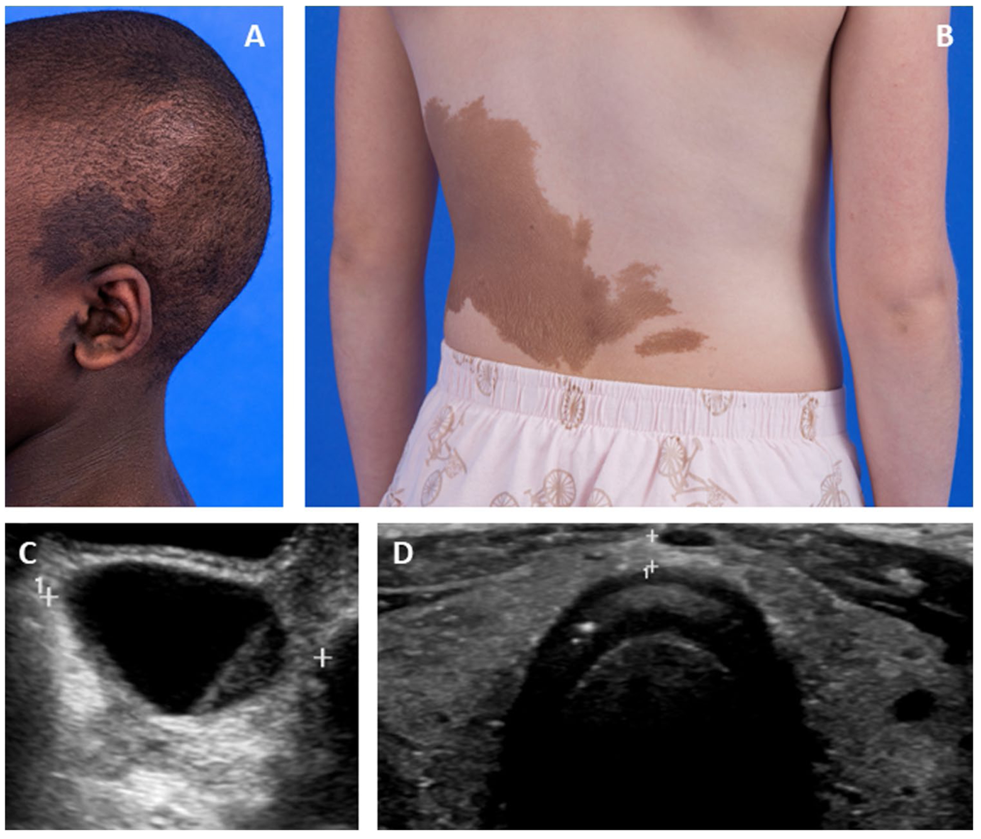
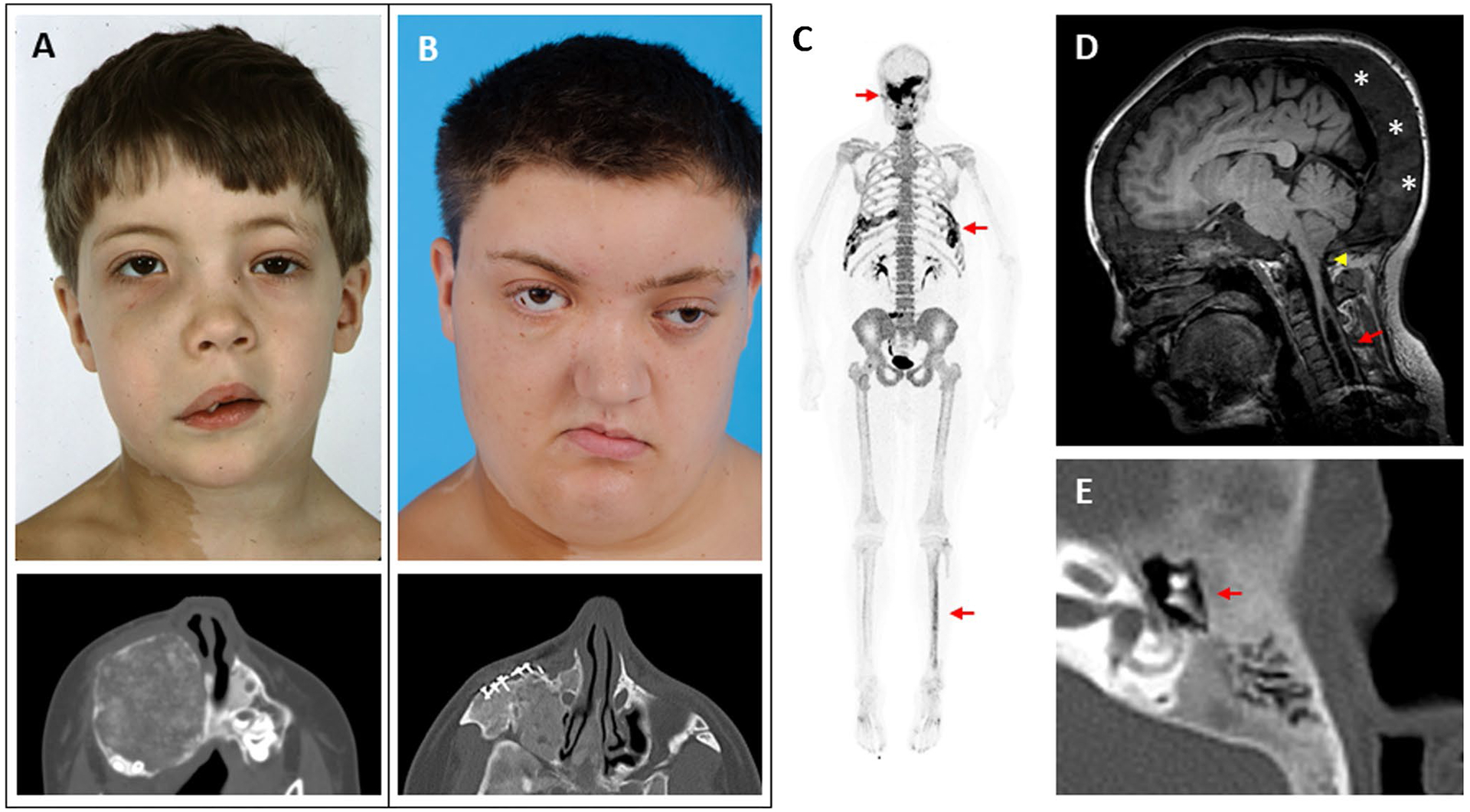
Recent findings: Fibrous dysplasia (FD) is a rare mosaic disorder in which normal bone and marrow are replaced with expansile fibro-osseous lesions. Disease presents along a broad spectrum and may be associated with extraskeletal features as part of McCune-Albright syndrome (MAS). The craniofacial skeleton is one of the most commonly impacted areas in FD, and its functional and anatomical complexities create unique challenges for diagnosis and management. This review summarizes current approaches to diagnosis and management in FD/MAS, with emphasis on the clinical and therapeutic implications for the craniofacial skeleton.

Keywords: Craniofacial fibrous dysplasia; Fibrous dysplasia; McCune Albright syndrome; Rare bone disease.


Conflict of interest statement

Conflict of interest The authors report no additional conflicts of interest.

Figures

Fig. 1
Fig. 1
Extraskeletal features of McCune-Albright syndrome. A Photograph showing typical hyperpigmented macules overlying a patient’s skull and ear. B Photograph showing typical hyperpigmented macules on a patient’s trunk. C Pelvic ultrasound image showing a typical ovarian cyst associated with precocious puberty. D Thyroid ultrasound image showing diffuse heterogeneity of both lobes
Fig. 2
Fig. 2
Clinical images of craniofacial fibrous dysplasia (FD). The left-hand panels show images from the same patient at ages 6 (A) and 18 years (B). Note the progression in facial asymmetry, including expansion of the right-sided face and orbital dystopia. The lower panels show axial computed tomography views of his right maxilla, including surgical implants from a reconstructive procedure performed at age 7. C 18F-NaF PET/CT scan performed in this patient at age 25 years. Areas of increased tracer uptake correspond to FD lesions in his skull, ribs, and tibia (red arrows). D Brain MRI from a 17-year-old girl with craniofacial FD and headaches. The cerebellar tonsils extend below the foramen magnum, consistent with a Chiari I malformation (yellow arrowhead). An associated syrinx has developed in the spinal cavity (red arrow). Note the diffuse expansion of the posterior cranium involved with FD (asterisks). E Computed tomography scan of the temporal bone in a 13-year-old patient with mild conductive hearing loss. The etpitympanum is diffusely involved with FD (red arrows), surrounding the ossicular chain
Fig. 3
Fig. 3
Histologic features of craniofacial fibrous dysplasia. A H&E section from a sphenoid bone shows diffuse fibrous tissue (ft) with prominent, irregularly situated osteoclasts (black arrows). B H&E section from a maxilla shows discontinuous, curvilinear trabeculae (tr) on a background on fibrous tissue (ft). Again noted are prominent osteoclasts (black arrows)

References

    1. Boyce AM, Florenzano P, de Castro LF, Collins MT. Fibrous Dysplasia / McCune-Albright Syndrome. In: Adam MP, Everman DB, Mirzaa GM, Pagon RA, Wallace SE, Bean LJH, Gripp KW, Amemiya A, editors. GeneReviews® [Internet]. Seattle (WA): University of Washington, Seattle; 2015. pp. 1993–2023. - PubMed
    1. Weinstein LS, Liu J, Sakamoto A, Xie T, Chen M. Minireview: GNAS: normal and abnormal functions. Endocrinology. 2004;145(12):5459–64. 10.1210/en.2004-0865. - DOI - PubMed
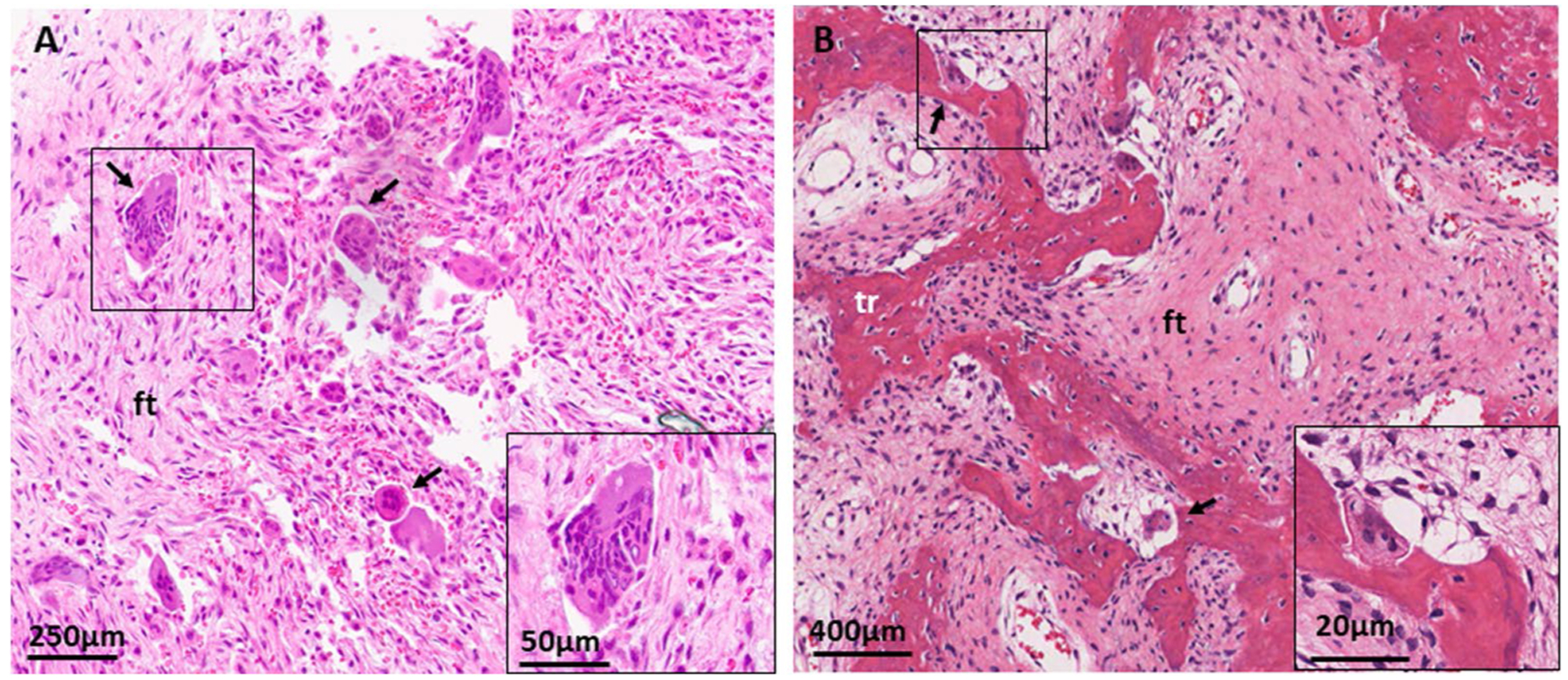
    1. Riminucci M, Liu B, Corsi A, et al. The histopathology of fibrous dysplasia of bone in patients with activating mutations of the Gs alpha gene: site-specific patterns and recurrent histological hallmarks. J Pathol. 1999;187(2):249–58. 10.1002/(sici)1096-9896(199901)187:2<249::Aid-path222>3.0.Co;2-j. - DOI - PubMed
    1. Riminucci M, Saggio I, Robey PG, Bianco P. Fibrous dysplasia as a stem cell disease. J Bone Miner Res. 2006;21(Suppl 2):P125–31. 10.1359/jbmr.06s224. - DOI - PubMed
    1. Collins MT, Singer FR, Eugster E. McCune-Albright syndrome and the extraskeletal manifestations of fibrous dysplasia. Orphanet J Rare Dis. 2012;7(Suppl 1):S4. 10.1186/1750-1172-7-s1-s4. - DOI - PMC - PubMed

Publication types

MeSH terms