

Impaired autophagy-accelerated senescence of alveolar type II epithelial cells drives pulmonary fibrosis induced by single-walled carbon nanotubes
- PMID: 36849924
- PMCID: PMC9970859
- DOI: 10.1186/s12951-023-01821-6
Impaired autophagy-accelerated senescence of alveolar type II epithelial cells drives pulmonary fibrosis induced by single-walled carbon nanotubes
Abstract
Background: The rapid increase in production and application of carbon nanotubes (CNTs) has led to wide public concerns in their potential risks to human health. Single-walled CNTs (SWCNTs), as an extensively applied type of CNTs, have shown strong capacity to induce pulmonary fibrosis in animal models, however, the intrinsic mechanisms remain uncertain.
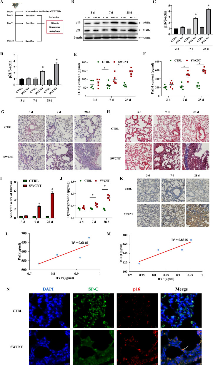
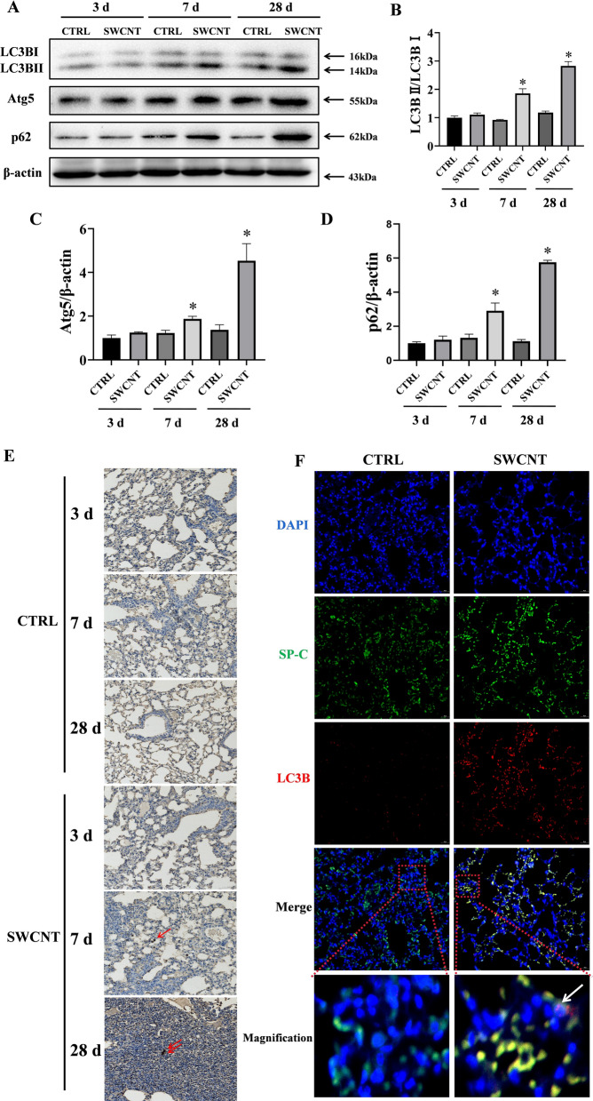
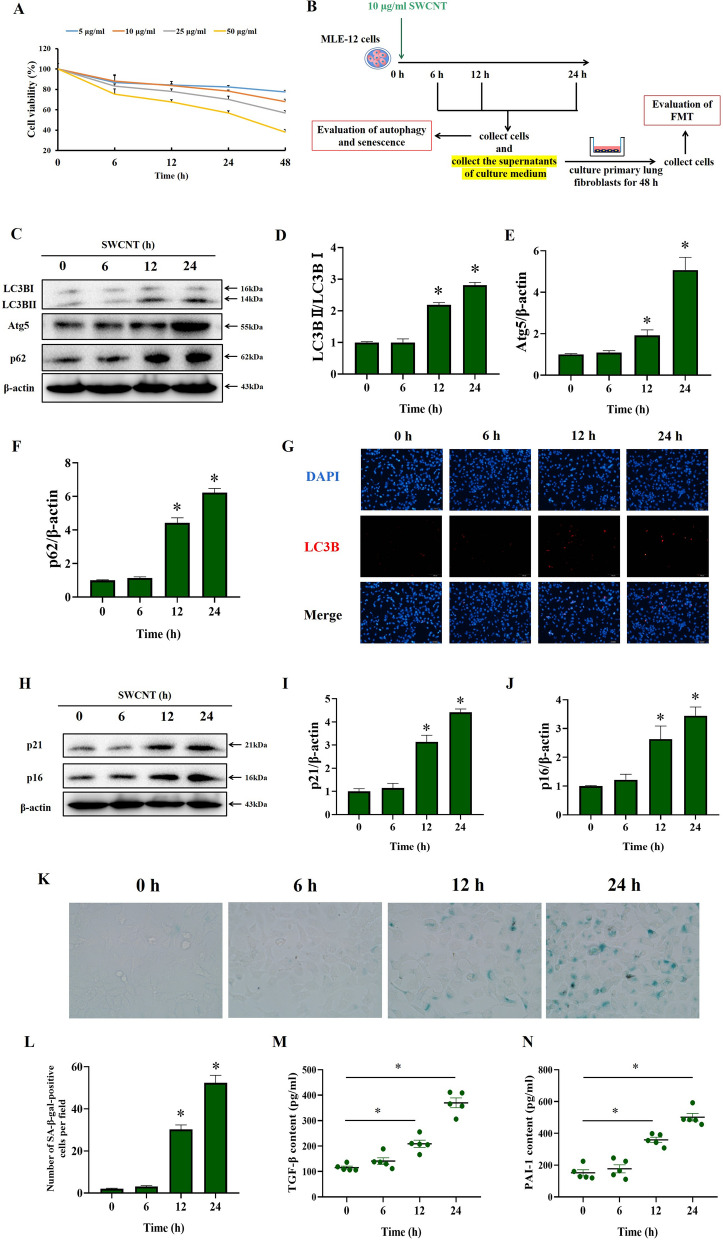
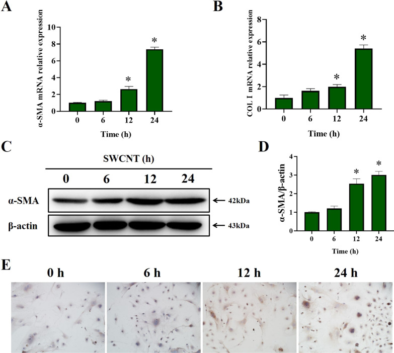
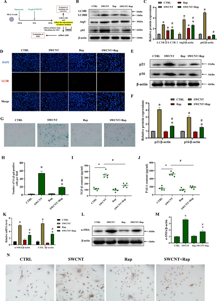
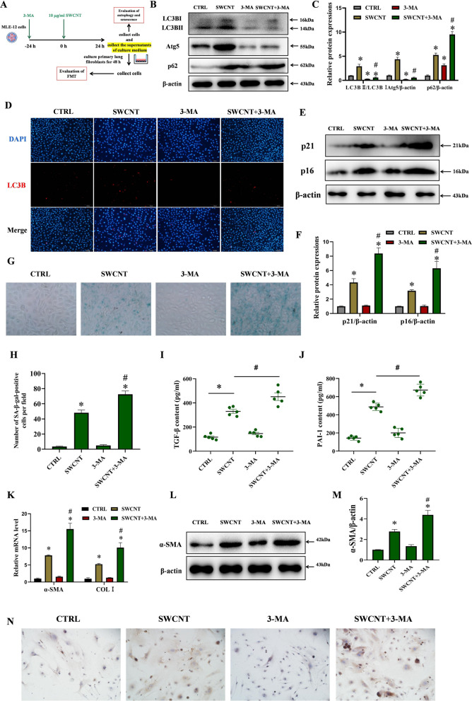
Results: In vivo experiments, we showed that accelerated senescence of alveolar type II epithelial cells (AECIIs) was associated with pulmonary fibrosis in SWCNTs-exposed mice, as well as SWCNTs-induced fibrotic lungs exhibited impaired autophagic flux in AECIIs in a time dependent manner. In vitro, SWCNTs exposure resulted in profound dysfunctions of MLE-12 cells, characterized by impaired autophagic flux and accelerated cellular senescence. Furthermore, the conditioned medium from SWCNTs-exposed MLE-12 cells promoted fibroblast-myofibroblast transdifferentiation (FMT). Additionally, restoration of autophagy flux with rapamycin significantly alleviated SWCNTs-triggered senescence and subsequent FMT whereas inhibiting autophagy using 3-MA aggravated SWCNTs-triggered senescence in MLE-12 cells and FMT.
Conclusion: SWCNTs trigger senescence of AECIIs by impairing autophagic flux mediated pulmonary fibrosis. The findings raise the possibility of senescence-related cytokines as potential biomarkers for the hazard of CNTs exposure and regulating autophagy as an appealing target to halt CNTs-induced development of pulmonary fibrosis.
Keywords: Alveolar type II epithelial cells; Autophagy; Carbon nanotube; Pulmonary fibrosis; Senescence.
© 2023. The Author(s).
Conflict of interest statement
The authors declare that they have no competing interest.
Figures
Similar articles
-
Carbon nanotubes promote alveolar macrophages toward M2 polarization mediated epithelial-mesenchymal transition and fibroblast-to-myofibroblast transdifferentiation.Nanotoxicology. 2021 Jun;15(5):588-604. doi: 10.1080/17435390.2021.1905098. Epub 2021 Apr 10. Nanotoxicology. 2021. PMID: 33840345
-
Carbon nanotubes and crystalline silica induce matrix remodeling and contraction by stimulating myofibroblast transformation in a three-dimensional culture of human pulmonary fibroblasts: role of dimension and rigidity.Arch Toxicol. 2018 Nov;92(11):3291-3305. doi: 10.1007/s00204-018-2306-9. Epub 2018 Sep 18. Arch Toxicol. 2018. PMID: 30229330
-
Effect of fiber length on carbon nanotube-induced fibrogenesis.Int J Mol Sci. 2014 Apr 29;15(5):7444-61. doi: 10.3390/ijms15057444. Int J Mol Sci. 2014. PMID: 24786100 Free PMC article.
-
A review of carbon nanotube toxicity and assessment of potential occupational and environmental health risks.Crit Rev Toxicol. 2006 Mar;36(3):189-217. doi: 10.1080/10408440600570233. Crit Rev Toxicol. 2006. PMID: 16686422 Review.
-
Myofibroblasts and lung fibrosis induced by carbon nanotube exposure.Part Fibre Toxicol. 2016 Nov 4;13(1):60. doi: 10.1186/s12989-016-0172-2. Part Fibre Toxicol. 2016. PMID: 27814727 Free PMC article. Review.
Cited by
-
Impact of immunosuppressive therapy on pulmonary perfusion in kidney transplant recipients after COVID-19 illness.Front Med (Lausanne). 2025 Jun 11;12:1562407. doi: 10.3389/fmed.2025.1562407. eCollection 2025. Front Med (Lausanne). 2025. PMID: 40568197 Free PMC article.
-
Senescence of alveolar epithelial progenitor cells: a critical driver of lung fibrosis.Am J Physiol Cell Physiol. 2023 Aug 1;325(2):C483-C495. doi: 10.1152/ajpcell.00239.2023. Epub 2023 Jul 17. Am J Physiol Cell Physiol. 2023. PMID: 37458437 Free PMC article. Review.
-
TMED3 promotes prostate cancer via FOXO1a and FOXO3a phosphorylation.Oncol Res. 2024 Dec 20;33(1):161-169. doi: 10.32604/or.2024.048054. eCollection 2025. Oncol Res. 2024. PMID: 39735675 Free PMC article.
-
Mechanisms and Therapeutic Potential of Myofibroblast Transformation in Pulmonary Fibrosis.J Respir Biol Transl Med. 2025 Mar;2(1):10001. doi: 10.70322/jrbtm.2025.10001. Epub 2025 Mar 7. J Respir Biol Transl Med. 2025. PMID: 40190620 Free PMC article.
-
Mechanism study of the effects of astragaloside IV and quercetin on idiopathic pulmonary fibrosis.J Nat Med. 2025 Jul;79(4):879-895. doi: 10.1007/s11418-025-01896-5. Epub 2025 Apr 30. J Nat Med. 2025. PMID: 40307659
References
-
- Behabtu N, Young CC, Tsentalovich DE, Kleinerman O, Wang X, Ma AW, Bengio EA, ter Waarbeek RF, de Jong JJ, Hoogerwerf RE, Fairchild SB, Ferguson JB, Maruyama B, Kono J, Talmon Y, Cohen Y, Otto MJ, Pasquali M. Strong, light, multifunctional fibers of carbon nanotubes with ultrahigh conductivity. Science. 2013;339(6116):182–186. doi: 10.1126/science.1228061. - DOI - PubMed
-
- Erdely A, Dahm M, Chen BT, Zeidler-Erdely PC, Fernback JE, Birch ME, Evans DE, Kashon ML, Deddens JA, Hulderman T, Bilgesu SA, Battelli L, Schwegler-Berry D, Leonard HD, McKinney W, Frazer DG, Antonini JM, Porter DW, Castranova V, Schubauer-Berigan MK. Carbon nanotube dosimetry: from workplace exposure assessment to inhalation toxicology. Part Fibre Toxicol. 2013;10(1):53. doi: 10.1186/1743-8977-10-53. - DOI - PMC - PubMed
-
- Beard JD, Erdely A, Dahm MM, de Perio MA, Birch ME, Evans DE, Fernback JE, Eye T, Kodali V, Mercer RR, Bertke SJ, Schubauer-Berigan MK. Carbon nanotube and nanofiber exposure and sputum and blood biomarkers of early effect among U.S. workers. Environ Int. 2018;116:214–228. doi: 10.1016/j.envint.2018.04.004. - DOI - PMC - PubMed
MeSH terms
Substances
Grants and funding
LinkOut - more resources
Full Text Sources
Medical
