. 2023 Feb 27;18(1):145.
doi: 10.1186/s13018-023-03572-4.

DEPDC1 and KIF4A synergistically inhibit the malignant biological behavior of osteosarcoma cells through Hippo signaling pathway

Affiliations

DEPDC1 and KIF4A synergistically inhibit the malignant biological behavior of osteosarcoma cells through Hippo signaling pathway

Mingming Yang et al. J Orthop Surg Res. .

Abstract

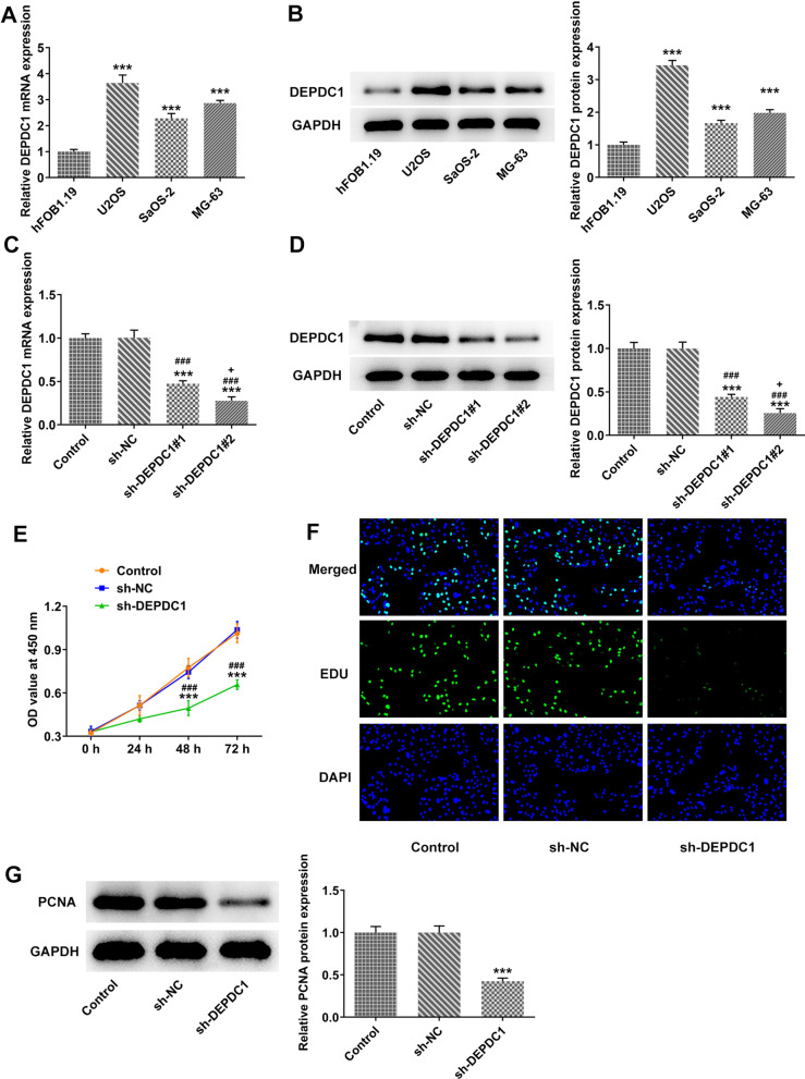
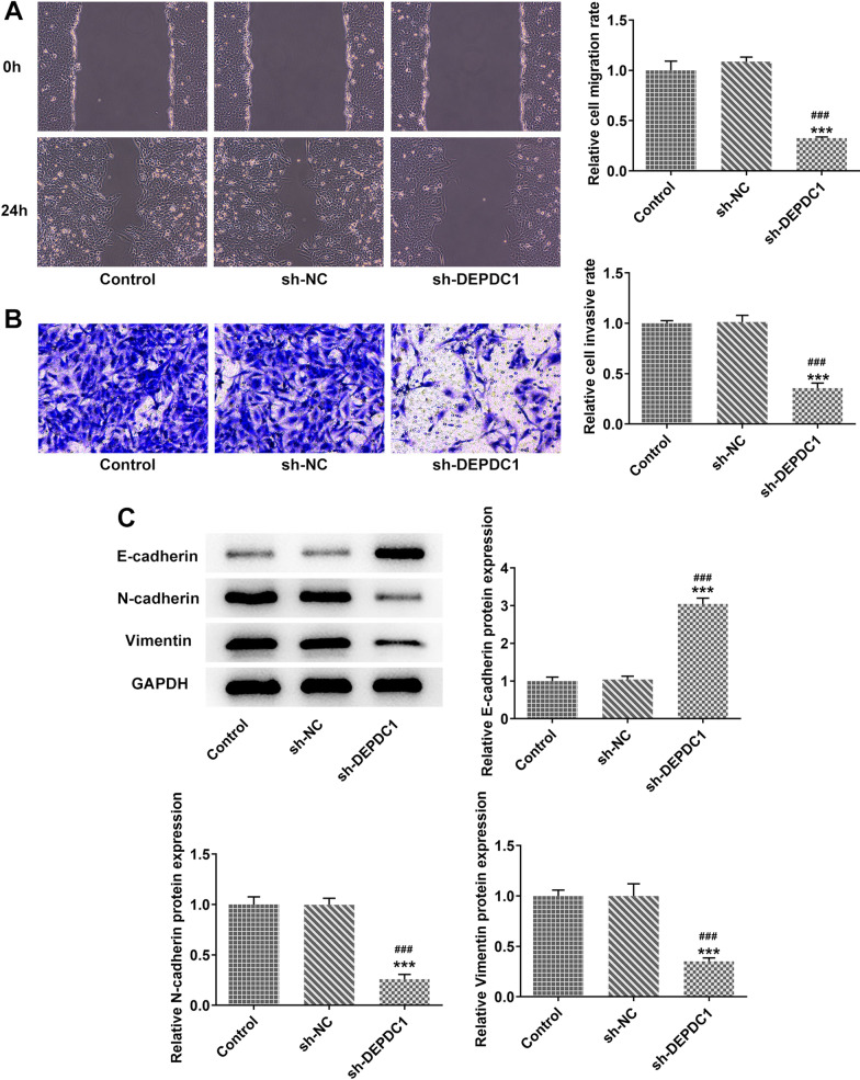
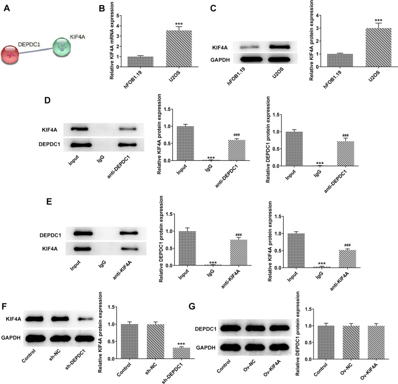
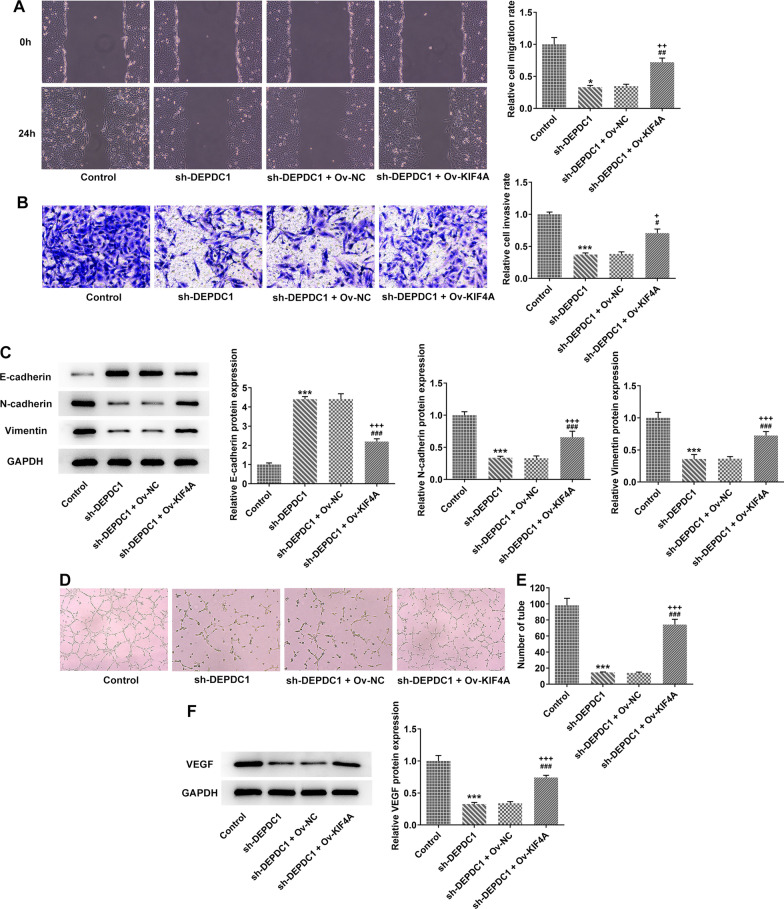
The treatment of osteosarcoma (OS) is still mainly surgery combined with systematic chemotherapy, and gene therapy is expected to improve the survival rate of patients. This study aimed to explore the effect of DEP domain 1 protein (DEPDC1) and kinesin super-family protein 4A (KIF4A) in OS and understand its mechanism. Th expression of DEPDC1 and KIF4A in OS cells was detected by RT-PCR and western blot. The viability, proliferation, invasion and migration of OS cells and tube formation of human umbilical vein endothelial cells (HUVECs) after indicated treatment were in turn detected by CCK-8 assay, EdU staining, wound healing assay, transwell assay and tube formation assay. The interaction between DEPDC1 and KIF4A was predicted by STRING and confirmed by co-immunoprecipitation. The expression of epithelial-mesenchymal transition (EMT)-related proteins, tube formation-related proteins and Hippo signaling pathway proteins was detected by western blot. As a result, the expression of DEPDC1 and KIF4A was all increased in U2OS cells. Down-regulation of DEPDC1 suppressed the viability, proliferation, invasion and migration of U2OS cells and tube formation of HUVECs, accompanied by the increased expression of E-cadherin and decreased expression of N-cadherin, Vimentin and VEGF. DEPDC1 was confirmed to be interacted with KIF4A. Upregulation of KIF4A partially reversed the effect of DEPDC1 interference on the above biological behaviors of U2OS cells. Down-regulation of DEPDC1 promoted the expression of p-LATS1 and p-YAP in Hippo signaling pathway, which was reversed by upregulation of KIF4A. In conclusion, down-regulation of DEPDC1 inhibited the malignant biological behavior of OS cells through the activation of Hippo signaling pathway, which could be reversed by upregulation of KIF4A.

Keywords: DEPDC1; Hippo signaling pathway; KIF4A; Osteosarcoma cells.


Conflict of interest statement

The authors declare that they have no competing interests.

Figures

Fig. 1
Fig. 1
DEPDC1 was highly expressed in OS cells, and down-regulation of DEPDC1 inhibited cell proliferation. A/B The expression of DEPDC1 in OS cells was detected by RT-qPCR and western blot. ***P < 0.001 versus hFOB1.19 group. C/D The transfection effects of sh-DEPDC1#1 and sh-DEPDC1#2 in U2OS cells was confirmed by RT-qPCR and western blot. E The viability of U2OS cells transfected with si-DEPDC1 was determined by CCK-8 assay. F The proliferation of U2OS cells transfected with sh-DEPDC1 was observed by EdU staining. G The expression of PCNA in U2OS cells transfected with sh-DEPDC1 was detected by western blot. ***P < 0.001 versus Control group. ###P < 0.001 versus sh-NC group. +P < 0.05 versus sh-DEPDC1#1
Fig. 2
Fig. 2
Down-regulation of DEPDC1 inhibited metastasis of OS cells. A The migration of U2OS cells transfected with sh-DEPDC1 was detected by wound healing assay. B The invasion of U2OS cells transfected with sh-DEPDC1 was detected by transwell assay. C Th expression of EMT-related proteins in U2OS cells transfected with sh-DEPDC1 was detected by western blot. ***P < 0.001 versus Control group. ###P < 0.001 versus sh-NC group
Fig. 3
Fig. 3
Down-regulation of DEPDC1 inhibited angiogenesis in OS cells. A/B The number of HUVECs tube formation in the supernatant of U2OS cells transfected with sh-DEPDC1 was analyzed by tube formation assay. C The expression of angiogenesis-related protein in U2OS cells transfected with sh-DEPDC1 was detected by western blot. ***P < 0.001 versus Control group. ###P < 0.001 versus sh-NC group
Fig. 4
Fig. 4
KIF4A was highly expressed in OS cells and interacted with DEPDC1. A The interaction between DEPDC1 and KIF4A was confirmed by STRING. B/C The expression of KIF4A in U2OS cells was detected by RT-qPCR and western blot. ***P < 0.001 versus hFOB1.19 group. D The expression of KIF4A and DEPDC1 in proteins incubating with anti-DEPDC1 was detected by western blot. E The expression of KIF4A and DEPDC1 in proteins incubating with anti-KIF4A was detected by western blot. ***P < 0.001 versus Input group. ###P < 0.001 versus Input group. F The expression of KIF4A in U2OS cells transfected with sh-DEPDC1 was detected by western blot. ***P < 0.001 versus Control group. G The expression of DEPDC1 in U2OS cells transfected with Ov-KIF4A was detected by western blot
Fig. 5
Fig. 5
Upregulation of KIF4A partially reversed the effect of DEPDC1 interference on proliferation of OS cells. A/B The expression of KIF4A in U2OS cells transfected with Ov-KIF4A was detected by RT-qPCR and western blot. ***P < 0.001 versus Control group. ###P < 0.001 versus Ov-NC group. C The viability of U2OS cells transfected with sh-DEPDC1 and Ov-KIF4A was determined by CCK-8 assay. D The proliferation of U2OS cells transfected with si-DEPDC1 and Ov-KIF4A was observed by EdU staining. E The expression of PCNA in U2OS cells transfected with sh-DEPDC1 and Ov-KIF4A was detected by western blot. ***P < 0.001 versus Control group. ###P < 0.001 versus sh-NC group. +++P < 0.001 versus si-DEPDC1 + Ov-NC group
Fig. 6
Fig. 6
Upregulation of KIF4A partially reversed the effect of DEPDC1 interference on metastasis and angiogenesis of OS cells. A The migration of U2OS cells transfected with sh-DEPDC1 and Ov-KIF4A was detected by wound healing assay. B The invasion of U2OS cells transfected with si-DEPDC1 and Ov-KIF4A was detected by transwell assay. C Th expression of EMT-related proteins in U2OS cells transfected with sh-DEPDC1 and Ov-KIF4A was detected by western blot. D/E The number of HUVECs tube formation in the supernatant of U2OS cells transfected with sh-DEPDC1 and Ov-KIF4A was analyzed by tube formation assay. F The expression of angiogenesis-related protein in U2OS cells transfected with si-DEPDC1 and Ov-KIF4A was detected by western blot. *P < 0.05 and ***P < 0.001 versus Control group. ##P < 0.01 and ###P < 0.001 versus sh-NC group. ++P < 0.01 and +++P < 0.001 versus sh-DEPDC1 + Ov-NC group
Fig. 7
Fig. 7
DEPDC1 and KIF4A synergistically regulated Hippo signaling pathway. Th expression of Hippo signaling pathway-related proteins in in U2OS cells transfected with sh-DEPDC1 and Ov-KIF4A was detected by western blot. ***P < 0.001 versus Control group. ###P < 0.001 versus sh-NC group. +++P < 0.001 versus sh-DEPDC1 + Ov-NC group

Similar articles

Cited by

References

    1. Torre LA, Bray F, Siegel RL, Ferlay J, Lortet-Tieulent J, Jemal A. Global cancer statistics, 2012. CA: Cancer J Clin. 2015;65:87–108. - PubMed
    1. Cepeda M, Sosa AJ, Mora G. Telangiectatic osteosarcoma in an infant. Boletin medico del Hospital Infantil de Mexico. 2017;74:60–64. - PubMed
    1. Siegel RL, Miller KD, Jemal A. Cancer statistics, 2020. CA: Cancer J Clin. 2020;70:7–30. - PubMed
    1. Geller DS, Gorlick R. Osteosarcoma: a review of diagnosis, management, and treatment strategies. Clin Adv Hematol Oncol: H&O. 2010;8:705–718. - PubMed
    1. Verma AH, Kumar TSS, Madhumathi K, Rubaiya Y, Ramalingan M, Doble M. Curcumin releasing eggshell derived carbonated apatite nanocarriers for combined anti-cancer, anti-inflammatory and bone regenerative therapy. J Nanosci Nanotechnol. 2019;19:6872–6880. doi: 10.1166/jnn.2019.16640. - DOI - PubMed