. 2023 Feb 27;25(1):30.
doi: 10.1186/s13075-023-03001-1.

TGF-β is elevated in hyperuricemic individuals and mediates urate-induced hyperinflammatory phenotype in human mononuclear cells

Collaborators, Affiliations

TGF-β is elevated in hyperuricemic individuals and mediates urate-induced hyperinflammatory phenotype in human mononuclear cells

Viola Klück et al. Arthritis Res Ther. .

Abstract

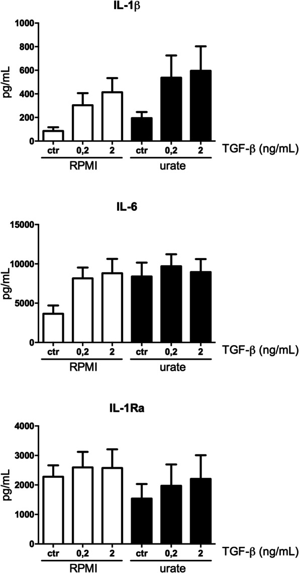
Background: Soluble urate leads to a pro-inflammatory phenotype in human monocytes characterized by increased production of IL-1β and downregulation of IL-1 receptor antagonist, the mechanism of which remains to be fully elucidated. Previous transcriptomic data identified differential expression of genes in the transforming growth factor (TGF)-β pathway in monocytes exposed to urate in vitro. In this study, we explore the role of TGF-β in urate-induced hyperinflammation in peripheral blood mononuclear cells (PBMCs).

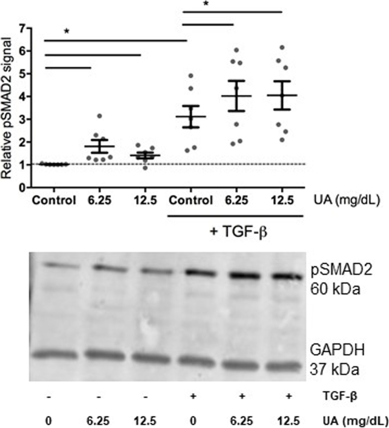
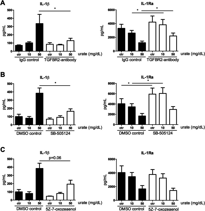
Methods: TGF-β mRNA in unstimulated PBMCs and protein levels in plasma were measured in individuals with normouricemia, hyperuricemia and gout. For in vitro validation, PBMCs of healthy volunteers were isolated and treated with a dose ranging concentration of urate for assessment of mRNA and pSMAD2. Urate and TGF-β priming experiments were performed with three inhibitors of TGF-β signalling: SB-505124, 5Z-7-oxozeaenol and a blocking antibody against TGF-β receptor II.

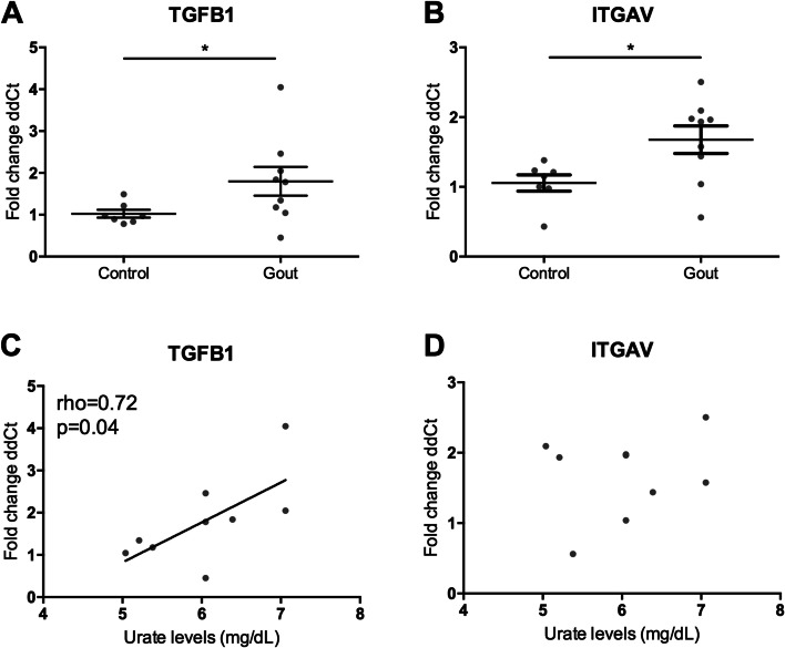
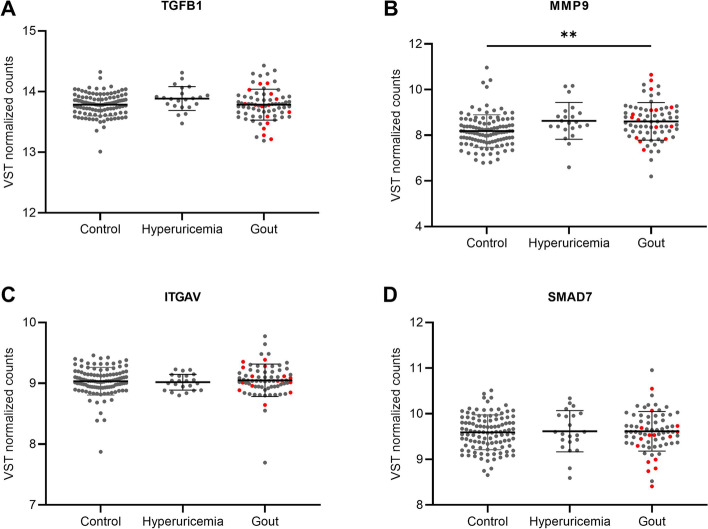
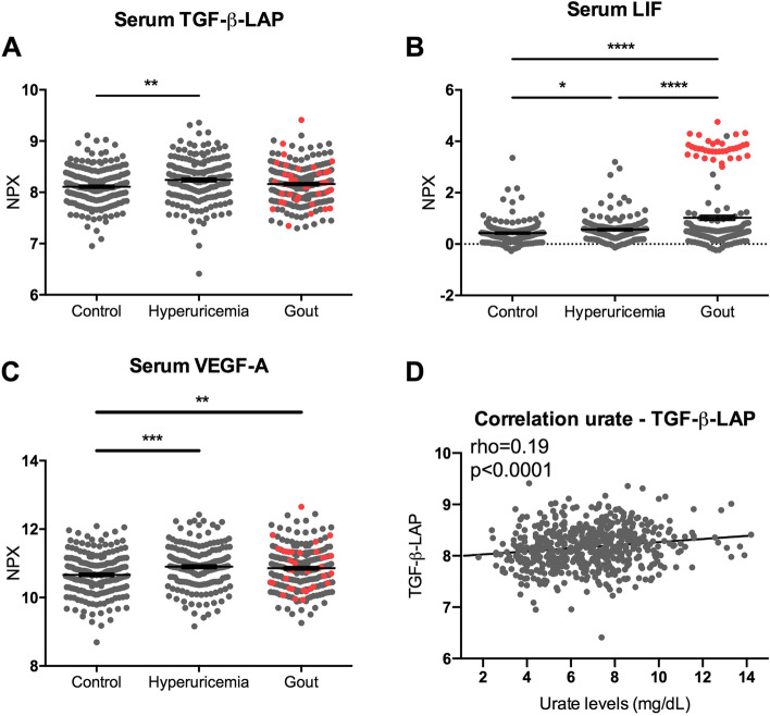
Results: TGF-β mRNA levels were elevated in gout patients compared to healthy controls. TGF-β-LAP levels in serum were significantly higher in individuals with hyperuricemia compared to controls. In both cases, TGF-β correlated positively to serum urate levels. In vitro, urate exposure of PBMCs did not directly induce TGF-β but did enhance SMAD2 phosphorylation. The urate-induced pro-inflammatory phenotype of monocytes was partly reversed by blocking TGF-β.

Conclusions: TGF-β is elevated in individuals with hyperuricemia and correlated to serum urate concentrations. In addition, the urate-induced pro-inflammatory phenotype in human monocytes is mediated by TGF-β signalling. Future studies are warranted to explore the intracellular pathways involved and to assess the clinical significance of urate-TGF-β relation.

Keywords: Hyperuricemia; Inflammation; Mononuclear leukocytes; TGF-β.


Conflict of interest statement

The authors declare that they have no competing interests.

Figures

Fig. 1
Fig. 1
TGF-β mRNA is upregulated in gout patients and correlates to serum urate levels in the discovery cohort. PBMCs from patients with gout and matched healthy controls (HC) were isolated and adhered to a flat-bottom plate, mRNA was isolated and compared to the mean dCT of healthy controls by Mann-Whitney U tests *p < 0.05 (A, B). Serum urate levels were correlated to relative mRNA expression levels and analysed by Spearman’s correlation (C, D)
Fig. 2
Fig. 2
mRNA expression of TGF-β and downstream targets in PBMCs from individuals with hyperuricemia or gout. PBMCs from individuals with normoruricemia (n = 110), hyperuricemia (n = 22) and gout (n = 72) of which 15 flaring (marked in red) were isolated and transcriptomics were analysed. Relative mRNA expression of TGFB1 (A), MMP9 (B), ITGAV (C) and SMAD7 (D) are shown. Lines represent means with SD. Means were compared by Welch ANOVA with Tamhane’s T2 multiple comparisons test. **p < 0.01
Fig. 3
Fig. 3
Serum TGF-β-LAP, LIF and VEGF-A levels are elevated in individuals with hyperuricemia and TGF-β-LAP correlates positively to serum urate. Serum proteins were analysed by Olink proteomics panel. Flaring gout patients (n = 36) are marked in red dots. NPX was shown for TGF-β-LAP (A), LIF (B) and VEGF-A (C). Means were compared by Welch ANOVA with Games-Howell’s multiple comparisons test. *p < 0.05, **p < 0.01, ***p < 0.001. Spearman correlation was used to analyse the correlation between serum TGF-β-LAP to urate levels (D). Serum LIF levels were significantly higher in flaring gout patients compared to intercritical gout patients (Welch’s t-test p < 0.0001)
Fig. 4
Fig. 4
Protein expression of pSMAD2 (Ser465/467) in urate primed monocytes. The monocytes were primed overnight with no, 6.25 or 12.5 mg/dL urate followed with a stimulation of 1 ng/mL TGFβ for 1 h. Cell lysates were used for western blotting. Relative pSMAD2 expression (n = 7) (A) and a representative blot (B) are shown. Wilcoxon signed-rank test was used to compare means. *p < 0.05
Fig. 5
Fig. 5
Both TGF-β and urate demonstrate pro-inflammatory effects in a priming model without a synergistic effect. Adherent monocytes isolated from healthy volunteers (n = 6) were treated with dose-ranging concentrations of recombinant TGF-β and/or urate (50 mg/dL) for 24 h after which cells were washed and stimulated with LPS (10 ng/mL) for 24 h. IL-1β (A), IL-6 (B) and IL-Ra (C) were measured in the supernatant after 48 h culture
Fig. 6
Fig. 6
Blocking TGF-β signalling pathway partly reverses urate priming effects. Adherent monocytes isolated from healthy volunteers (An = 10; B-C n = 6) were treated with dose-ranging concentrations of urate (50 mg/dL) in the presence or absence of a TGF-β inhibitor for 24 h after which cells were washed and stimulated with LPS (10 ng/mL) for 24 h. IL-1β and IL-Ra were measured in the supernatant after 48 h culture. TGF-β inhibitors: a blocking antibody against TGF-β receptor II with mouse IgG1 as the isotype control (10 μg/mL), SB-505124 (5 μM) and 5Z-7-oxozeaenol (100nM) both with DMSO as solvent control. Wilcoxon signed rank test was applied to compare means. *p < 0.05

Similar articles

Cited by

References

    1. Shiozawa A, Szabo SM, Bolzani A, Cheung A, Choi HK. Serum uric acid and the risk of incident and recurrent gout: a systematic review. J Rheumatol. 2017;44(3):388–396. doi: 10.3899/jrheum.160452. - DOI - PubMed
    1. Joosten LA, Netea MG, Mylona E, Koenders MI, Malireddi RK, Oosting M, et al. Engagement of fatty acids with Toll-like receptor 2 drives interleukin-1beta production via the ASC/caspase 1 pathway in monosodium urate monohydrate crystal-induced gouty arthritis. Arthritis Rheum. 2010;62(11):3237–3248. doi: 10.1002/art.27667. - DOI - PMC - PubMed
    1. So A, Dumusc A, Nasi S. The role of IL-1 in gout: from bench to bedside. Rheumatology (Oxford) 2018;57(suppl_1):i12–ii9. - PubMed
    1. Dalbeth N, Gosling AL, Gaffo A, Abhishek A. Gout. Lancet. 2021;397(10287):1843–1855. doi: 10.1016/S0140-6736(21)00569-9. - DOI - PubMed
    1. Kuo CF, Grainge MJ, Zhang W, Doherty M. Global epidemiology of gout: prevalence, incidence and risk factors. Nat Rev Rheumatol. 2015;11(11):649–662. doi: 10.1038/nrrheum.2015.91. - DOI - PubMed

Publication types