. 2023 Feb 11;11(2):417.
doi: 10.3390/vaccines11020417.

Toxicological Assessments of a Pandemic COVID-19 Vaccine-Demonstrating the Suitability of a Platform Approach for mRNA Vaccines

Affiliations

Toxicological Assessments of a Pandemic COVID-19 Vaccine-Demonstrating the Suitability of a Platform Approach for mRNA Vaccines

Cynthia M Rohde et al. Vaccines (Basel). .

Abstract

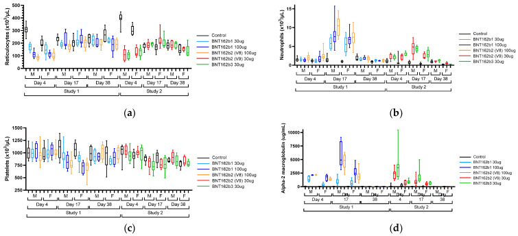
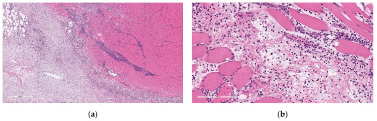
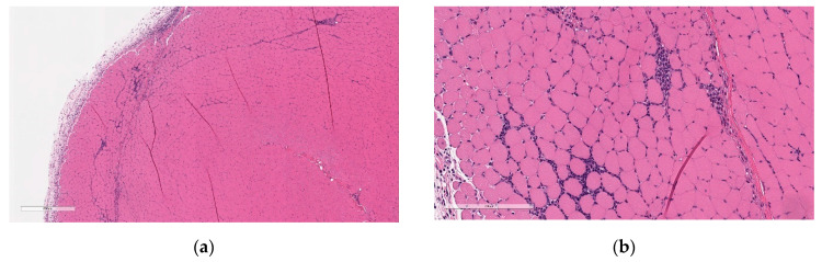
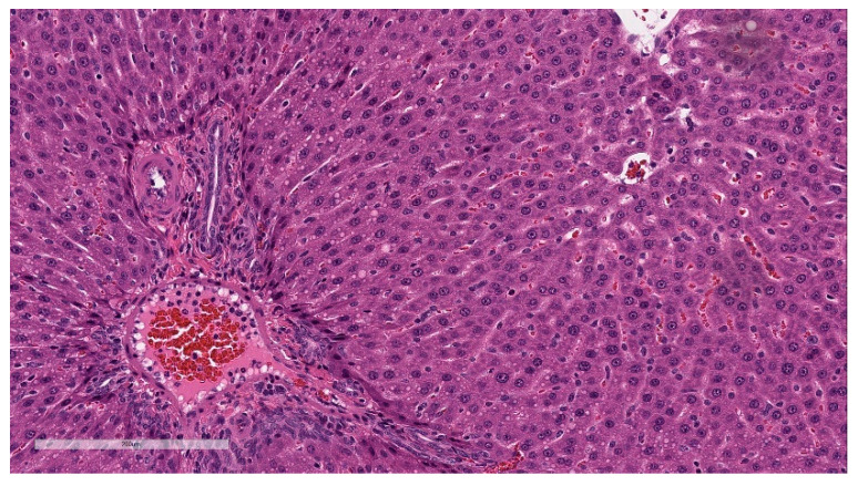
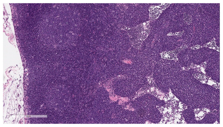
The emergence of SARS-CoV-2 at the end of 2019 required the swift development of a vaccine to address the pandemic. Nonclinical GLP-compliant studies in Wistar Han rats were initiated to assess the local tolerance, systemic toxicity, and immune response to four mRNA vaccine candidates encoding immunogens derived from the spike (S) glycoprotein of SARS-CoV-2, encapsulated in lipid nanoparticles (LNPs). Vaccine candidates were administered intramuscularly once weekly for three doses at 30 and/or 100 µg followed by a 3-week recovery period. Clinical pathology findings included higher white blood cell counts and acute phase reactant concentrations, lower platelet and reticulocyte counts, and lower RBC parameters. Microscopically, there was increased cellularity (lymphocytes) in the lymph nodes and spleen, increased hematopoiesis in the bone marrow and spleen, acute inflammation and edema at the injection site, and minimal hepatocellular vacuolation. These findings were generally attributed to the anticipated immune and inflammatory responses to the vaccines, except for hepatocyte vacuolation, which was interpreted to reflect hepatocyte LNP lipid uptake, was similar between candidates and resolved or partially recovered at the end of the recovery phase. These studies demonstrated safety and tolerability in rats, supporting SARS-CoV-2 mRNA-LNP vaccine clinical development.

Keywords: BNT162b1; BNT162b2; BNT162b3; COVID-19 mRNA vaccine; nonclinical safety; rat; toxicity.


Conflict of interest statement

CMR, MG, SC, LR, and YC are employees and/or shareholders of Pfizer, Inc. RSS was a Pfizer employee and/or shareholder of Pfizer stock at the time the studies were conducted. CL, JD, ABV, AM, and US are employees and/or shareholders of BioNTech SE.

Figures

Figure 1
Figure 1
Overview of experimental design. Male and female Wistar Han rats (15/sex/group) were administered 3 once weekly intramuscular injections (formula image
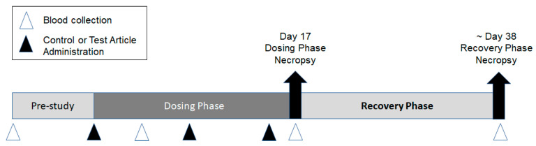
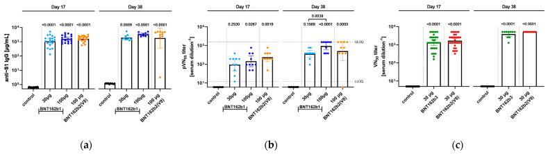
) of control (saline or vehicle), BNT162b1 (30 or 100 µg), BNT162b2 V8 (100 µg), BNT162b2 V9 (30 µg), or BNT162b3 (30 µg). Animals were euthanized two days after the last dose, Day 17 (10 animals/sex/group), and approximately 3 weeks later (Day 38). Blood (Δ) was collected for serology analysis prior to dose initiation and at each necropsy. Blood was collected for clinical pathology assessments on Day 3 and at each necropsy.
Figure 2
Figure 2
Edema incidence and severity in male (M) and female (F) Wistar Han rats immunized intramuscularly (IM) with (a) BNT162b2 V9 (30 µg), (b) BNT162b2 V8 (100 µg), (c) BNT162b1 (30 µg or 100 µg), and (d) BNT162b3 (30 µg), or buffer control. Edema measurements from Dosing Phase Days 2, 9, 16, and 21 for BNT162b2 V9 (30 µg) and BNT162b3 (30 µg). Edema measurements from Dosing Phase Days 2, 10, 16, and 21 for BNT162b2 V8 (100 µg) and BNT162b1 (30 µg or 100 µg).
Figure 3
Figure 3
Erythema incidence and severity in male (M) and female (F) Wistar Han rats immunized intramuscularly (IM) with (a) BNT162b2 V8 (100 µg), (b) BNT162b2 V9 (30 µg), (c) BNT162b3 (30 µg), and (d) BNT162b1 (30 µg or 100 µg) or buffer control. Erythema measurements from Dosing Phase Days 2, 3/5 *, 9, 14 *, and 16. * Days 3/5 and 14 are only available for Study 1.
Figure 3
Figure 3
Erythema incidence and severity in male (M) and female (F) Wistar Han rats immunized intramuscularly (IM) with (a) BNT162b2 V8 (100 µg), (b) BNT162b2 V9 (30 µg), (c) BNT162b3 (30 µg), and (d) BNT162b1 (30 µg or 100 µg) or buffer control. Erythema measurements from Dosing Phase Days 2, 3/5 *, 9, 14 *, and 16. * Days 3/5 and 14 are only available for Study 1.
Figure 4
Figure 4
Reticulocyte counts (a), neutrophil counts (b), platelet counts (c), and alpha-2 macroglobulin (d) concentrations in male (M) and female (F) Wistar Han rats immunized intramuscularly (IM) with BNT162b1 (30 µg or 100 µg), BNT162b2 V8 (100 µg), BNT162b2 V9 (30 µg), BNT162b3 (30 µg), or buffer control. Blood samples were collected on Dosing Phase Days 4, 17, and 38. Box-whisker plots show group medians (middle line), 25th and 75th percentiles (box), and min and max (lower and upper whiskers).
Figure 5
Figure 5
Histopathologic features of Injection site inflammation and edema at the end of the dosing phase. (a) Injection site sections of BNT162b2 (V9) treated animals euthanized 2 days after the third dose (Day 17) showed inflammatory cells admixed with abundant pale eosinophilic fluid (edema) infiltrating and expanding subcutaneous tissue and connective tissue of skeletal muscle. (b) Higher magnification of (a) showing.
Figure 6
Figure 6
Histopathologic features of Injection site inflammation at the end of recovery. (a) Injection site sections of BNT162b2 (V9) treated animals euthanized after 3-week recovery showed fewer inflammatory cells infiltrating and expanding subcutaneous tissue and spaces around skeletal myofibers. (b) Higher magnification of (a) showing inflammatory cells (fewer plasma cells and lymphocytes) surrounding myofibers. Data are scanned from Study 2.
Figure 7
Figure 7
Histopathologic features of the liver at the end of the dosing phase. Liver sections of BNT162b2 (V9) treated animals euthanized 2 days after the third dose (Day 17) showed hepatocytes containing small clear round membrane-bound structures within the cytoplasm (inset). Data are scanned from Study 2.
Figure 8
Figure 8
Histopathologic features of draining lymph node at the end of the dosing phase. Lymph node sections of BNT162b2 (V9) vaccinated animal showing prominent germinal centers (GC) and inflammatory cells (predominately plasma cells) expanding sinuses (inset). Data are scanned from Study 2.
Figure 9
Figure 9
Wistar Han rats were immunized intramuscularly (IM) with BNT162b1, BNT162b2, or BNT162b3 vaccine candidates or buffer control. Analysis of serum samples was performed 17 days [n = 20] or 38 days [(a,b): n = 10; (c): n =5] after first immunization. Geometric means of each group ± 95% confidence interval (CI) are included, and p-values are given to control or determine if significance occurred as indicated (One-Way ANOVA of mixed-effect analysis using Tukey’s multiple comparisons test). (a), S1-specific IgG levels in sera of rats determined by ELISA. (b), 90% pseudovirus neutralization (pVN90) titers in sera of rats immunized with the different BNT162b vaccine candidates. (c), 50% SARS-CoV-2 neutralization (VN50) titer in sera of rats immunized with different BNT162b vaccine candidates.

References

    1. Zhu N., Zhang D., Wang W., Li X., Yang B., Song J., Zhao X., Huang B., Shi W., Lu R., et al. A Novel Coronavirus from Patients with Pneumonia in China, 2019. N. Engl. J. Med. 2020;382:727–733. doi: 10.1056/NEJMoa2001017. - DOI - PMC - PubMed
    1. Dolgin E. The tangled history of mRNA vaccines. Nature. 2021;597:318–324. doi: 10.1038/d41586-021-02483-w. - DOI - PubMed
    1. Wallis J., Shenton D.P., Carlisle R.C. Novel approaches for the design, delivery and administration of vaccine technologies. Clin. Exp. Immunol. 2019;196:189–204. doi: 10.1111/cei.13287. - DOI - PMC - PubMed
    1. Aldosari B.N., Alfagih I.M., Almurshedi A.S. Lipid Nanoparticles as Delivery Systems for RNA-Based Vaccines. Pharmaceutics. 2021;13:206. doi: 10.3390/pharmaceutics13020206. - DOI - PMC - PubMed
    1. Hou X., Zaks T., Langer R., Dong Y. Lipid nanoparticles for mRNA delivery. Nat. Rev. Mater. 2021;6:1078–1094. doi: 10.1038/s41578-021-00358-0. - DOI - PMC - PubMed

LinkOut - more resources