Review
. 2023 Feb 16;11(2):458.
doi: 10.3390/vaccines11020458.

Emerging Trends in Nano-Driven Immunotherapy for Treatment of Cancer

Affiliations
Review

Emerging Trends in Nano-Driven Immunotherapy for Treatment of Cancer

Gayathri Kandasamy et al. Vaccines (Basel). .

Abstract

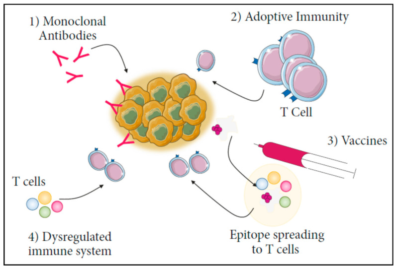
Despite advancements in the development of anticancer medications and therapies, cancer still has the greatest fatality rate due to a dismal prognosis. Traditional cancer therapies include chemotherapy, radiotherapy, and targeted therapy. The conventional treatments have a number of shortcomings, such as a lack of selectivity, non-specific cytotoxicity, suboptimal drug delivery to tumour locations, and multi-drug resistance, which results in a less potent/ineffective therapeutic outcome. Cancer immunotherapy is an emerging and promising strategy to elicit a pronounced immune response against cancer. Immunotherapy stimulates the immune system with cancer-specific antigens or immune checkpoint inhibitors to overcome the immune suppressive tumour microenvironment and kill the cancer cells. However, delivery of the antigen or immune checkpoint inhibitors and activation of the immune response need to circumvent the issues pertaining to short lifetimes and effect times, as well as adverse effects associated with off-targeting, suboptimal, or hyperactivation of the immune system. Additional challenges posed by the tumour suppressive microenvironment are less tumour immunogenicity and the inhibition of effector T cells. The evolution of nanotechnology in recent years has paved the way for improving treatment efficacy by facilitating site-specific and sustained delivery of the therapeutic moiety to elicit a robust immune response. The amenability of nanoparticles towards surface functionalization and tuneable physicochemical properties, size, shape, and surfaces charge have been successfully harnessed for immunotherapy, as well as combination therapy, against cancer. In this review, we have summarized the recent advancements made in choosing different nanomaterial combinations and their modifications made to enable their interaction with different molecular and cellular targets for efficient immunotherapy. This review also highlights recent trends in immunotherapy strategies to be used independently, as well as in combination, for the destruction of cancer cells, as well as prevent metastasis and recurrence.

Keywords: cancer therapy; immunotherapy; nanoparticles; tumour microenvironment; tumour recurrence.


Conflict of interest statement

The authors declare no conflict of interest.

Figures

Figure 2
Figure 2
Currently available immunotherapy techniques. (1) Monoclonal antibodies, (2) Adoptive immunity, (3) Vaccines, and (4) Dysregulated immune system. These approaches are useful in evoking an immune response against cancers [22]. CC BY 4.0.
Figure 3
Figure 3
Schematic depiction of the self-assembly of PyroR and its mechanism of photodynamic treatment (PDT) causing immunogenic cell death (ICD) and macrophage polarisation. Pyro and R848 could interact noncovalently between themselves to form PyroR. PyroR could inhibit initial tumour growth by PDT after passively accumulating at the tumour site and polarise macrophages from M2 to M1 phenotype to secrete cytokines. Further, the PDT aids immunotherapy by triggering immunological cascades, such as ICD activation, CRT and HMGB1 release, DC maturation, and lymph node migration. ICD cascades and M1 macrophage polarisation activate T cells to curb metastatic tumours [23]. Reproduced with permissions from Xiayun Chen et al., Chemical Engineering Journal, Elsevier, 2022.
Figure 1
Figure 1
Recently explored nanoparticles for cancer immunotherapy using different strategies and modifications.
Figure 4
Figure 4
Different types of microneedles employed in the controlled delivery of immunotherapeutic drugs [82]. Reproduced with permissions from Hamed Amani et al., Journal of controlled release, Elsevier, 2021.
Figure 5
Figure 5
Schematic description of the mechanism of rolling microelectrode array (RoMEA) (a) RoMEA’s minimally invasive method of continuous electroporation in a vast region of the target tissue by rolling on the appropriate place. (b) Overall design of RoMEA device. Anode (red) and cathode (black) are connected by two nearby microneedle blades. (c) Microneedle electroporation. (d) Stimulation of RoMEA by electric field (50 V). (e) The RoMEA prototype [90]. Reproduced with permissions from Tongren Yeng et al., Nano Today, Elsevier, 2021.
Figure 6
Figure 6
Development of a DNA nanocarrier co-encapsulating CpG and doxorubicin for chemoimmunotherapy. The nanoparticle administration offered superior therapeutic outcome due to the synergistic action of the activated antigen presenting cells and cytotoxicity of the chemotherapeutic agent [96]. Reproduced with permissions from Qian Wang et al., ACS Applied Nanomaterials, American Chemical Society, 2022.
Figure 7
Figure 7
Schematic representation of drug loading of exosomes through the use of culturing, electroporation, sonication, extrusion, freeze-and-thaw cycles, and saponin-assisted loading. Exosome surface modification through chemical alteration, electrostatic interaction, and EV membrane for enhanced targeting efficiency to cancer cells [110]. Reproduced with permissions from Ya-Nan Pi et al., Biochemical Pharmacology, Elsevier, 2021.
Figure 8
Figure 8
Schematic representation of the in vivo experimental setup for optogenetic perioperative immunotherapy using hydrogel implants loaded with far-red light-controlled immunomodulatory engineered [125]. Copyright CC BY 4.0.
Figure 9
Figure 9
Schematic depiction of the bioconjugation and coating reactions on TMV: (A) alkyne labelling of TMV through amidation of the interior glutamate residues of TMV with propargyl amine; (B) azide labelling of 1V209 through amidation of the carboxylic group with aminooxy-PEG1-azide; (C) 1V209-TMVproduction by copper-mediated azide-alkyne cycloaddition (CuAAC); and (D) polydopamine coating of the prepared 1V209 (Tris buffer, pH 8.5) using oxidative polymerization [134]. The results were analyzed with One-way ANOVA followed by Dunnett′s post-test: * p < 0.0001 vs. medium; # p = 0. Reproduced with permissions from Christian Isalomboto Nkanga et al., Nanomedicine: Nanotechnology, Biology and Medicine, Elsevier, 2022.
Figure 10
Figure 10
Representation of M13 phage-based vaccine platform design. (a) Development of the hybrid M13 phage vaccine (b,c) loaded with tumour antigens. Schematic diagram representing the stages of antitumour immune response induced by HMP@Ag vaccine. Following subcutaneous administration in mice, DCs internalized the HMP@Ag vaccine for antigen release and cross-presentation for DC maturation (d). Mature DCs move to lymph nodes where CD8+ T lymphocytes specific for the antigen get activated and expanded (e). Combination of M13 phage-based vaccination with ICB therapy inhibits both primary and metastatic cancers and elicit a neoantigen-based CTL response, as well as suppresses tumour recurrence following surgery (f) [135]. Reproduced with permissions from Xue Dong et al., Biomaterials, Elsevier, 2023.
Figure 11
Figure 11
(A) Tumour cells (a) were cultured and engrafted into mouse model (b1). (b2) The cell membrane was extracted and mixed with an oncolytic adenovirus serotype 5, with a 24-base-pairs deletion, carrying -CpG islands (i.e., A5-Δ24-CpG). (c) The virus was wrapped with the cell mem brane using the process of extrusion to obtain ExtraCRAd. (d) The established tumours were treated with multiple intratumoral injections of ExtraCRAd. (B) Cryo-transmission electron microscopy (TEM) images of virus, lipid cancer membrane vesicles, and ExtraCRAd (C) Median tumour growth [138]. The results were analyzed with a two-way ANOVA, and Dunnet’s post-test comparison, and the levels of significance were * p < 0.05, *** p < 0.001, **** p < 0.0001. Copyright CC BY 4.0.
Figure 12
Figure 12
Scheme depicting the upconversion optogenetic system (UCNs@FA and NIR light) and engineered NIR-light-responsive bacteria (EcN@EL222-TNF) [141]. Reproduced with permissions from Huizhuo Pan et al., Chemical Engineering Journal, Elsevier, 2021.
Figure 13
Figure 13
CuS-OMVs for combinational cancer therapy involving PTT and immunotherapy. (a) Schematic representation of the preparation of CuS-OMVs. (b) Schematic representation of PTT and antitumour immunity generated by CuS-OMVs following NIR–II light exposure. CuS-OMVs target tumours effectively and cause cytotoxicity in tumour cells due to a combination of ICD, DC maturation and CD8+ T-cell activation in response to NIR-II light exposure. CuS-OMVs act as immune adjuvants that support DC maturation and repolarize TAMs from M2 to M1 phenotype to inhibit tumour growth and metastasis [146]. Reproduced with permissions from Jiaqi Qin et al., Nano Today, Elsevier, 2022.
Figure 14
Figure 14
Targeting stiffness in primary and metastatic tumour niches. (A) Lysyl oxidase (LOX) inhibitor therapy prevents tumour metastasis by destructing the premetastatic niche, thereby inhibiting cancer cells from colonization. (B) Antiangiogenic therapy for the treatment of metastatic cancer by drugs that target the renin-angiotensin system (RAS), and inactivating metastasis-associated fibroblasts (MAFs), which stimulate angiogenesis in the metastatic niche. (C) Tissue-softening techniques combined with CTL-targeting immunotherapies, such as immune checkpoint inhibitors and chimeric antigen receptor-T (CAR-T) cells, the infiltration of cytotoxic T lymphocytes (CTLs) increases. (D) Recruitment of CTLs into tumour lesions is accompanied by the degradation of tumour ECM by the fusion protein (TNF-CSG), which binds to laminin-nidogen complexes in tumour ECM. (E) Heparanase, an enzyme that breaks down the ECM, is expressed by CAR-T cells, which increases their capacity to penetrate the ECM [156]. Reproduced with permissions from Jeongeun Hyun et al., Trends in Molecular Medicine, Elsevier, 2022.

References

    1. Gowd V., Ahmad A., Tarique M., Suhail M., Zughaibi T.A., Tabrez S., Khan R. Advancement of Cancer Immunotherapy Using Nanoparticles-Based Nanomedicine. Semin. Cancer Biol. 2022;86:624–644. doi: 10.1016/j.semcancer.2022.03.026. - DOI - PubMed
    1. Buabeid M.A., Arafa E.S.A., Murtaza G. Emerging Prospects for Nanoparticle-Enabled Cancer Immunotherapy. J. Immunol. Res. 2020;2020:9624532. doi: 10.1155/2020/9624532. - DOI - PMC - PubMed
    1. Aikins M.E., Xu C., Moon J.J. Engineered Nanoparticles for Cancer Vaccination and Immunotherapy. Accounts Chem. Res. 2020;53:2094–2105. doi: 10.1021/acs.accounts.0c00456. - DOI - PMC - PubMed
    1. Muluh T.A., Chen Z., Li Y., Xiong K., Jin J., Fu S., Wu J. Enhancing Cancer Immunotherapy Treatment Goals by Using Nanoparticle Delivery System. Int. J. Nanomed. 2021;16:2389–2404. doi: 10.2147/IJN.S295300. - DOI - PMC - PubMed
    1. Lim S., Park J., Shim M.K., Um W., Yoon H.Y., Ryu J.H., Lim D.K., Kim K. Recent Advances and Challenges of Repurposing Nanoparticle-Based Drug Delivery Systems to Enhance Cancer Immunotherapy. Theranostics. 2019;9:7906–7923. doi: 10.7150/thno.38425. - DOI - PMC - PubMed

LinkOut - more resources