. 2023 Jan 27;15(2):363.
doi: 10.3390/v15020363.

Establishing a New Platform to Investigate the Efficacy of Oncolytic Virotherapy in a Human Ex Vivo Peritoneal Carcinomatosis Model

Affiliations

Establishing a New Platform to Investigate the Efficacy of Oncolytic Virotherapy in a Human Ex Vivo Peritoneal Carcinomatosis Model

Jana Koch et al. Viruses. .

Abstract

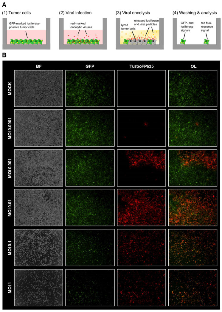
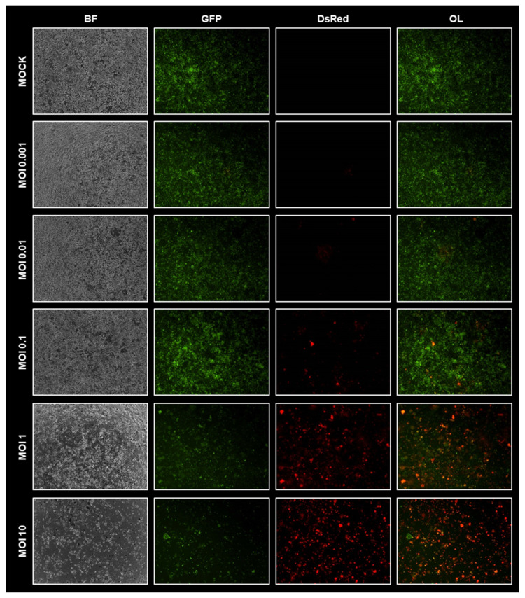
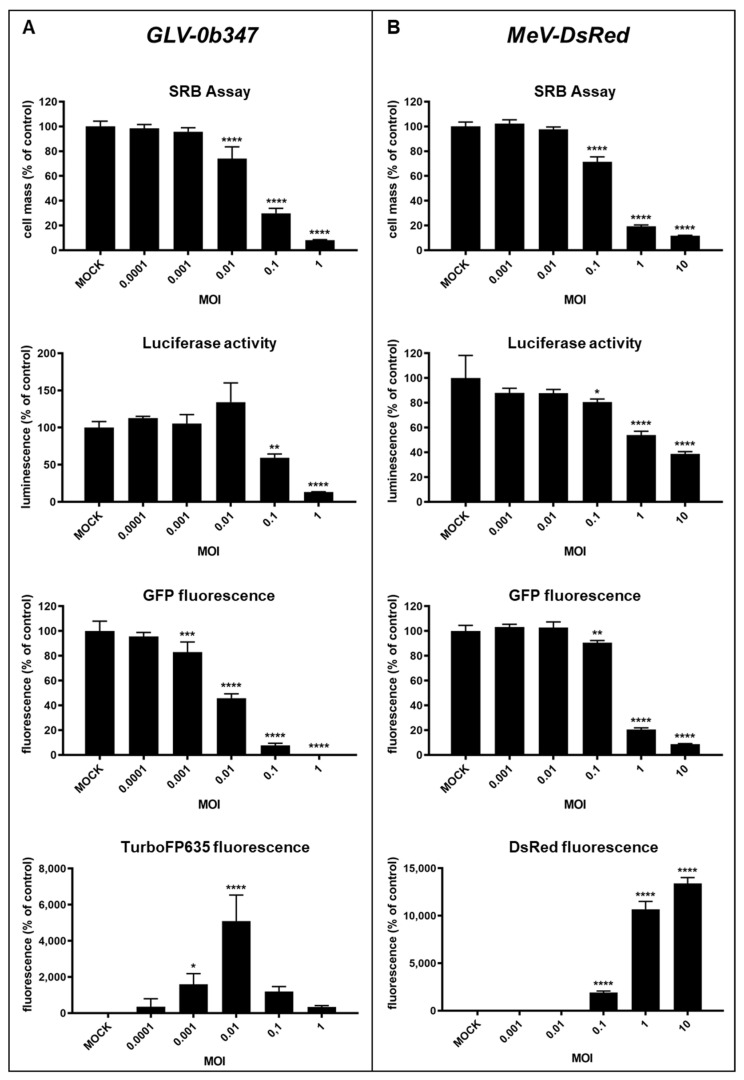
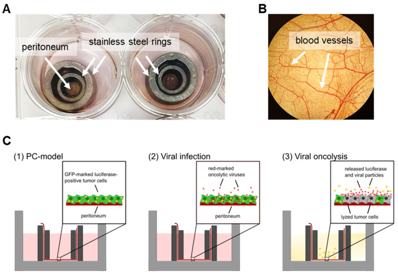
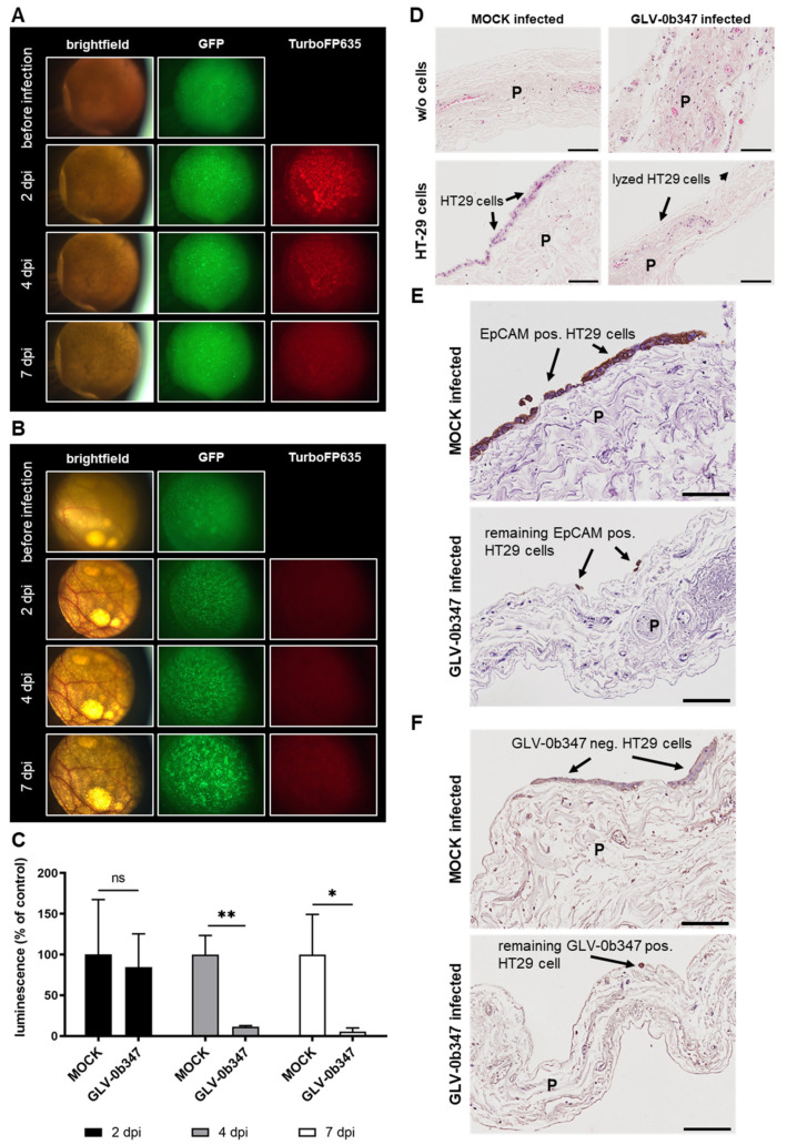
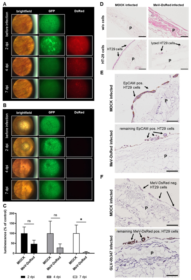
Oncolytic virotherapy constitutes a promising treatment option for many solid cancers, including peritoneal carcinomatosis (PC), which still represents a terminal stage of many types of tumors. To date, the in vitro efficacy of oncolytic viruses is mostly tested in 2D-cultured tumor cell lines due to the lack of realistic 3D in vitro tumor models. We have investigated the feasibility of virotherapy as a treatment option for PC in a human ex vivo peritoneum co-culture model. Human HT-29 cancer cells stably expressing marker genes GFP and firefly luciferase (GFP/luc) were cultured on human peritoneum and infected with two prototypic oncolytic viruses (GLV-0b347 and MeV-DsRed). Both viral constructs were able to infect HT-29 cells in patient-derived peritoneum with high tumor specificity. Over time, both GFP signal and luciferase activity decreased substantially, thereby indicating successful virus-induced oncolysis. Furthermore, immunohistochemistry stainings showed specific virotherapeutic infections of HT-29 cells and effective tumor cell lysis in infected co-cultures. Thus, the PC model established here provides a clinically relevant screening platform to evaluate the therapeutic efficacy of virotherapeutic compounds and also to investigate, in an autologous setting, the immunostimulatory potential of oncolytic viruses for PC in a unique human model system superior to standard 2D in vitro models.

Keywords: ex vivo peritoneal culture model; oncolytic measles vaccine virus; oncolytic vaccinia virus; peritoneal carcinomatosis; virotherapy.


Conflict of interest statement

The authors declare no conflict of interest.

Figures

Figure 1
Figure 1
Virotherapeutic treatment of GFP/luc-labeled human HT-29 tumor cells in cell culture with GLV-0b347. (A) Schematic illustration of the three-step virotherapeutic process and associated detection capabilities: (1) GFP/luc-labeled HT-29 tumor cells were seeded into a 24-well cell culture plate. Successful plating of the cells can be verified by the determination of GFP fluorescence. (2) The treatment of HT-29 cells with oncolytic viruses encoding a red-fluorescent marker protein. The successful infection of the tumor cells can be verified by the determination of red fluorescence. (3)/(4) The viral oncolysis can be determined by a decrease in GFP as well as in luciferase activity. Over time, enhanced red fluorescence indicates an increasing number of tumor cells being infected by the red-fluorescence marker gene encoding virotherapeutic compounds. (B) The fluorescence images of HT-29 GFP/luc-labeled cells at 72 h postinfection (hpi) with GLV-0b347 at different multiplicities of infection (MOIs), as depicted. BF, brightfield; OL, overlay of GFP and TurboFP635 signal.
Figure 2
Figure 2
Virotherapeutic treatment of GFP/luc-labeled human HT-29 tumor cells in cell culture with MeV-DsRed. Fluorescence images of HT-29 GFP/luc-labeled cells at 72 h postinfection (hpi) with MeV-DsRed at different multiplicities of infection (MOIs), as depicted. BF, brightfield; OL, overlay of GFP and DsRed signal.
Figure 3
Figure 3
Comparison of different detection options for the oncolytic activity of virotherapeutic compounds GLV-0b347 (A, images to the left) and MeV-DsRed (B, images to the right) in HT-29 GFP/luc tumor cells. HT-29 GFP/luc cells were infected with GLV-0b347 (A) or MeV-DsRed (B) at different multiplicities of infection (MOIs) ranging from 0.0001 to 1 for GLV-0b347, from 0.001 to 10 for MeV-DsRed, or remained uninfected (MOCK). At 72 h postinfection (hpi), remaining tumor cell masses were determined by either (i) SRB viability assays, (ii) the measurement of the luciferase activity, or (iii) the quantification of the GFP or red-fluorescence intensity. Each measurement was calculated relative to the MOCK control. The mean ± SD of at least two independent experiments performed in triplicate is shown. ANOVA test relative to MOCK-infected control: * p < 0.05, ** p < 0.01, *** p < 0.001, and **** p < 0.0001.
Figure 4
Figure 4
Human ex vivo peritoneum model and schematic illustration of the three-step virotherapeutic process in co-cultures with GFP/luc-labeled human HT-29 tumor cells. (A) Photographic image of the human ex vivo peritoneal model cultivated between stainless steel rings in a 24-well plate. (B) Photographic image of the peritoneum in the ex vivo model through a light microscope. (C) (1) Preparation of co-cultures of the peritoneum from noncancer patients and human GFP/luc-labeled HT-29 tumor cells. Successful plating of the cells can be verified by fluorescence microscopy. (2) Virotherapeutic treatment of co-cultures with oncolytic viruses carrying a red-fluorescent marker protein. Successful infection of the tumor cells can be verified by the determination of red fluorescence via fluorescence microscopy. (3) Viral oncolysis can be determined by a decrease in GFP and red fluorescence as well as by a decrease in luciferase activity.
Figure 5
Figure 5
Virotherapeutic treatment of PC models with recombinant vaccinia virus GLV-0b347. The fluorescence images of GLV-0b347-infected co-cultures ((A); 1.5 × 10⁶ plaque-forming units (PFU)) or MOCK-infected (B) co-cultures consisting of the peritoneum of noncancer patients and adherently growing GFP/luc-labeled human HT-29 tumor cells at days 2, 4, and 7 postinfection (dpi); original magnification 4×. (C) Luciferase activity of GFP/luc–HT-29 cells growing on peritoneum at 2, 4, and 7 dpi, either GLV-0b347-infected or MOCK-infected. Each measurement is normalized to the MOCK control. The mean ± SD of one experiment performed in triplicates is shown. t-test relative to MOCK-infected control: * p < 0.05 and ** p < 0.01. ns; not significant. (D) The hematoxylin and eosin staining of human peritoneal tissue with and w/o co-culture of GFP/luc-labeled HT-29 cells at 7 dpi, either GLV-0b347-infected or MOCK-infected. (E) The EpCAM staining of peritoneal tissue with the co-culture of HT-29 cells at 7 dpi, either GLV-0b347-infected or MOCK-infected. (F) The vaccinia virus staining of peritoneal tissue with co-culture of HT-29 cells at 7 dpi, either GLV-0b347-infected or MOCK-infected. Experiments were conducted with the peritoneal tissue from different patients and show representative data from at least three different experiments. P; peritoneum. Black arrows indicate intact or infected and lysed HT-29 cells on the surface of the peritoneum. Scale bars represent 100 μm.
Figure 6
Figure 6
Virotherapeutic treatment of PC models with recombinant measles vaccine virus MeV-DsRed. The fluorescence images of MeV-DsRed-infected co-cultures ((A); 1.5 × 10⁶ plaque-forming units (PFU)) or MOCK-infected (B) co-cultures consisting of the peritoneum of noncancer patients and GFP/luc-labeled human HT-29 tumor cells at days 2, 4, and 7 postinfection (dpi); original magnification 4×. (C) The luciferase activity of GFP/luc–HT-29 cells growing on peritoneum at 2, 4, and 7 dpi, either MeV-DsRed-infected or MOCK-infected. Each measurement is normalized to the MOCK control. The mean ± SD of one experiment performed in triplicates is shown. t-test relative to MOCK-infected control: * p < 0.05. ns; not significant. (D) The hematoxylin and eosin staining of human peritoneal tissue with and w/o co-culture of GFP/luc-labeled HT-29 tumor cells at 7 dpi with MeV-DsRed or MOCK infection. (E) The EpCAM staining of peritoneal tissue with co-culture of HT-29 cells at 7 dpi with MeV-DsRed or MOCK infection. (F) The MeV staining of peritoneal tissue with co-culture of HT-29 cells at 7 dpi with MeV-DsRed or MOCK infection. The experiments were conducted with peritoneal tissue from different patients and show representative data from at least three different experiments. P; peritoneum. Black arrows indicate intact or infected and lysed HT-29 tumor cells on the surface of the peritoneum. Scale bars represent 100 μm.

Similar articles

Cited by

References

    1. Van Der Wal J.B.C., Jeekel J. Biology of the peritoneum in normal homeostasis and after surgical trauma. Color. Dis. 2007;9((Suppl. 2)):9–13. doi: 10.1111/j.1463-1318.2007.01345.x. - DOI - PubMed
    1. Mikula-Pietrasik J., Uruski P., Tykarski A., Ksiazek K. The peritoneal “soil” for a cancerous “seed”: A comprehensive review of the pathogenesis of intraperitoneal cancer metastases. Cell Mol. Life Sci. 2018;75:509–525. doi: 10.1007/s00018-017-2663-1. - DOI - PMC - PubMed
    1. Liu T.C., Galanis E., Kirn D. Clinical trial results with oncolytic virotherapy: A century of promise, a decade of progress. Nat. Clin. Pract. Oncol. 2007;4:101–117. doi: 10.1038/ncponc0736. - DOI - PubMed
    1. Kaufman H.L., Kohlhapp F.J., Zloza A. Oncolytic viruses: A new class of immunotherapy drugs. Nat. Rev. Drug Discov. 2015;14:642–662. doi: 10.1038/nrd4663. - DOI - PMC - PubMed
    1. Kloker L.D., Yurttas C., Lauer U.M. Three-dimensional tumor cell cultures employed in virotherapy research. Oncolytic Virother. 2018;7:79–93. doi: 10.2147/OV.S165479. - DOI - PMC - PubMed

Publication types