. 2023 Feb 10;15(2):494.
doi: 10.3390/v15020494.

Caffeic Acid Has Antiviral Activity against Ilhéus Virus In Vitro

Affiliations

Caffeic Acid Has Antiviral Activity against Ilhéus Virus In Vitro

Marielena Vogel Saivish et al. Viruses. .

Abstract

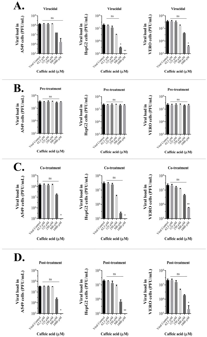
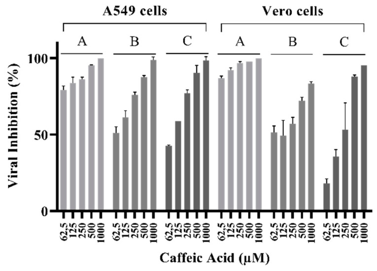
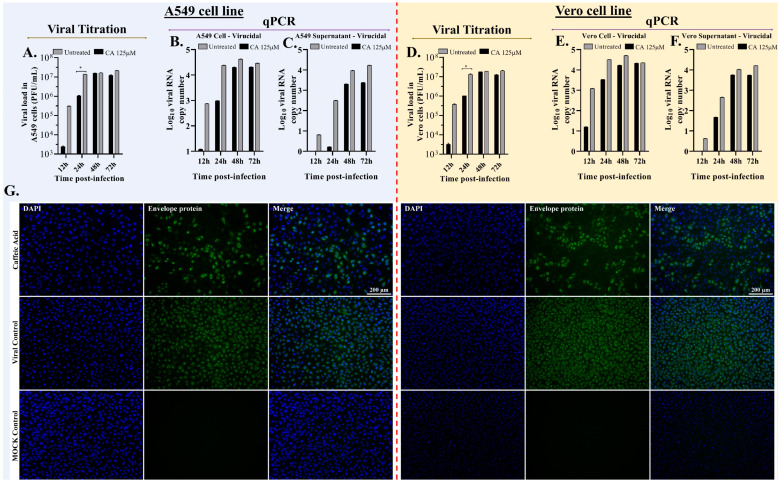
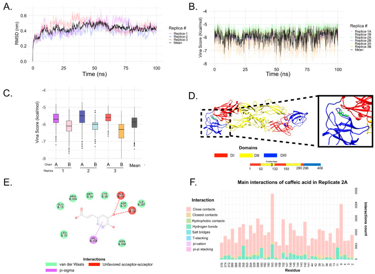
Ilhéus virus (ILHV) is a neglected mosquito-borne flavivirus. ILHV infection may lead to Ilhéus fever, an emerging febrile disease like dengue fever with the potential to evolve into a severe neurological disease characterized by meningoencephalitis; no specific treatments are available for this disease. This study assessed the antiviral properties of caffeic acid, an abundant component of plant-based food products that is also compatible with the socioeconomic limitations associated with this neglected infectious disease. The in vitro activity of caffeic acid on ILHV replication was investigated in Vero and A549 cell lines using plaque assays, quantitative RT-PCR, and immunofluorescence assays. We observed that 500 µM caffeic acid was virucidal against ILHV. Molecular docking indicated that caffeic acid might interact with an allosteric binding site on the envelope protein.

Keywords: Ilhéus virus; antiviral activity; caffeic acid; in silico analysis; inhibition.


Conflict of interest statement

The authors declare no conflict of interest.

Figures

Figure 1
Figure 1
Scheme of the antiviral assay at different stages of virus infection.
Figure 2
Figure 2
Caffeic acid’s effect on viral yield is concentration-dependent. ILHV production was measured in the presence of several dilutions of the tested compound in A549, HepG2, and Vero cells, with initial inoculum of MOI 1 under (A) virucidal, (B) pre-treatment, (C) co-treatment, or (D) post-treatment conditions 48 hpi. PFU infectivity titration of ILHV is shown in the left vertical axis. Error bars represent standard deviations. Values are the mean ± standard error obtained from three independent experiments. Asterisks indicate statistical significance between the control and each group as determined by Kruskall–Wallis test with Dunn’s multiple comparisons test. (* p < 0.05, ** p < 0.005).
Figure 3
Figure 3
Plaque reduction potential of caffeic acid (62.5–1000 μM) on A549 or Vero cells infected with ILHV at 50 PFU/well. (A) Virucidal assay, incubating ILHV with caffeic acid at 37 °C for 1 h followed by addition to cell culture. (B) Co-treatment assay, simultaneously adding virus and caffeic acid to cell culture. (C) Post-treatment assay, infecting cells by incubation at 37 °C for 1 h followed by removal of virus and addition of caffeic acid post-infection. Viral control corresponds to 0% viral inhibition.
Figure 4
Figure 4
Virucidal assay of caffeic acid (CA). (A) A549 culture supernatants were harvested at the indicated times, and ILHV production was measured in the presence of 125 μM dilution of CA in cells infected with MOI 1 by viral titration and plaque-forming unit assay. (B) Total RNA viral quantification in cell content of A549 cells harvested at the indicated times; ILHV production was measured using real-time PCR in cell content in the presence of 125 μM dilution CA in cells infected with MOI 1. (C) A549 culture supernatants were harvested at the indicated times; ILHV production was measured in the presence of 125 μM dilution CA in cells infected with MOI 1 by real-time RT-PCR. Total RNA viral quantification in culture supernatants of A549 cells. (D) Vero culture supernatants were harvested at the indicated times; ILHV production was measured in the presence of 125 μM dilution of CA in cells infected with MOI 1, by viral titration and plaque-forming unit assay. (E) Total RNA viral quantification in cell contents of Vero cells harvested at the indicated times; ILHV production was measured in cell content in the presence of 125 μM dilution CA in cells infected with MOI 1 by real-time RT-PCR. (F) Vero culture supernatants were harvested at the indicated times; ILHV production was measured in the presence of 125 μM dilution CA in cells infected with MOI 0.1 by real-time RT-PCR. Total RNA viral quantification in culture supernatants of Vero cells. (G) ILHV was cultured in A549 or Vero cells with CA at 125 μM. The cells were challenged with MOI 0.1 of virus for 24 h. Mock (uninfected cells) and ILHV-infected cells without CA (viral control MOI 0.1) were used as controls. Expression of the ILHV envelope was detected using IFA, staining the 4G2 antibody with the 488 ALEXA antibody. (* p < 0.05).
Figure 5
Figure 5
Antiviral activity of caffeic acid against ILHV investigated under different treatment conditions. (A,D,G,J) Culture supernatants were harvested at the indicated times; ILHV production was measured in the presence of 125 µM dilution CA in cells infected with MOI 1 by plaque-forming assay. (B,C,E,F,H,I,K,L) Culture supernatants and cell total RNA were harvested at the indicated time points. ILHV production was measured in the presence of 125 µM dilution of CA in cells infected with MOI 1, using real-time RT-PCR. (A) Viral load in co-treated A549 cells. (B) Viral RNA copy number in co-treated A549 cells content. (C) Viral RNA copy number in supernatant co-treated A549 cells. (D) Viral load in co-treated Vero cells. (E) Viral RNA copy number in co-treated Vero cells content. (F) Viral RNA copy number in supernatant co-treated Vero cells. (G) Viral load in post-treated A549 cells. (H) Viral RNA copy number in post-treated A549 cells content. (I) Viral RNA copy number in supernatant post-treated A549 cells. (J) Viral load in post-treated Vero cells. (K) Viral RNA copy number in post-treated Vero cells content. (I) Viral RNA copy number in supernatant post-treated Vero cells. Degree of significance indicated on the figure as follows: * p < 0.05.
Figure 6
Figure 6
Immunofluorescence assay. Immunoflourescent images of ILHV-infected A549 or Vero cells under pre-treatment, co-treatment and post-treatment conditions with CA at 125 μM. The cells were challenged with MOI 0.1 of virus. Mock and ILHV-infected cells without CA were used as controls. Expression of the ILHV envelope protein was detected by IFA, staining 4G2 antibody with 488 ALEXA antibody.
Figure 7
Figure 7
MD and ensemble docking analysis. (A) RMSD behavior throughout the 100 ns trajectory for all replicates. Replicates are shown as transparent colored lines, and the average for the five replicates is represented as a black line. (B) Vina score from caffeic acid docking in each frame for all trajectories. Details of values (mean, max, min, etc.) for RMSD and Vina score are presented in Tables S2 and S3. (C) Box plot of Vina Score distribution. Replicate 3B performed better (lower Vina Score values) than the other replicates/chains. (D) Caffeic acid (green line) binding between DI/DIII domains (red and blue ribbons, respectively). This is a complex retrieved from replica 2, chain A. (E) 2D diagram of interaction of protein-ligand complex from panel (D). This figure was created using Discovery Studios. (F) Main interaction residues of protein E, replicate 2A. Note that most of the residues are interacting in DI (residues 1–51; 132–192; 280–294) and DIII (residues 296–406). The interaction count below 1000 was not plotted. This analysis of the protein–ligand interaction was conducted using the BINANA algorithm and a Python script developed in-house. Analysis of the main interaction residues from all replicates and chains can be seen in Figure S4.

References

    1. Sips G.J., Wilschut J., Smit J.M. Neuroinvasive Flavivirus Infections. Rev. Med. Virol. 2012;22:69–87. doi: 10.1002/rmv.712. - DOI - PubMed
    1. Smith D.R. Waiting in the Wings: The Potential of Mosquito Transmitted Flaviviruses to Emerge. Crit. Rev. Microbiol. 2017;43:405–422. doi: 10.1080/1040841X.2016.1230974. - DOI - PubMed
    1. Immunologists A.A. of The Virus of Ilhéus Encephalitis: Isolation, Serological Specificity and Transmission. J. Immunol. 1947;55:61–67. - PubMed
    1. Johnson B.W., Cruz C., Felices V., Espinoza W.R., Manock S.R., Guevara C., Olson J.G., Kochel T.J. Ilheus Virus Isolate from a Human, Ecuador. Emerg. Infect. Dis. 2007;13:956–958. doi: 10.3201/eid1306.070118. - DOI - PMC - PubMed
    1. Venegas E.A., Aguilar P.V., Cruz C., Guevara C., Kochel T.J., Vargas J., Halsey E.S. Ilheus Virus Infection in Human, Bolivia. Emerg. Infect. Dis. 2012;18:516–518. doi: 10.3201/eid1803.111486. - DOI - PMC - PubMed

Publication types

LinkOut - more resources