. 2023 Feb 12;15(2):511.
doi: 10.3390/v15020511.

Coxsackievirus A6 Infection Causes Neurogenic Pathogenesis in a Neonatal Murine Model

Affiliations

Coxsackievirus A6 Infection Causes Neurogenic Pathogenesis in a Neonatal Murine Model

Qiang Sun et al. Viruses. .

Abstract

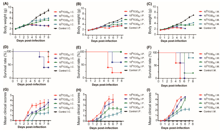
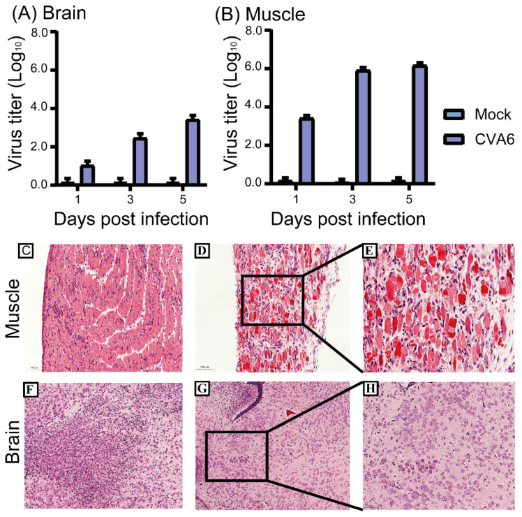
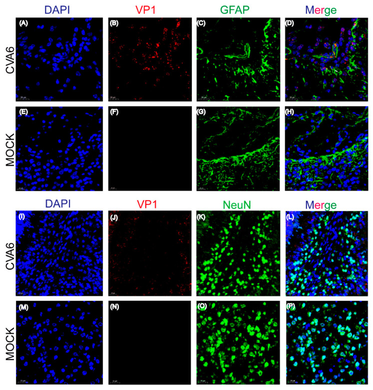
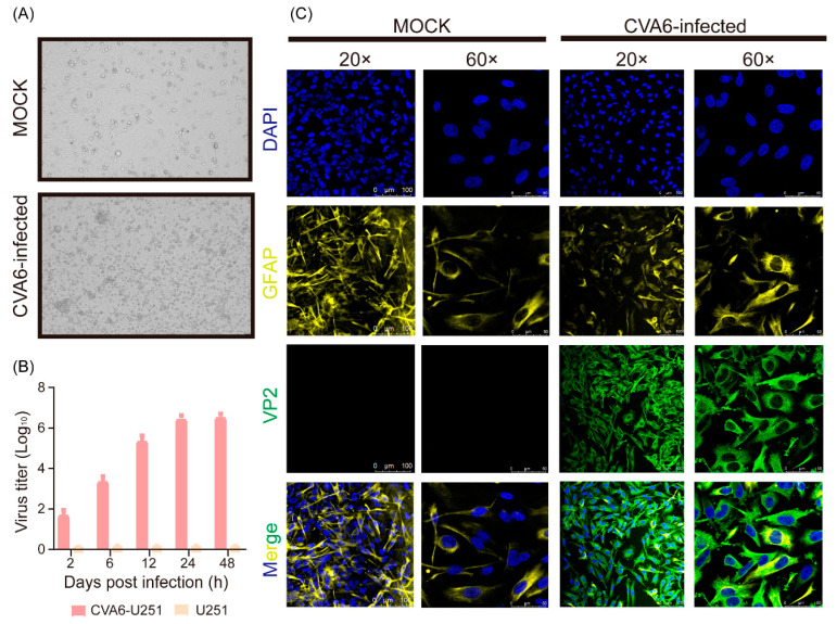
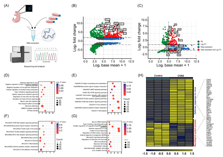
Coxsackievirus A6 (CVA6), a member of species A enterovirus, is associated with outbreaks of hand-foot-and-mouth disease and causes a large nationwide burden of disease. However, the molecular pathogenesis of CVA6 remains unclear. In the present study, we established a suckling Institute of Cancer Research (ICR) mouse infection model to explore the neural pathogenicity of CVA6. Five-day-old mice infected with CVA6 strain F219 showed lethargy and paralysis, and died 5 or 6 days after infection via IM injection. Cerebral edema and neuronal cell swelling were observed in the infected brain tissue, and we found that the CVA6 VP1 antigen could co-localize with GFAP-positive astrocytes in infected mouse brain using an immunofluorescence assay. CVA6 strain F219 can also infect human glioma (U251) cells. Transcriptome analysis of brain tissues from infected mice and infected U251 cells showed that significantly differentially expressed genes were enriched in antiviral and immune response and neurological system processes. These results indicate that CVA6 could cause neural pathogenesis and provide basic data for exploring the mechanism of how host-cell interactions affect viral replication and pathogenesis. Importance: Coxsackievirus A6 (CVA6) surpasses the two main pathogens, enterovirus 71 (EV-A71) and coxsackievirus A16 (CVA16), which are the leading pathogens causing HFMD in many provinces of China. In our study, CVA6 infection caused neurogenic pathogenesis in a neonatal murine model, manifesting as cerebral edema and neuronal cell swelling, CVA6 VP1 antigen could co-localize with GFAP-positive astrocytes in the infected mouse brain. Based on CVA6-infected brain tissue and U251 cell transcriptome analysis, we found upregulated antiviral and immune response-related genes such as Zbp1, Usp18, Oas2, Irf7, Ddx60, Ifit3, Ddx58, and Isg15, while the neurological system process-related genes were downregulated, including Fcrls, Ebnrb, Cdk1, and Anxa5.

Keywords: RNA-Seq; animal model; coxsackievirus A6; pathogenesis.


Conflict of interest statement

The authors declare no conflict of interest.

Figures

Figure 1
Figure 1
Body weight, survival rate, and clinical score of suckling mice in inoculation route and F-virus challenge dose experiments. Five-day-old ICR mice were infected with strain F219 at 103, 104, 105, and 106 TCID50 by intracranial injection (A,D,G), intraperitoneal injection (B,E,H), and intramuscular injection (C,F,I), respectively (n = 7 per group); control mice were inoculated with cell culture maintenance medium (MM). Body weight (AC), survival rate (DF) and clinical scores (GI) of 5-day-old mice inoculated via IC, IP, and IM were monitored daily. Clinical scores of CVA6-infected and control mice. 0, no disease; 1, ruffled fur; 2, weight loss; 3, single limb paralysis; 4, hindlimb paralysis; 5, moribund or dead.
Figure 2
Figure 2
Virus titers in tissues and histopathological changes of 5-day-old mice. Intramuscular injection of the CVA6 strain F219 was performed in 5-day-old ICR mice. Mice were then sacrificed after 1, 3, and 5 days, and virus titers were measured in the brain and muscle tissue (A,B). Histopathological analysis showing muscle fiber rupture (D,E), cerebral edema, and neuronal cell swelling (G,H); no changes were observed in control tissues (C,F). The magnification of (C,D,F,G) is × 200, and the magnification of (E,H) is ×400. The scale bar for (C,D,F,G) is 100 μm, and the scale bar for (E,H) is 50 μm.
Figure 3
Figure 3
Co-localization of viral antigens with astrocyte marker GFAP in infected brain tissues. Suckling mice were injected with CVA6 or cell culture maintenance medium. Brain tissue sections of suckling mice were fixed and immunostained with DAPI (Blue), VP1 (Red), and astrocyte marker GFAP (Green) (AH); immunostaining with DAPI (Blue), VP1 (Red), and neuron marker NeuN (Green) (IP); Magnifications: (A,B) ×400. Scale bar = 20 μm.
Figure 4
Figure 4
CVA6 infects U251 cells. Cytopathic effect of virus on U251 at 24 h post infection (A); cells were inoculated with CVA6 at 37 °C, and harvested at 2, 6, 12, 24, and 48 h post-inoculation. Virus titers were determined on RD cells (B); U251 cells infected with CVA6 and control were fixed and immunostained with DAPI (Blue), astrocyte marker (GFAP, Yellow), and VP2 (Green). The representative images were acquired using fluorescence confocal microscopy. Magnifications: (A,B), ×200 × 600. Scale bar = 100 μm (C).
Figure 5
Figure 5
Transcriptome analysis of the brain tissues and U251 cells infected with CVA6. Total RNA was extracted from U251 cells and brain tissues of suckling mice and analyzed by sequencing (A). Hiplot Enhanced MA v0.1.0 analysis showing the comparison of the RNA-Seq of differential gene expression of infected U251 cells (B) and that of infected suckling mice brain tissue (C). GO and KEGG pathway enrichment analysis of the RNA-Seq of differentially expressed genes in the infected U251 cells (D,E) and that of infected brain tissue (F,G). The dot plot displays the enrichment score values. Visualization of differentially expressed genes in the brain tissue (H).

References

    1. Lefkowitz E.J., Dempsey D.M., Hendrickson R.C., Orton R.J., Siddell S.G., Smith D.B. Virus taxonomy: The database of the International Committee on Taxonomy of Viruses (ICTV) Nucleic Acids Res. 2018;46:D708–D717. doi: 10.1093/nar/gkx932. - DOI - PMC - PubMed
    1. Song Y., Zhang Y., Han Z., Xu W., Xiao J., Wang X., Wang J., Yang J., Yu Q., Yu D., et al. Genetic recombination in fast-spreading coxsackievirus A6 variants: A potential role in evolution and pathogenicity. Virus Evol. 2020;6:veaa048. doi: 10.1093/ve/veaa048. - DOI - PMC - PubMed
    1. Ji T., Han T., Tan X., Zhu S., Yan D., Yang Q., Song Y., Cui A., Zhang Y., Mao N., et al. Surveillance, epidemiology, and pathogen spectrum of hand, foot, and mouth disease in mainland of China from 2008 to 2017. Biosaf. Health. 2019;1:32–40. doi: 10.1016/j.bsheal.2019.02.005. - DOI
    1. Hussain K., Chawla S., Muir D., Roberts N. Clinical manifestations of coxsackievirus A6 what a dermatologist needs to know. Int. J. Dermatol. 2022 doi: 10.1111/ijd.16478. - DOI - PubMed
    1. Li Y., Xiong T., Meng Y., Zhao R., Yu G., Liu J., Xiao J., Wang K., Wu T., Wei S., et al. Risk factors for severe hand, foot, and mouth disease infected with Coxsackievirus A6: A hospital-based case-control study. J. Med. Virol. 2020;92:3144–3150. doi: 10.1002/jmv.26343. - DOI - PubMed

Publication types

LinkOut - more resources