. 2023 Feb 21:16:707-721.
doi: 10.2147/JIR.S396049. eCollection 2023.

DNase I and Sivelestat Ameliorate Experimental Hindlimb Ischemia-Reperfusion Injury by Eliminating Neutrophil Extracellular Traps

Affiliations

DNase I and Sivelestat Ameliorate Experimental Hindlimb Ischemia-Reperfusion Injury by Eliminating Neutrophil Extracellular Traps

Chun-Lian Wang et al. J Inflamm Res. .

Abstract

Purpose: Neutrophil extracellular traps (NETs) play an important role in ischemia-reperfusion injury (IRI) of the hindlimb. The aim of this study was to investigate the effect of recombinant DNase I and sivelestat in eliminating NETs and their effects on IRI limbs.

Patients and methods: An air pump was used to apply a pressure of 300 mmHg to the root of the right hindlimb of the rat for 2 h and then deflated to replicate the IRI model. The formation of NETs was determined by the detection of myeloperoxidase (MPO), neutrophil elastase (NE), and histone H3 in the skeletal muscles of the hindlimbs. Animals were administered 2.5 mg/kg bw/d DNase I, 15 or 60 mg/kg bw/d sivelestat by injection into the tail vein or intramuscularly into the ischemic area for 7d. Elimination of NETs, hindlimb perfusion, muscle fibrosis, angiogenesis and motor function were assessed.

Results: DNase I reduced NETs, attenuated muscle fibrosis, promoted angiogenesis in IRI area and improved limb motor function. Local administration of DNase I improved hindlimb perfusion more than intravenous administration. Sivelestat at a dose of 15 mg/kg bw/d increased perfusion, counteracted skeletal muscle fibrosis, promoted angiogenesis and enhanced motor function. However, sivelestat at a dosage of 60 mg/kg bw/d had an adverse effect on tissue repair, especially when injected locally.

Conclusion: Both DNase I and moderate doses of sivelestat can eliminate IRI-derived NETs. They improve hindlimb function by improving perfusion and angiogenesis, preventing muscle fibrosis. Appropriate administration mode and dosage is the key to prevent IRI by elimination of NETs. DNase I is more valid when administered topically and sivelestat is more effective when administered intravenously. These results will provide a better strategy for the treatment of IRI in clinical.

Keywords: DNase I; IRI; NETs; hindlimb; ischemia-reperfusion injury; neutrophil extracellular traps; sivelestat.


Conflict of interest statement

The authors declare that there is no conflict of interest that could be perceived as prejudicing the impartiality of the research reported or any financial or other potential conflicts of interest.

Figures

Figure 1
Figure 1
Experimental procedure. An air pump was used to apply a pressure of 300 mmHg to the root of the right hindlimb of rats for 2 h. Releasing the pressure resulted in hindlimb ischemic reperfusion injury (IRI). The extracellular traps (NETs) released by neutrophils lead to tissue damage in the ischemic reperfusion area. The DNA digestion enzyme recombinant DNase I and the neutrophil elastase inhibitor sivelestat are used to eliminate the components of NETs. Blood flow, motor function, angiogenesis, and muscle fibrosis in the ischemic limbs are then tested.
Figure 2
Figure 2
Compare of blood perfusion of the ischemic limb and contralateral nonischemic limb after IRI modeling. Determined and photographed with a laser image-speckle interferometer. The yellow arrow indicates the poor perfusion.
Figure 3
Figure 3
Effects of ischemia-reperfusion injury on muscle morphology and neutrophil Extracellular Traps (NETs) formation. (A) Hematoxylin-eosin staining of gastrocnemius muscle sections. IRI resulted in abnormal muscle bundle morphology and many inflammatory cells infiltration. (B) Masson’s trichrome staining of gastrocnemius muscle sections. After IRI, necrotic skeletal muscle fibers are replaced by fibrous tissue. Scale bar = 50 μm. (C) Detection of NETs after IRI in the hindlimbs of rats. Between muscle fibers, neutrophil elastase (NE, n=5), myeloperoxidase (MPO, n=10), and histone H3 (n=10) stained positively, indicating the presence of NETs. Scale bar = 100 μm. Data are expressed as mean ± standard deviation, ***p < 0.001.
Figure 4
Figure 4
Myeloperoxidase (MPO) levels 24 h after ischemia-reperfusion injury in rat hindlimb muscles. MPO decreased after administration of DNAse-I or sivelestat. n=5, data are expressed as mean ± standard deviation, *P < 0.05, **P < 0.01. Scale bar = 100 μm.
Figure 5
Figure 5
Histone H3 levels 24 h after ischemia-reperfusion injury in rat hindlimb muscles. H3 decreased after administration of DNAse-I or sivelestat. n=5, data are expressed as mean ± standard deviation, *P < 0.05, ***P < 0.001. Scale bar = 100 μm.
Figure 6
Figure 6
Neutrophil elastase (NE) levels 24 h after ischemia-reperfusion injury in rat hindlimb muscles. NE decreased after administration of DNAse-I or Sivelestat. n=5, data are expressed as mean ± standard deviation, *P < 0.05, **P < 0.01. Scale bar = 100 μm.
Figure 7
Figure 7
Blood perfusion in the hindlimbs of rats 7 d after ischemia-reperfusion injury in the hindlimbs. Blood circulation in the hindlimbs of rats was determined by laser speckle contrast imaging technique. Intramuscular DNase I, intramuscular and intravenous sivelestat at a dose of 15mg/kg bw/d were beneficial for restoring blood flow in the ischemic limbs. n=3, data are expressed as mean ± standard deviation, *P < 0.05, **P < 0.01.
Figure 8
Figure 8
Hindlimb motor scores within 14 d after ischemia-reperfusion injury of hindlimb muscles in rats. Sorted by rate of recovery of motor function: DNAse (im) > sivelestat (60mg.iv) > sivelestat (15mg.iv) > sivelestat (15mg.im) > DNAse (iv) > IRI> sivelestat (60mg.im). n=3, data are expressed as mean ± standard deviation.
Figure 9
Figure 9
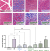
Masson’s trichrome staining of the ischemic area 14 d after ischemia-reperfusion injury in rat hindlimb muscles. Muscle fibers are stained red, collagen is stained blue, and nuclei are stained dark brown. Necrotic myofibers are replaced by fibrin hyperplasia after ischemia-reperfusion, showing large blue areas. Intravenous injection of 15 mg sivelestat most markedly reduced collagen fibrillar hyperplasia. Scale bar = 100 μm. n=3, data are expressed as mean ± standard deviation, *P < 0.05, **P < 0.01, ***P < 0.001.
Figure 10
Figure 10
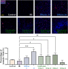
Immunofluorescence staining of CD31 in the ischemic area 14 d after ischemia-reperfusion injury of rat hindlimb muscles. Angiogenesis in the IRI region was assessed by labeling vascular endothelial cells with anti-CD31 antibody (red fluorescence). Single or clustered CD31-positive signals were counted as vascular vessels. Both DNase I and sivelestat treatment contributed to angiogenesis, with local injection of DNase I have the most obvious effect. Cell nuclei were stained with DAPI (blue fluorescence). Scale bar = 100 μm. n=3, data are expressed as mean ± standard deviation, *P < 0.05, **P < 0.01, ***P < 0.001.

References

    1. Heilmann C, Schmoor C, Siepe M, et al. Controlled reperfusion versus conventional treatment of the acutely ischemic limb: results of a randomized, open-label, multicenter trial. Circ Cardiovasc Interv. 2013;6(4):417–427. doi:10.1161/CIRCINTERVENTIONS.112.000371 - DOI - PubMed
    1. Yang B, Fung A, Pac-Soo C, Ma D. Vascular surgery-related organ injury and protective strategies: update and future prospects. Br J Anaesth. 2016;117(Suppl 2):ii32–ii43. doi:10.1093/bja/aew211 - DOI - PubMed
    1. Leurcharusmee P, Sawaddiruk P, Punjasawadwong Y, Chattipakorn N, Chattipakorn SC. The possible pathophysiological outcomes and mechanisms of tourniquet-induced ischemia-reperfusion injury during total knee arthroplasty. Oxid Med Cell Longev. 2018;2018:8087598. doi:10.1155/2018/8087598 - DOI - PMC - PubMed
    1. Yassin MM, Harkin DW, Barros D’Sa AA, Halliday MI, Rowlands BJ. Lower limb ischemia-reperfusion injury triggers a systemic inflammatory response and multiple organ dysfunction. World J Surg. 2002;26(1):115–121. doi:10.1007/s00268-001-0169-2 - DOI - PubMed
    1. Gillani S, Cao J, Suzuki T, Hak DJ. The effect of ischemia reperfusion injury on skeletal muscle. Injury. 2012;43(6):670–675. doi:10.1016/j.injury.2011.03.008 - DOI - PubMed