. 2023 Jun;29(6):1667-1677.
doi: 10.1111/cns.14130. Epub 2023 Feb 27.

Srs11-92, a ferrostatin-1 analog, improves oxidative stress and neuroinflammation via Nrf2 signal following cerebral ischemia/reperfusion injury

Affiliations

Srs11-92, a ferrostatin-1 analog, improves oxidative stress and neuroinflammation via Nrf2 signal following cerebral ischemia/reperfusion injury

Yuhua Chen et al. CNS Neurosci Ther. 2023 Jun.

Abstract

Aim: Ferroptosis is increasingly becoming to be considered as an important mechanism of pathological cell death during stroke, and specific exogenous ferroptosis inhibitors have the ability to reverse cerebral ischemia/reperfusion injury. However, research on Srs11-92 (AA9), a ferrostatin-1 (Fer-1) analog, in preclinical studies is limited.

Methods: In the middle cerebral artery occlusion-reperfusion (MCAO/R) mice model or oxygen-glucose deprivation/reperfusion (OGD/R) cell model, Fer-1, AA9, and/or ML385 were administered, and brain infarct size, neurological deficits, neuronal damage, oxidative stress, and neuroinflammation were determined after the damage, in vitro and in vivo.

Results: Fer-1 and AA9 improved brain infarct size, neuronal damage, and neurological deficits in mice model of MCAO/R, and inhibited the overloaded iron deposition, ROS accumulation, and neuroinflammation response: it also increased the expression of GPx4, Nrf2, and HO-1 and suppressed the expression of HMGB1 and NF-κB p65 in the epicenter of injured hippocampal formation. However, Nrf2 inhibitor ML385 reversed the neuroprotective effect of AA9, including the oxidative stress and neuroinflammation. In vitro studies showed that AA9 relieved OGD/R-induced neuronal oxidative stress and neuroinflammation via the Nrf2 pathway, which was impaired by ML385 in primary neurons.

Conclusion: The findings imply that Fer-1 analog AA9 may be suitable for further translational studies for the protection of neuronal damage via Nrf2 signal pathway-mediated oxidative stress and neuroinflammation in stroke and others neurological diseases.

Keywords: Srs11-92; ferroptosis; ischemic stroke; neuroinflammation; oxidative stress.


Conflict of interest statement

The authors declare that they have no competing interests.

Figures

FIGURE 1
FIGURE 1
AA9 decreases iron accumulation and improves motor behavior after MCAO. (A) At 24 h after MCAO, Perl's Prussian blue staining was performed to measure the iron deposition in injured brain tissues (black arrow indicated the iron deposition). (B) The iron concentration in the brain tissues on 1 day after MCAO. (C) Measurement of GSH/GSSG ratio, SOD level, ROS accumulation, and MDA level. (D) Fer‐1 and AA9 were administered through intraperitoneal injection immediately after MCAO, and all mice were conducted twice neurological test before surgery and 24 and 72 h after MCAO, including neurological deficit score and Garcia score. Data are presented as the mean ± SEM, n = 6. *p < 0.05, vs. sham group; #p < 0.05, vs. MCAO group. Scale bar: 50 μm.
FIGURE 2
FIGURE 2
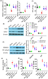
AA9 enhances GPx4 level and Nrf2 expression and inhibits inflammation response after MCAO. At 24 h after MCAO, qRT‐PCR analysis (A) and western blot analysis (B) of the key molecules of ferroptosis GPx4 and Nrf2 levels in the epicenter of injured tissues. (C) GPx activity in the epicenter of injured tissues at 24 h after MCAO. (D) Representative western blot results of HMGB1 and NF‐κB p65 expression in the epicenter of injured brain tissues at 24 h after MCAO, and quantitative analysis of protein expression. ELISA analysis of HMGB1 activation (E) and pro‐inflammatory factors TNF‐α and IL‐1β levels (F) in the epicenter of injured brain tissues at 24 h after MCAO. Data are presented as the mean ± SEM (n = 5). *p < 0.05, vs. sham group; #p < 0.05, vs. MCAO group.
FIGURE 3
FIGURE 3
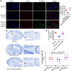
Nrf2 inhibition reduces the neuroprotection of AA9 after MCAO. (A) TUNEL/NeuN staining analysis of injured hippocampal formation at 24 h after MCAO. Dead cells labeled with TUNEL in dentate gyrls with green fluorescence. Nuclei were stained with DAPI and neurons were stained with NeuN. Scale bar: 20 μm. (B) At 24 h after MCAO, Perl's Prussian blue staining was performed to measure the iron deposition in injured brain tissues. (C) Nissl staining analysis of the neuronal damage at 24 h after MCAO. (D) The neurological deficit score analysis of motor behavior at 24 and 72 h after MCAO. Data are presented as the mean ± SEM (n = 5). *p < 0.05, vs. MCAO group; #p < 0.05, vs. MCAO + AA9 group.
FIGURE 4
FIGURE 4
Nrf2 as a regulator of AA9 reverses GPx4 activity and neuroinflammation after MCAO. (A) respective western blot results of protein expression in the epicenter of injured brain tissues at 24 h after MCAO, and quantitative analysis of protein expression. (B) The iron concentration in the brain tissues on 1 day after MCAO. GPx activity(C), HMGB1activation (D), and IL‐1β level (E) in the epicenter of injured tissues at 24 h after MCAO. (F) Nrf2/HMGB1 co‐staining analysis of protein expression at 24 h after MCAO. Nuclei were stained with DAPI. Scale bar: 20 μm. Data are presented as the mean ± SEM (n = 5). *p < 0.05, vs. Sham group; #p < 0.05, vs. MCAO group; &p < 0.05, vs. MCAO + AA9 group.
FIGURE 5
FIGURE 5
AA9 improves OGD‐induced oxidative stress and inflammation in primary neurons. (A) CCK8 analysis of cell viability. (B) The oxidative stress of GSH/GSSG ratio, SOD level, ROS accumulation, and MDA level in cells were detected. (C) ELISA analysis of pro‐inflammatory factors TNF‐α and IL‐1β levels in cell culture medium supernatant. (D) Western blot results of Nrf2, HO‐1, HMGB1, and NF‐κB p65 expression, and quantitative analysis of protein expression. Data are presented as the mean ± SEM (n = 3). *p < 0.05, vs. control group; #p < 0.05, vs. OGD group.
FIGURE 6
FIGURE 6
ML385 exacerbates OGD‐induced oxidative stress and inflammation in primary neurons. After OGD treatment, cells were immediately treated with AA9 (1 μM), ML385 (1 μM), or AA9 + ML385 for 24 h, or not. (A) CCK8 analysis of cell viability. (B) The oxidative stress of GSH/GSSG ratio and ROS accumulation in cells were detected. (C) ELISA analysis of pro‐inflammatory factor IL‐1β levels in cell culture medium supernatant. (D) Western blot results of HMGB1 and NF‐κB p65 expression, and quantitative analysis of protein expression. Data are presented as the mean ± SEM (n = 3). *p < 0.05, vs. OGD group; #p < 0.05, vs. AA9 group.

References

    1. Virani SS, Alonso A, Aparicio HJ, et al. Heart disease and stroke Statistics‐2021 update: a report from the American Heart Association. Circulation. 2021;143(8):e254‐e743. - PubMed
    1. Spence JD. China stroke statistics 2019: a wealth of opportunities for stroke prevention. Stroke Vasc Neurol. 2020;5(3):240‐241. - PMC - PubMed
    1. Li L, Scott CA, Rothwell PM, Oxford Vascular Study . Trends in stroke incidence in high‐income countries in the 21st century: population‐based study and systematic review. Stroke. 2020;51(5):1372‐1380. - PMC - PubMed
    1. Datta A, Sarmah D, Mounica L, et al. Cell death pathways in ischemic stroke and targeted pharmacotherapy. Transl Stroke Res. 2020;11(6):1185‐1202. - PubMed
    1. Hughes RE, Tadi P, Bollu PC. TPA therapy. StatPearls. StatPearls Publishing; 2021. - PubMed

Publication types

MeSH terms