. 2024 Mar;30(3):e14136.
doi: 10.1111/cns.14136. Epub 2023 Feb 27.

Vascular stability of brain arteriovenous malformations after partial embolization

Affiliations

Vascular stability of brain arteriovenous malformations after partial embolization

Yingkun He et al. CNS Neurosci Ther. 2024 Mar.

Abstract

Introduction: Brain arteriovenous malformation (bAVM) might have a higher risk of rupture after partial embolization, and previous studies have shown that some metrics of vascular stability are related to bAVM rupture risk.

Objective: To analyze vascular stability of bAVM in patients after partial embolization.

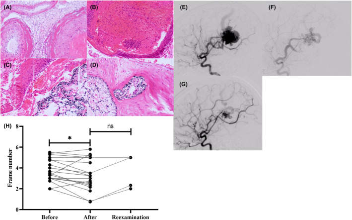
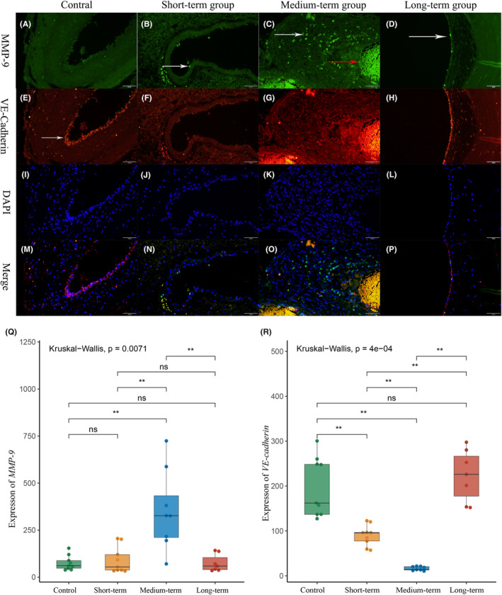
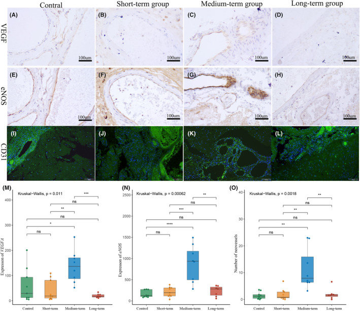
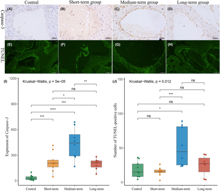
Methods: Twenty-four patients who underwent partial embolization were classified into the short-term, medium-term, and long-term groups, according to the time interval between partial embolization and surgery. The control group consisted of 9 bAVM patients who underwent surgery alone. Hemodynamic changes after partial embolization were measured by angiogram. The inflammatory infiltrates and cell-cell junctions were evaluated by MMP-9 and VE-cadherin. At the protein level, the proliferative and apoptotic events of bAVMs were analyzed by immunohistochemical staining of VEGFA, eNOS, and caspase-3. Finally, neovascularity and apoptotic cells were assessed by CD31 staining and TUNEL staining.

Results: Immediately after partial embolization, the blood flow velocity of most bAVMs increased. The quantity of MMP-9 in the medium-term group was the highest, and VE-cadherin in the medium-term group was the lowest. The expression levels of VEGFA, eNOS, and neovascularity were highest in the medium-term group. Similarly, the expression level of caspase-3 and the number of apoptotic cells were highest in the medium-term group.

Conclusion: The biomarkers for bAVM vascular stability were most abnormal between 1 and 28 days after partial embolization.

Keywords: angiogenesis; brain arteriovenous malformation; intracerebral hemorrhage; partial embolization.


Conflict of interest statement

None.

Figures

FIGURE 1
FIGURE 1
Results of H&E staining and bAVM nidal blood flow analysis. (A) Image of bAVM tissues, HE staining 100×. (B–D) Tissues image of bAVM after partial embolization at different times. (E–G) DSA image of patient no. 18. (E) When the draining vein is visualized, the peripheral vessels have entered the capillary phase. (F) The image just after partial embolization. When the draining vein is visualized, the peripheral vessels remain in the early arterial phase. (G) The image of 85 days after partial embolization. When the draining vein is visualized, the peripheral vessels have entered the late arterial phase. (H) The paired test results for hemodynamic change.
FIGURE 2
FIGURE 2
Inflammatory infiltration and cell junction integrity of bAVM malformation vessels after partial embolization, 200×. (A–D) MMP‐9 and biomarkers of inflammatory cells. (A) There was almost no inflammatory infiltration in the control group. (B–D) The vascular inflammatory infiltration in the short‐term and long‐term groups, with sporadic inflammatory cells attached to the vascular surface(white arrow). (C) A large number of inflammatory cells infiltrating the vessel wall in the metaphase group (white arrow) and the red arrow was non‐specific staining of red blood cells. (E–H) VE‐cadherin and biomarkers of cell junction. (E) The typical bAVM interendothelial cell junctions, with endothelial cell stacks and less continuous interendothelial cell junctions (white arrow). (F, G) Interendothelial cell junction almost disappeared in the short‐term and medium‐term groups, but it was relatively intact in the long‐term group (H). (I–L) DAPI and fluorescence of 4',6‐diamidino‐2‐phenylindole; (M–P) merge, merge of MMP‐P, VE‐cadherin, and DAPI. (Q, R) Quantitation of MMP‐9 and VE‐cadherin levels.*p<0.05; **p<0.01; ***p<0.005; ****p<0.001
FIGURE 3
FIGURE 3
Increased expression of proliferation‐associated proteins in bAVMs after partial embolization (VEGFA, eNOS 200X; CD31, 100×). (A–D) VEGFA level of each group. (E–H) eNOS level of each group. (I–L) The neovessels increasing of bAVM after partial embolization. Green is the endothelial cell marker CD31 and DAPI is blue. (I, J) There were few neovessels in the control group and short‐term group. (K) Numerous neovessels are composed of a single layer of endothelial cells (yellow arrow). (L) The mature small vessel with an intact structure (red arrow) around the malformed vessel (white arrow). (M, N) The quantification of the mean IOD of VEGFA and eNOS. *p<0.05; **p<0.01; ***p<0.005; ****p<0.001. (O) The quantification of the mean neovessels number in each group. *p<0.05; **p<0.01; ***p<0.005; ****p<0.001
FIGURE 4
FIGURE 4
Apoptosis in bAVM tissues after partial embolization (Caspase‐3,200X; Tunel stain, 100×). (A–D) Caspase‐3 level of each group. (A, B) In the control group and short‐term group, caspase‐3 was found only in vascular endothelial cells. (C, D) After 24 h, caspase‐3 was highly expressed not only in vascular endothelial cells in bAVM tissue but also in vascular smooth muscle cells and fibroblasts. (E–H) Typical pictures of TUNEL staining of each group. (E, F) As same as caspase‐3, in the control and short‐term groups, TUNEL‐positive cells were mainly located in the endothelium. (G, H) TUNEL‐positive cells were expressed in both endothelium and vessel wall in the medium‐term and long‐term groups. (I) Quantification of the mean IOD of caspase‐3. (J) The numbers of the mean TUNEL‐positive cells in each group. *p<0.05; **p<0.01; ***p<0.005; ****p<0.001

References

    1. Chen CJ, Ding D, Derdeyn CP, et al. Brain arteriovenous malformations: a review of natural history, pathobiology, and interventions. Neurology. 2020;95(20):917‐927. - PubMed
    1. Nerva JD, Kim LJ, Barber J, et al. Outcomes of multimodality therapy in pediatric patients with ruptured and unruptured brain arteriovenous malformations. Neurosurgery. 2016;78(5):695‐707. - PubMed
    1. Sure U, Butz N, Siegel AM, Mennel HD, Bien S, Bertalanffy H. Treatment‐induced neoangiogenesis in cerebral arteriovenous malformations. Clin Neurol Neurosurg. 2001;103(1):29‐32. - PubMed
    1. Lv X, Wu Z, Li Y, Yang X, Jiang C. Hemorrhage risk after partial endovascular NBCA and ONYX embolization for brain arteriovenous malformation. Neurol Res. 2012;34(6):552‐556. - PubMed
    1. Laakso A, Dashti R, Seppänen J, et al. Long‐term excess mortality in 623 patients with brain arteriovenous malformations. Neurosurgery. 2008;63(2):244‐253. discussion 253–255. - PubMed

Publication types

MeSH terms