. 2023 Jan-Dec:32:9636897231154574.
doi: 10.1177/09636897231154574.

Hypermethylation-Mediated lncRNA MAGI2-AS3 Downregulation Facilitates Malignant Progression of Laryngeal Squamous Cell Carcinoma via Interacting With SPT6

Affiliations

Hypermethylation-Mediated lncRNA MAGI2-AS3 Downregulation Facilitates Malignant Progression of Laryngeal Squamous Cell Carcinoma via Interacting With SPT6

Jiantao Wang et al. Cell Transplant. 2023 Jan-Dec.

Abstract

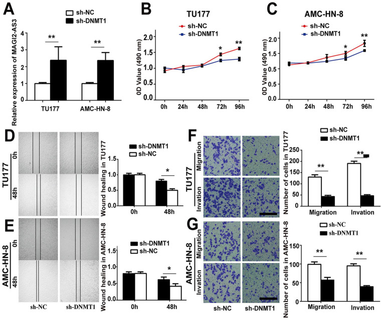
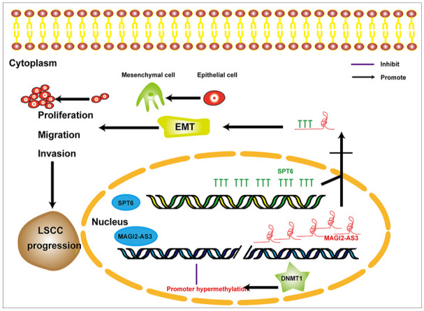
Long noncoding RNAs (lncRNAs) have an effect on the occurrence and progression of a considerable number of diseases, especially cancer. Existing research has suggested that MAGI2 antisense RNA 3 (MAGI2-AS3) takes on a critical significance in the development of hepatocellular carcinoma and lung cancer. However, the functions of MAGI2-AS3 in laryngeal squamous cell carcinoma (LSCC) remain unclear. In this study, MAGI2-AS3 expression level in LSCC tissue and cell lines was detected, and the effect of MAGI2-AS3 overexpressed on LSCC phenotypes and the possible influence mechanisms were examined. MAGI2-AS3 was downregulated in the tissues of LSCC patients versus non-tumor tissues, and it was correlated with advanced TNM (tumor, node, metastasis) stage and lymph node metastases, as indicated by the results of this study. MAGI2-AS3 inhibited the proliferation, migration, and invasion of LSCC cells in vitro and in vivo. Furthermore, the hypermethylation level of the MAGI2-AS3 promoter region was indicated by bisulfite genomic sequencing and methylation-specific polymerase chain reaction, such that MAGI2-AS3 expression was downregulated. Besides, MAGI2-AS3 promoter hypermethylation was regulated by DNA methyltransferase 1 (DNMT1), and MAGI2-AS3 expression was reversed by 5-Aza-2'-deoxycytidine (5-Aza). Moreover, the result of the RNA pull-down experiment suggested that 38 proteins were enriched in the MAGI2-AS3 group versus the control group in TU177 cells. To be specific, SPT6 (ie, a conserved protein) was enriched by fold change >10. SPT6 knockdown reduced the antitumor effect of MAGI2-AS3 in TU177 and AMC-HN-8 cells. Meanwhile, SPT6 overexpression inhibited the proliferation, metastasis, and invasion of TU177 and AMC-HN-8 cells. As revealed by the above findings, DNMT1-regulated MAGI2-AS3 promoter hypermethylation led to downregulated MAGI2-AS3 expression, such that the presence and progression of LSCC were inhibited in an SPT6 binding-dependent manner.

Keywords: DNA methylation; MAGI2-AS3; SPT6; biomarker; laryngeal squamous cell carcinoma.


Conflict of interest statement

The author(s) declared no potential conflicts of interest with respect to the research, authorship, and/or publication of this article.

Figures

Figure 1.
Figure 1.
MAGI2-AS3 was downregulated in LSCC. (A) Heatmap diagram of lncRNA expression in four pairs of LSCC tissues and matched non-tumor tissues. (B) Relative expression of MAGI2-AS3 in various tumor types in the GEPIA data set. (C) Relative expression levels of MAGI2-AS3 in 61 pairs of LSCC tissues and matched non-tumor tissues. (D) Relative expression levels of MAGI2-AS3 in LSCC cells. (E) Relative expression of MAGI2-AS3 in different subgroups. (F, J) ROC analysis of the prognostic sensitivity and specificity for LSCC patients and general people. Data are presented as mean ± SD from three independent experiments. *P < 0.05 and **P < 0.01. MAGI2-AS3: MAGI2 antisense RNA 3; LSCC: laryngeal squamous cell carcinoma; lncRNAs: long noncoding RNAs; LN: lymph node; GEPIA: Gene Expression Profiling Interactive Analysis; ROC: receiver operating characteristic; ACC: adrenocortical carcinoma; BLCA: bladder urothelial carcinoma; BRCA: breast invasive carcinoma; CESC: cervical squamous cell carcinoma and endocervical adenocarcinoma; CHOL: cholangiocarcinoma; COAD: colon adenocarcinoma; DLBC: lymphoid neoplasm diffuse large B cell lymphoma; ESCA: esophageal carcinoma; GBM: glioblastoma multiforme; HNSCC: head and neck squamous cell carcinoma; KICH: kidney chromophobe; KIRC: kidney renal clear cell carcinoma; KIRP: kidney renal papillary cell carcinoma; LAML: acute myeloid leukemia; LGG: brain lower grade glioma; LIHC: liver hepatocellular carcinoma; LUAD: lung adenocarcinoma; LUSC: lung squamous cell carcinoma; MESO: mesothelioma; OV: ovarian serous cystadenocarcinoma; PAAD: pancreatic adenocarcinoma; PCPG: pheochromocytoma and paraganglioma; PRAD: prostate adenocarcinoma; READ: rectum adenocarcinoma; SARC: sarcoma; SKCM: skin cutaneous melanoma; STAD: stomach adenocarcinoma; TGCT: testicular germ cell tumors; THCA: thyroid carcinoma; THYM: thymoma; UCEC: uterine corpus endometrial carcinoma; UCS: uterine carcinosarcoma; UVM: uveal melanoma.
Figure 2.
Figure 2.
Functional analysis of MAGI2-AS3 in TU177 and AMC-HN-8 cells. (A) The expression levels of MAGI2-AS3 in TU177 and AMC-HN-8 transfected with pcDNA3.1-MAGI2-AS3 and pcDNA3.1. (B, C) Proliferation of TU177 and AMC-HN-8 cells transfected with pcDNA3.1-MAGI2-AS3 was examined by the MTS assay. (D) Overexpression of MAGI2-AS3 significantly suppresses cell growth in a colony formation assay. (E, F) The effect of MAGI2-AS3 on migration was assessed using a scratch assay. (G, H) Migration and invasion abilities of the TU177 and AMC-HN-8 cell lines, following overexpression of MAGI2-AS3 in the transwell assay. Scale bar, 100 µm. Data are presented as mean ± SD from three independent experiments. *P < 0.05, **P < 0.01. MAGI2-AS3: MAGI2 antisense RNA 3; oe-MAGI2-AS3: pcDNA3.1-MAGI2-AS3; oe-NC: pcDNA3.1 vector. AMC-HN-8: Asian Medical Center-Head and Neck cancer-8.
Figure 3.
Figure 3.
MAGI2-AS3 suppressed EMT in vitro. (A, B) Expression levels of MAGI2-AS3 in nuclear and cytoplasmic fractions of TU177 and AMC-HN-8 cells were detected by qPCR. GAPDH: cytoplasmic control; U6: nuclear control. (C, D) Overexpression of MAGI2-AS3 increased the expression of EMT-associated markers detected by the qPCR method. (E, F) Relative protein expression levels of E-cadherin, N-cadherin, and Vimentin were assessed by Western blotting analysis in TU177/overexpression-MAGI2-AS3, AMC-HN-8/overexpression-MAGI2-AS3, and control cells. (G, H) After TU177 and AMC-HN-8 cells were administrated with different doses (2.5, 5, 7.5, 10 μmol/L) of 5-Aza-dC, N-cadherin and Vimentin were detected by Western blotting. Data are presented as mean ± SD from three independent experiments. *P < 0.05, **P < 0.01. qPCR: quantitative polymerase chain reaction; EMT: epithelial–mesenchymal transition; AMC-HN-8: Asian Medical Center-Head and Neck cancer-8; MAGI2-AS3: MAGI2 antisense RNA 3.
Figure 4.
Figure 4.
Methylation mechanisms of inactivation of MAGI2-AS3 in LSCC. (A) Location, MAGI2-AS3 cited from UCSC. (B) Schematic structure of MAGI2-AS3 CpG islands predicted by MethPrimer. (C) Schematic of cytosine changed after bisulfite DNA modification. (D) Relative expression levels of MAGI2-AS3 in 5-Aza-dC-treated two LSCC cell lines. (F) High-resolution mapping of the methylation status of every CpG site in MAGI2-AS3 CpG islands in two LSCC cell lines and two pairs of LSCC tissues obtained by BGS. Each CpG site is shown at the top row as an individual number. The color of circles for each CpG site represents the percentage of methylation. (E, G) The methylation status of three regions of MAGI2-AS3 detected by MSP analysis in two LSCC lines with or without 5-Aza-dC treatment and LSCC tissues versus matched non-tumor tissues. (H) Relative expression of MAGI2-AS3 in tumor tissues with and without methylation of the three regions. Data are presented as mean ± SD from three independent experiments. *P < 0.05, **P < 0.01. MAGI2-AS3: MAGI2 antisense RNA 3; oe-MAGI2-AS3: pcDNA3.1-MAGI2-AS3; oe-NC: pcDNA3.1 vector. LSCC: laryngeal squamous cell carcinoma; USSC: University of California Santa Cruz; BGS: bisulfite genomic sequencing; MSP: methylation-specific polymerase; AMC-HN-8: Asian Medical Center-Head and Neck cancer-8.
Figure 5.
Figure 5.
Functional analysis of DNMT1 in TU177 and AMC-HN-8 cells. (A) TU177 and AMC-HN-8 cells were transfected with sh-DNMT1 or pGenecil-1 vector, and then MAGI2-AS3 expression was obtained by qPCR. (B, C) TU177 and AMC-HN-8 cells were transfected with sh-DNMT1 or pGenecil-1 vector; cell proliferation was detected by the MTS assay. (D, E) Proliferative capacity of TU177 and AMC-HN-8 cells transfected with sh-DNMT1 or pGenecil-1 vector was assessed by the wound-healing assay. (F, G) Effect of DNMT1 on the migration and invasion of TU177 and AMC-HN-8 cells was assessed by transwell migration and invasion assays. Scale bar, 100 µm. Data are presented as mean ± SD of three independent experiments. *sh-NC versus sh-DNMT1. *P < 0.05, **P < 0.01. MAGI2-AS3: MAGI2 antisense RNA 3; DNMT1: DNA methyltransferase 1; AMC-HN-8: Asian Medical Center-Head and Neck cancer-8; qPCR: quantitative polymerase chain reaction.
Figure 6.
Figure 6.
MAGI2-AS3 induced antitumor effects in LSCC cells by interacting with SPT6. (A) Schematic of the MS2-12X-dependent RNA pull-down experiments. (B) MAGI2-AS3-binding proteins by mass spectrometry analysis. (C) SPT6 identified specifically bind to MAGI2-AS3 by Western blotting. (D) RIP was performed to confirm the interaction of SPT6 and MAGI2-AS3. (E) The expression levels of SPT6 in the TU177 knockdown of SPT6. (F, G) TU177 and AMC-HN-8 cells were transfected with oe-MAGI2-AS3 or cotransfected with oe-MAGI2-AS3 and sh-SPT6. Cell proliferation was obtained by the MTS assay. (H) Colony formation assays were performed to assess the proliferative ability of TU177 and AMC-HN-8 cells transfected with oe-MAGI2-AS3 or cotransfected with oe-MAGI2-AS3 and sh-SPT6. Effects of oe-MAGI2-AS3 and sh-SPT6 on the migration and invasion of TU177 and AMC-HN-8 cells were assessed by the (I, J) wound-healing assay and (K, L) transwell migration and invasion assays. Scale bar, 100 µm. Data are presented as mean ± SD of three independent experiments. *Vector versus oe-MAGI2-AS3; #oe-MAGI2-AS3 versus oe-MAGI2-AS3+sh-SPT6; *# Vector versus oe-MAGI2-AS3+sh-SPT6. *P < 0.05, **P < 0.01; #P < 0.05, ##P < 0.01; *#P < 0.01. MAGI2-AS3: MAGI2 antisense RNA 3; LSCC: laryngeal squamous cell carcinoma; SPT6: Suppressor Of Ty 6 Homolog; RIP: RNA Immunoprecipitation; AMC-HN-8: Asian Medical Center-Head and Neck cancer-8; GFP: green fluorescent protein.
Figure 7.
Figure 7.
SPT6 regulated the proliferation, migration, and invasion of LSCC cells. (A) The expression levels of SPT6 in TU177 overexpressed SPT6. (B) The colony formation assay was performed for the proliferation of SPT6 regulating TU177 and AMC-HN-8 cells. (C, D) The wound-healing assay was performed for the migration of SPT6 regulating TU177 and AMC-HN-8 cells. (E, F) The transwell assay was performed for invasion and migration of SPT6 regulating TU177 and AMC-HN-8 cells. Scale bar, 100 µm. Data are presented as mean ± SD of three independent experiments. *sh-NC versus sh-SPT6; #oe-NC versus oe-SPT6. *P < 0.05, **P < 0.01; #P < 0.05, ##P < 0.01; *#P < 0.01. SPT6: Suppressor Of Ty 6 Homolog; LSCC: laryngeal squamous cell carcinoma; AMC-HN-8: Asian Medical Center-Head and Neck cancer-8.
Figure 8.
Figure 8.
MAGI2-AS3 inhibited the tumor growth of LSCC cells in vivo. (A) The expression levels of MAGI2-AS3 in TU177 stably overexpressed MAGI2-AS3 (lenti-MAGI2-AS3). (B, D) Nude mice were subcutaneously injected with negative control (lenti-Blast), and lenti-MAGI2-AS3 stably transfected TU177 cells. After 28 days, tumors were dissected and imaged. (C) Starting from day 7 after injection, the tumor volume was examined every 3–4 days, and the tumor growth curve was plotted. (E) The tumor weight was obtained on the day the mice were killed. Data represent mean ± SD (n = 10 each group). (F, H) The expression levels of MAGI2-AS3, SPT6, and Ki67 in xenograft tumors were obtained by qPCR. (I) H&E staining revealed the structure of xenograft tumors derived from oe-NC and oe-MAGI2-AS3 LSCC cells. Scale bar, 50 μm. (J) Changes in Ki67, N-cadherin, and Vimentin expression in xenograft tumors were detected by IHC staining. Scale bar, 50 μm. Data are presented as mean ± SD from three independent experiments. *P < 0.05 and **P < 0.01. LSCC: laryngeal squamous cell carcinoma; SPT6: Suppressor Of Ty 6 Homolog; AMC-HN-8: Asian Medical Center-Head and Neck cancer-8; IHC: immunohistochemistry; qPCR: quantitative polymerase chain reaction; MAGI2-AS3: MAGI2 antisense RNA 3.
Figure 9.
Figure 9.
A proposed scheme of the roles and molecular mechanisms of MAGI2-AS3 in LSCC progression is presented. LSCC: laryngeal squamous cell carcinoma; EMT: epithelial-mesenchymal transition; SPT6: Suppressor Of Ty 6 Homolog; DNMT1: DNA methyltransferase1; MAGI2-AS3: MAGI2 antisense RNA 3.

References

    1. Steuer CE, El-Deiry M, Parks JR, Higgins KA, Saba NF. An update on larynx cancer. CA Cancer J Clin. 2017;67(1):31–50. - PubMed
    1. Siegel RL, Miller KD, Jemal A. Cancer statistics, 2019. CA Cancer J Clin. 2019;69(1):7–34. - PubMed
    1. Zhao S, Zhang X, Chen S, Zhang S. Natural antisense transcripts in the biological hallmarks of cancer: powerful regulators hidden in the dark. J Exp Clin Cancer Res. 2020;39(1):187. - PMC - PubMed
    1. Peng WX, Koirala P, Mo YY. LncRNA-mediated regulation of cell signaling in cancer. Oncogene. 2017;36(41):5661–67. - PMC - PubMed
    1. Magistri M, Faghihi MA, St Laurent G, 3rd, Wahlestedt C. Regulation of chromatin structure by long noncoding RNAs: focus on natural antisense transcripts. Trends Genet. 2012;28(8):389–96. - PMC - PubMed

Publication types