. 2023 Apr 24;8(8):e166811.
doi: 10.1172/jci.insight.166811.

Potently neutralizing human mAbs against the zoonotic pararubulavirus Sosuga virus

Affiliations

Potently neutralizing human mAbs against the zoonotic pararubulavirus Sosuga virus

Helen M Parrington et al. JCI Insight. .

Abstract

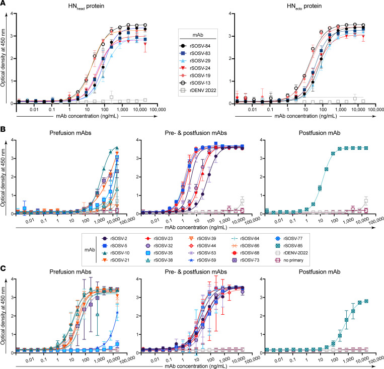
Sosuga virus (SOSV) is a recently discovered paramyxovirus with a single known human case of disease. There has been little laboratory research on SOSV pathogenesis or immunity, and no approved therapeutics or vaccines are available. Here, we report the discovery of human mAbs from the circulating memory B cells of the only known human case and survivor of SOSV infection. We isolated 6 mAbs recognizing the functional attachment protein hemagglutinin-neuraminidase (HN) and 18 mAbs against the fusion (F) protein. The anti-HN mAbs all targeted the globular head of the HN protein and could be organized into 4 competition-binding groups that exhibited epitope diversity. The anti-F mAbs can be divided into pre- or postfusion conformation-specific categories and further into 8 competition-binding groups. The only Ab in the panel that did not display neutralization activity was the single postfusion-specific anti-F mAb. Most of the anti-HN mAbs were more potently neutralizing than the anti-F mAbs, with mAbs in 1 of the HN competition-binding groups possessing ultrapotent (<1 ng/mL) half-maximal inhibitory virus neutralization values. These findings provide insight into the molecular basis for human Ab recognition of paramyxovirus surface proteins and the mechanisms of SOSV neutralization.

Keywords: Adaptive immunity; Immunoglobulins; Immunology; Infectious disease.


Conflict of interest statement

Conflict of interest: JEC has served as a consultant for Luna Labs USA, Merck Sharp & Dohme, Emergent Biosolutions, and GlaxoSmithKline. He is a member of the Scientific Advisory Board of Meissa Vaccines, a former member of the Scientific Advisory Board of Gigagen (Grifols), and founder of IDBiologics. The laboratory of JEC received sponsored research agreements from AstraZeneca, Takeda, and IDBiologics during the conduct of the study. Vanderbilt University has applied for patents (U.S. Provisional Patent Application Serial No. 63/036,756) for some of the Abs in this paper.

Figures

Figure 1
Figure 1. Cotransfection of cDNAs encoding SOSV F and HN proteins causes robust syncytia formation in cell culture monolayers.
Representative field of view (10× objective) of transfected Vero cell culture monolayers. Nuclei were stained with DAPI (blue) and SOSV proteins were stained with a polyclonal mix of 6 anti-SOSV mAbs (3 anti-HN and 3 anti-F) or mouse anti-FLAG Ab with goat anti-human IgG with Alexa Fluor 488 dye or goat anti-mouse IgG with Alexa Fluor 488 dye Abs as secondary Abs. A total of 10–12 fields of view were imaged for each of the transfection conditions and the average area of fluorescent cells was determined for each field. (A) Syncytia-producing transfections: cotransfection of SOSV F-WT + SOSV HN-WT, cotransfection of SOSV F-FLAG + SOSV HN-FLAG, or cotransfection of SOSV F-FLAG + SOSV HN-Flag constructs stained with anti-FLAG Abs. (B) Nonsyncytia-producing transfections: cDNA-encoding SOSV F-WT or SOSV HN-WT were transfected individually. (C) Controls: mock transfection or VSV G-WT transfection. Scale bar: 300 μm. (D) Average area of fluorescently stained clusters (cells or syncytia). A 1-way ANOVA with Tukey’s multiple comparison with a P value threshold of < 0.05; ****P < 0.0001 and ***P < 0.001.
Figure 2
Figure 2. SOSV HN and F soluble protein designs.
Soluble versions of the SOSV HN and F proteins were generated by removing the cytoplasmic tail and transmembrane domains and adding a human CD5 or mouse IL-2 signal peptide. To aid in purification, a 6-His tag or StrepII tags were added to the carboxy (F) or amino (HN) terminal ends. (A) HNecto design includes a longer portion of the stalk region starting at residue 75, while the HNhead construct is composed of nearly only the globular head domain of the HN protein. (B) Additional modifications were necessary to include in the SOSV F prefusion (preF-tHS) construct, which included removal of the furin cleavage site and creation of a stabilizing disulfide bond by point mutations to cysteines at 206 and 223. The postfusion construct of SOSV F (postF-tHS) more closely resembles the WT sequence but with replacement of the cytoplasmic and transmembrane domains with a GCNt trimerization domain (also in preF-tHS).
Figure 3
Figure 3. Representative curves in ELISA for binding of rSOSV mAbs to soluble glycoprotein antigens.
Shown here is a single representative set of data of the 3 biological replicates that were performed. Curves show the average of 3 technical replicates plotted with SD. The anti-F rmAbs are divided into 3 subsets: prefusion, pre- and postfusion, or postfusion, although all the rmAbs were tested simultaneously. (A) Anti-HN rSOSV mAbs against HNecto and HNhead proteins. (B) Anti-F rSOSV mAbs against postfusion SOSV F. (C) Anti-F rSOSV mAbs against SOSV prefusion F construct.
Figure 4
Figure 4. Competition-binding assay for anti-HN mAbs.
Unlabeled primary (first) mAb was bound to antigen-coated plates in saturating conditions (10 μg/mL) with secondary Abs added at a final concentration of 100 ng/mL for HN-mAbs and 500 ng/mL for F-mAbs according to the grid layout shown. Data were converted to percent binding relative to the maximal uncompeted binding of the second Ab (lacking a primary mAb). Assays were repeated in triplicate with quadruplicate technical replicates. A representative assay for each antigen/mAb set tested is shown. (A) Binding data of mAbs against HNecto as antigen. (B) Binding data of mAbs against HNhead as antigen. (C) Binding data for pre- and postfusion anti-F mAbs (rSOSV-2, 5, 23, 32, 44, 53, 68, and 77) and the postfusion mAb (rSOSV-85) against prefusion F protein. (D) Binding data for prefusion F-specific mAbs (rSOSV-10, 21, 35, 38, 39, 59, 64, 66, and 73) against prefusion F antigen.
Figure 5
Figure 5. Neutralization assay of SOSV mAbs against live virus.
SOSV mAbs were tested for inhibition of authentic rSOSV-ZsG in quadruplicate on Vero-E6 cell culture monolayers. (A) Neutralization data for anti-F mAbs. Data are grouped according to the pattern of antigen reactivity: prefusion F, pre- and postfusion F, or postfusion F protein. (B) Neutralization data for the HN-specific mAbs.

Similar articles

Cited by

References

    1. Plemper RK, Lamb RA. Paramyxoviridae: The Viruses and Their Replication. In: Howley PM, et al, eds. Fields Virology Volume 1: Emerging Viruses. Wolters Kluwer; 2021;503–555.
    1. Baker KS, et al. Co-circulation of diverse paramyxoviruses in an urban African fruit bat population. J Gen Virol. 2012;93(pt 4):850–856. doi: 10.1099/vir.0.039339-0. - DOI - PMC - PubMed
    1. Drexler JF, et al. Bats host major mammalian paramyxoviruses. Nat Commun. 2012;3(1):796–796. doi: 10.1038/ncomms1796. - DOI - PMC - PubMed
    1. Pernet O, et al. Evidence for henipavirus spillover into human populations in Africa. Nat Commun. 2014;5(1):5342. doi: 10.1038/ncomms6342. - DOI - PMC - PubMed
    1. Philbey AW, et al. An apparently new virus (family Paramyxoviridae) infectious for pigs, humans, and fruit bats. Emerg Infect Dis. 1998;4(2):269–271. doi: 10.3201/eid0402.980214. - DOI - PMC - PubMed

Publication types