. 2023 Feb 28;18(2):e0282246.
doi: 10.1371/journal.pone.0282246. eCollection 2023.

HR LC-MS/MS metabolomic profiling of Yucca aloifolia fruit and the potential neuroprotective effect on rotenone-induced Parkinson's disease in rats

Affiliations

HR LC-MS/MS metabolomic profiling of Yucca aloifolia fruit and the potential neuroprotective effect on rotenone-induced Parkinson's disease in rats

Dalia E Ali et al. PLoS One. .

Abstract

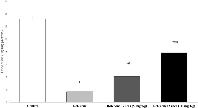
Yucca aloifolia L. fruit (Yucca or Spanish bayonet, family Asparagaceae) is recognized for its purplish red color reflecting its anthocyanin content, which has a powerful antioxidant activity. This study aimed to investigate yucca (YA) fruit extract's protective effect on Parkinson's disease (PD). In vitro study, the anti-inflammatory activity of yucca fruit extracts was explored by measuring tumor necrosis factor receptor 2 (TNF-R2) and nuclear factor kappa B (NF-KB) to choose the most effective extract. Afterward, a detailed in vivo investigation of the protective effect of the most active extract on rotenone-induced PD was performed on male albino Wister rats. First, the safety of the extract in two different doses (50 and 100 mg/kg in 0.9% saline orally) was confirmed by a toxicological study. The rats were divided into four groups: 1) normal control (NC); 2) rotenone group; and third and fourth groups received 50 and 100 mg/kg yucca extract, respectively. The neurobehavioral and locomotor activities of the rats were tested by rotarod, open field, and forced swim tests. Striatal dopamine, renal and liver functions, and oxidative stress markers were assessed. Western blot analysis of brain tissue samples was performed for p-AMPK, Wnt3a, and β-catenin. Histopathological examination of striatal tissue samples was performed by light and electron microscopy (EM). The metabolites of the active extract were characterized using high-resolution LC-MS/MS, and the results showed the prevalence of anthocyanins, saponins, phenolics, and choline. Biochemical and histopathological tests revealed a dose-dependent improvement with oral Yucca extract. The current study suggests a possible neuroprotective effect of the acidified 50% ethanol extract (YA-C) of the edible Yucca fruit, making it a promising therapeutic target for PD.


Conflict of interest statement

The authors have declared that no competing interests exist.

Figures

Fig 1
Fig 1. The effect of different fruit extracts on anti-inflammatory markers (TNF-R2 and NF-K B).
Fig 2
Fig 2. BPC of Yucca aloifolia fruit extract in negative ion mode.
Fig 3
Fig 3. BPC of Yucca aloifolia fruit extract in positive ion mode.
Fig 4
Fig 4. HPLC chromatogram of YC against rutin and gallic acid standards.
Fig 5
Fig 5. Photomicrographs of rat striatum (H & E, 100).
(A) Normal neurons with some blood vessels as a control (black arrows). (B) Low dose Yucca (50 mg/kg) and (C) high dose yucca (100 mg/kg): perineuronal vacuolation (red arrows), blood vessel (black arrows).
Fig 6
Fig 6. Effect of rotenone and its cotreatment with yucca extract on oxidative and antioxidative stress markers.
A, B: cotreatment of yucca extract with rotenone decreased MDA and nitric oxide. C, D: increased GSH and SOD in rats’ striatum. Data are represented as the mean ± SEM of eight animals in each group. (*) Significantly differs (P < 0.05) compared to control group; (#) significantly differs (P < 0.05) compared to rotenone alone treated group; (≡) significantly differs (P < 0.05) compared to rotenone + yucca (50mg/kg) treated group.
Fig 7
Fig 7. Effect of rotenone and its cotreatment with yucca extract on striatal Dopamine (μg/mg protein).
Data are represented as the mean ± SEM of eight animals in each group. (*) Significantly differs (P< 0.05) compared to control group; (#) significantly differs (P< 0.05) compared to rotenone alone treated group; (≡) significantly differs (P< 0.05) compared to rotenone + yucca (50mg/kg) treated group.
Fig 8
Fig 8
(A) The protein expression of p-AMPK, β-catenin and Wnt3a determined by western blot analysis. (B-D): the relative band intensities of p-AMPK, β-catenin and Wnt3a adjusted to the expression of β-actin. Co-treatment of yucca extract with rotenone increased protein expression of (B) p-AMPK, (C) β-catenin (D) Wnt3a. Data are represented as the mean ± SEM of eight animals in each group. (*) Significantly differs (P< 0.05) compared to control group; (#) significantly differs (P< 0.05) compared to rotenone alone treated group; (≡) significantly differs (P< 0.05) compared to rotenone + yucca (50mg/kg) treated group.
Fig 9
Fig 9. Photomicrographs of rat striatum (H & E, 100).
(A) normal neurons with some blood vessels as a control (black arrows). (B) Rotenone (positive control group): epineuronal vacuolation (v), dilated blood vessel congested (star), and vacuolated neuropil (blue arrow) pyknotic neuclei neuron (green arrows). (C) Rotenone + Yucca 50mg/kg: pyknotic (green arrows) and vascularization remain (black arrow). (D) Rotenone + yucca 100 mg/kg: nearly normal tissue with a small number of blood vessels (black arrow).
Fig 10
Fig 10. Electron microscopic pictures showing morphological changes in mitochondria of the striatal neurons of rats treated with different doses of yucca alone or with rotenone.
(A) The control group shows normal mitochondria (M) with their double membranes (arrows), regular cristae and healthy matrix. (B) A micrograph of the rotenone group showing a disintegrated mitochondrion (M), with discontinuity of its membranes (arrow). The cristae are distorted (arrowhead) and the matrix shows hypointensity (*) and vacuolization (V). (C) A micrograph of the rotenone + low dose of yucca group shows mild improvement. The mitochondrial membranes are less disrupted, but the matrix still shows vacuoles (V). (D) A micrograph of the rotenone + high dose of yucca group showing a marked improvement of the mitochondria (M), with continuous membranes (arrows), regular lamellar cristae (arrowhead), and apparently normal matrix.

Similar articles

Cited by

References

    1. Li P, Feng D, Yang D, Li X, Sun J, Wang G, et al.. Protective effects of anthocyanins on neurodegenerative diseases. J Trends in Food Science Technology. 2021;117:205–17.
    1. Wang T, Li C, Han B, Wang Z, Meng X, Zhang L, et al.. Neuroprotective effects of Danshensu on rotenone-induced Parkinson’s disease models in vitro and in vivo. J BMC complementary medicine therapies. 2020;20(1):1–10. doi: 10.1186/s12906-019-2738-7 - DOI - PMC - PubMed
    1. Henriques JF, Serra D, Dinis TC, Almeida LM. The anti-neuroinflammatory role of anthocyanins and their metabolites for the prevention and treatment of brain disorders. J International Journal of Molecular Sciences. 2020;21(22):8653. doi: 10.3390/ijms21228653 - DOI - PMC - PubMed
    1. Singh S, Mishra A, Mohanbhai SJ, Tiwari V, Chaturvedi RK, Khurana S, et al.. Axin-2 knockdown promote mitochondrial biogenesis and dopaminergic neurogenesis by regulating Wnt/β-catenin signaling in rat model of Parkinson’s disease. J Free Radical Biology Medicine. 2018;129:73–87. - PubMed
    1. Zhao J, Yue W, Zhu MJ, Sreejayan N, Du M. AMP-activated protein kinase (AMPK) cross-talks with canonical Wnt signaling via phosphorylation of β-catenin at Ser 552. J Biochemical biophysical research communications. 2010;395(1):146–51. - PMC - PubMed