. 2023 Feb 28;14(1):1130.
doi: 10.1038/s41467-022-35606-6.

Immunogenicity and protection of a variant nanoparticle vaccine that confers broad neutralization against SARS-CoV-2 variants

Affiliations

Immunogenicity and protection of a variant nanoparticle vaccine that confers broad neutralization against SARS-CoV-2 variants

James Logue et al. Nat Commun. .

Abstract

SARS-CoV-2 variants have emerged with elevated transmission and a higher risk of infection for vaccinated individuals. We demonstrate that a recombinant prefusion-stabilized spike (rS) protein vaccine based on Beta/B.1.351 (rS-Beta) produces a robust anamnestic response in baboons against SARS-CoV-2 variants when given as a booster one year after immunization with NVX-CoV2373. Additionally, rS-Beta is highly immunogenic in mice and produces neutralizing antibodies against WA1/2020, Beta/B.1.351, and Omicron/BA.1. Mice vaccinated with two doses of Novavax prototype NVX-CoV2373 (rS-WU1) or rS-Beta alone, in combination, or heterologous prime-boost, are protected from challenge. Virus titer is undetectable in lungs in all vaccinated mice, and Th1-skewed cellular responses are observed. We tested sera from a panel of variant spike protein vaccines and find broad neutralization and inhibition of spike:ACE2 binding from the rS-Beta and rS-Delta vaccines against a variety of variants including Omicron. This study demonstrates that rS-Beta vaccine alone or in combination with rS-WU1 induces antibody-and cell-mediated responses that are protective against challenge with SARS-CoV-2 variants and offers broader neutralizing capacity than a rS-WU1 prime/boost regimen alone. Together, these nonhuman primate and murine data suggest a Beta variant booster dose could elicit a broad immune response to fight new and future SARS-CoV-2 variants.


Conflict of interest statement

N.P., B.Z., S.M., H.Z., A.D.P., J.-H.T., A.R., M.G.-X., M.M., A.M.G., M.J.M., G.M.G., and G.S. are employees of Novavax. J.L., R.M.J., M.E.M., R.E.H., S.M.W., H.H., and M.F. were supported in part by funds from Novavax. M.F. is on the Scientific Advisory Board of Aikido Pharma which has no role in this research. The remaining authors declare no competing interests.

Figures

Fig. 1
Fig. 1. Characterization of SARS-CoV-2 recombinant spike protein construct based on Beta variant.
A Linear diagram of the full-length SARS-CoV-2 spike (S) protein based on the protein sequence of the Beta variant. Structural elements include the cleavable signal sequence (SS, white), N-terminal domain (NTD, blue), receptor binding domain (RBD, green), subdomains 1 and 2 (SD1 and SD2, cyan), S2 cleavage site (S2ʹ), fusion peptide (FP, red), heptad repeat 1 (HR1, yellow), central helix (CH, orange), heptad repeat 2 (HR2, purple), transmembrane domain (TM, black), and cytoplasmic tail (CT, white). Amino acid changes from the prototype rS protein sequence (rS-WU1) are shown in black text underneath the linear diagram. The native furin cleavage site was mutated (RRAR to QQAQ) to resist proteolytic cleavage and two proline mutations were also introduced to increase stability; these mutations are noted in red text underneath the linear diagram. B Reduced SDS-PAGE gel with Coomassie blue staining of purified full-length rS-Beta construct (left panel) showing the main protein product at the expected molecular weight of ~170 kD. Western blot using a spike-specific primary antibody confirming the identity of the main protein product (center panel). Scanning densitometry results are shown in the right panel. C Negative stain transmission electron microscopy and 2D class averaging of rS-Beta. 2D images of rS-Beta showed well-defined lightbulb-shaped intact prefusion spike trimeric particles. Trimers exhibited as a nanoparticle with PS-80 micelles as indicated with a cyan arrow in the top panel. Class average images showed a good fit of the rS-Beta trimer with a cryo-EM solved the structure of the prefusion SARS-CoV-2 trimeric spike protein ectodomain (PDB ID 6VXX) overlaid on the 2D image (top panel). The bottom panel shows two rS-Beta trimers anchored into a PS-80 micelle.
Fig. 2
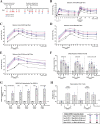
Fig. 2. Immunogenicity of one or two booster rS-Beta doses approximately 1 year after immunization with rS-WU1 in olive baboons.
A A small cohort of baboons (N = 2–3/group) were originally immunized with 1 µg, 5 µg, or 25 µg rS-WU1 with 50 µg Matrix-M adjuvant or unadjuvanted 25 µg rS-WU1 on day 0 and 21 (week 0 and 3, respectively). Approximately 1 year later, all animals were boosted with one or two doses of 3 µg rS-Beta with 50 µg Matrix-M adjuvant on day 318 and 339 (week 45 and 48, respectively). B Anti-S (WU1) IgG titers were measured throughout the course of the study. Individual animals’ titers are shown over time, different colored symbols and lines represent different dose groups for the initial rS-WU1 immunization series. Sera collected pre-rS-Beta boost (study week 43) as well as 7, 21, 35, 81, 137, 193, and 300 days after the first rS-Beta boost were analyzed to determine C anti-rS-WU1 (same data displayed in panel B), D anti-rS-Beta, and E anti-rS-Omicron BA.1 IgG titers by ELISA (n = 3). F Antibody titers capable of disrupting the interaction between rS-WU1, rS-Beta, or rS-Omicron and the hACE2 receptor by ELISA (n = 3) (gray bars represent means), and G antibody titers capable of neutralizing SARS-CoV-2 variants USA-WA1, Beta, Alpha, Delta, and Omicron with a PRNT assay (n = 3) (gray bars represent geometric means). H The presence of multifunctional CD4 + T cells positive for three Th1 cytokines (IFN-γ, IL-2, and TNF-α) was evaluated with intracellular cytokine staining after stimulation with rS-WU1 or rS-Beta (n = 2, gray bars represent means).
Fig. 3
Fig. 3. Antibody-mediated immunity induced upon immunization with SARS-CoV-2 rS based on Wuhan-Hu-1 or Beta variant in BALB/c laboratory mice.
A Groups of mice (N = 20/group) were immunized in a prime/boost regimen on days 0 and 14 with combinations of SARS-CoV-2 rS based on Beta or Wuhan-Hu-1. Mice were either primed and boosted with rS-Beta, with rS-WU1, primed with rS-WU1 and boosted with rS-Beta, or primed and boosted with bivalent rS-WU1 + rS-Beta. Antigen doses were 1 µg rS for each monovalent immunization, or 1 µg rS for each bivalent immunization (2 µg rS total). All antigen doses were administered with 5 µg Matrix-M adjuvant. A control group received a formulation buffer (Placebo). Sera and tissues were collected at the timepoints listed in the diagram. B Anti-SARS-CoV-2 S IgG serum titers were measured in sera collected on day 21 using an ELISA to measure antibody titers against the Wuhan-Hu-1 spike protein (left panel) or Beta spike protein (right panel) (n = 20). Bars indicate the geometric mean titer (GMT) and error bars represent 95% confidence interval (CI) for each group. Individual animal titers are indicated with colored symbols. C ELISA was also used to determine the functional antibody titers in sera collected on day 21 capable of disrupting binding between the SARS-CoV-2 receptor hACE2 and Wuhan-Hu-1 spike protein (left panel) or Beta spike protein (right panel) (n = 20). Bars indicate the geometric mean titer (GMT) and error bars represent 95% confidence interval (CI) for each group. Individual animal titers are indicated with colored symbols. D SARS-CoV-2 neutralization antibody titers in sera collected on day 32 from n = 5 animals/group were determined using ny microneutralization. Sera were evaluated for neutralization of SARS-CoV-2 USA-WA1, Beta, or Omicron (n = 5 samples per group, run in duplicate). Bars indicate the geometric mean titer (GMT) and error bars represent 95% confidence interval (CI) for each group. Individual animal titers are indicated with symbols. Statistical significance was calculated by performing one-way ANOVA with Tukey’s post hoc test on log10-transformed data; significant differences among groups are indicated with asterisks and P values in panels B and C.
Fig. 4
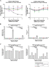
Fig. 4. Protective efficacy of immunization with SARS-CoV-2 rS based on Wuhan-Hu-1 or Beta against challenge with live SARS-CoV-2 Beta or Alpha virus in BALB/c laboratory mice.
Mice were immunized in a prime/boost regimen on days 0 and 14 with combinations of SARS-CoV-2 rS based on Beta or Wuhan-Hu-1. Mice were either prime/boosted with rS-Beta, primed/boosted with rS-WU1, primed with rS-WU1 and boosted with rS-Beta, or prime/boosted with bivalent rS-WU1 + rS-Beta. Antigen doses were 1 µg rS for each monovalent immunization or 1 µg rS for each construct upon bivalent immunization (2 µg rS total). All immunizations were administered with 5 µg Matrix-M adjuvant. A control group received formulation buffer (Placebo, N = 5). Immunized mice (N = 10/group) were challenged with SARS-CoV-2 Beta (left panels) or Alpha (right panels). For 4 days after challenge, mice were weighed daily and their percentage weight loss was calculated relative to their initial body weight. A Mean percentage body weight loss is shown with symbols and error bars represent standard error of the mean. Student’s t test was used to calculate significance of differences between each immunization group and the placebo group groups. ns nonsignificant; *P < 0.05; **P < 0.001, ***P < 0.005; ****P < 0.0001. Colors indicate the respective immunization group being compared; differences between two or more immunization groups and placebo group are shown in black. B Half of the mice were sacrificed at 2 days post-challenge and lung tissue was subjected to a plaque formation assay to determine lung viral titers, the remaining mice were sacrificed at 4 days post-challenge. Mean and standard deviation of lung titer are graphed. C Levels of SARS-CoV-2 subgenomic RNA were also determined in lung tissue and expressed as fold change in RNA relative to the mean in the respective Placebo group on day 2 post-challenge (n = 5 mice per group run in duplicate). Horizontal bars represent group mean fold change from N = 5 mice at each timepoint and error bars represent standard deviation. For B, C, mixed-effects analysis was used to compare differences in viral loads from lung homogenates between vaccinated groups and the placebo control group; **P ≤ 0.01, ***P ≤ 0.001, ****P ≤ 0.0001.
Fig. 5
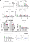
Fig. 5. Cell-mediated immunity upon immunization with rS-WU1 or rS-Beta regimens in BALB/c laboratory mice.
A Groups of mice (N = 8/group) were immunized in a prime/boost regimen on days 0 and 21 with various combinations of SARS-CoV-2 rS based on Beta or Wuhan-Hu-1. Mice were either primed and boosted with rS-Beta, primed and boosted with rS-WU1, primed with rS-WU1 and boosted with rS-Beta, or primed and boosted with bivalent rS-WU1 + rS-Beta. Antigen doses were 1 µg rS for each monovalent immunization, or 1 µg rS for each construct upon bivalent immunization (2 µg rS total). All immunizations were administered with 5 µg Matrix-M adjuvant. A control group received formulation buffer (Placebo, n = 5). B, C Spleens were harvested on day 28 for cell collection. Splenocytes were stimulated with rS-WU1 or rS-Beta, then subjected to ELISpot assay in triplicate to determine IFN-γ-positive cells as a representative Th1 cytokine (B) and IL-5-positive cells as a representative Th2 cytokine (n = 8) (C). Data from panels B and C were used to calculate the Th1/Th2 balance of responses to immunization (D). E The numbers of multifunctional CD4 + T cells that stained positively for three Th1 cytokines (IFN-γ, IL-2, and TNF-α) using intracellular cytokine staining were quantified and expressed as the number of triple cytokine-positive cells per 106 CD44hiCD62Low effector memory CD4 + T cells (n = 8). F T-follicular helper cells were quantified by determining the percentage of PD-1 + CXCR5 + cells among all CD4 + T cells (n = 8). G Germinal center formation was evaluated by determining the percentage of GL7 + CD95 + cells among CD19 + B cells using flow cytometry (n = 8). Gray bars represent means and error bars represent standard deviation. Individual animal data are shown with colored symbols. An example of the gating strategy is shown in the right panel. Mean and standard deviation are graphed in B-G. Differences among experimental groups were evaluated by one-way ANOVA with Tukey’s post hoc test (data in panel B were log10-transformed before analysis). P values <0.05 were considered statistically significant; ****P < 0.0001.
Fig. 6
Fig. 6. Cross-neutralizing antibody responses to spike nanoparticle-based vaccines in BALB/c laboratory mice.
A Groups of mice (N = 20/group) were immunized in a prime/boost regimen on days 0 and 14 with 1 µg of SARS-CoV-2 rS based on Wuhan-Hu-1 (WU1), Beta, Alpha, Gamma, Delta, or Delta Plus rS with 5 μg of Matrix-M adjuvant. Sera were collected at the timepoints listed in the diagram. B SARS-CoV-2 neutralization antibody titers in sera collected on day 21 from n = 20 animals/group were determined using a CPE assay. Sera were evaluated for their ability to neutralize SARS-CoV-2 USA-WA1, Alpha, Beta, Gamma, Mu, Delta, Delta Plus, or Omicron BA.1 variants (n = 2 per sample). Data points indicate pooled serum titer from each group and connect lines connecting similar vaccines. C Functional antibody titers capable of disrupting binding between the SARS-CoV-2 receptor hACE2 and Wuhan-Hu-1, Alpha, Beta, Gamma, Delta, Delta Plus, Omicron BA.1 spike proteins listed in the figure were measured in sera collected on day 21. Data points indicate geometric mean titer from each group (n = 20/group).

References

    1. Bermingham A, et al. Severe respiratory illness caused by a novel coronavirus, in a patient transferred to the United Kingdom from the Middle East, September 2012. Eur. Surveill. Bull. Eur. Sur Mal. Transm. Eur. Commun. Dis. Bull. 2012;17:20290. - PubMed
    1. Nicholls JM, et al. Lung pathology of fatal severe acute respiratory syndrome. Lancet. 2003;361:1773–1778. doi: 10.1016/S0140-6736(03)13413-7. - DOI - PMC - PubMed
    1. Peiris JSM, et al. Coronavirus as a possible cause of severe acute respiratory syndrome. Lancet. 2003;361:1319–1325. doi: 10.1016/S0140-6736(03)13077-2. - DOI - PMC - PubMed
    1. Stadler K, et al. SARS-beginning to understand a new virus. Nat. Rev. Microbiol. 2003;1:209–218. doi: 10.1038/nrmicro775. - DOI - PMC - PubMed
    1. Nicholls J, Dong XP, Jiang G, Peiris M. SARS: clinical virology and pathogenesis. Respirology. 2003;8(Suppl):S6–S8. doi: 10.1046/j.1440-1843.2003.00517.x. - DOI - PMC - PubMed

Publication types

Supplementary concepts