Complete sequencing of a cynomolgus macaque major histocompatibility complex haplotype
- PMID: 36854669
- PMCID: PMC10078292
- DOI: 10.1101/gr.277429.122
Complete sequencing of a cynomolgus macaque major histocompatibility complex haplotype
Abstract
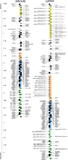
Macaques provide the most widely used nonhuman primate models for studying the immunology and pathogenesis of human diseases. Although the macaque major histocompatibility complex (MHC) region shares most features with the human leukocyte antigen (HLA) region, macaques have an expanded repertoire of MHC class I genes. Although a chimera of two rhesus macaque MHC haplotypes was first published in 2004, the structural diversity of MHC genomic organization in macaques remains poorly understood owing to a lack of adequate genomic reference sequences. We used ultralong Oxford Nanopore and high-accuracy Pacific Biosciences (PacBio) HiFi sequences to fully assemble the ∼5.2-Mb M3 haplotype of an MHC-homozygous, Mauritian-origin cynomolgus macaque (Macaca fascicularis). The MHC homozygosity allowed us to assemble a single MHC haplotype unambiguously and avoid chimeric assemblies that hampered previous efforts to characterize this exceptionally complex genomic region in macaques. The high quality of this new assembly is exemplified by the identification of an extended cluster of six Mafa-AG genes that contains a recent duplication with a highly similar ∼48.5-kb block of sequence. The MHC class II region of this M3 haplotype is similar to the previously sequenced rhesus macaque haplotype and HLA class II haplotypes. The MHC class I region, in contrast, contains 13 MHC-B genes, four MHC-A genes, and three MHC-E genes (vs. 19 MHC-B, two MHC-A, and one MHC-E in the previously sequenced haplotype). These results provide an unambiguously assembled single contiguous cynomolgus macaque MHC haplotype with fully curated gene annotations that will inform infectious disease and transplantation research.
© 2023 Karl et al.; Published by Cold Spring Harbor Laboratory Press.
Figures


Similar articles
-
Major histocompatibility complex haplotyping and long-amplicon allele discovery in cynomolgus macaques from Chinese breeding facilities.Immunogenetics. 2017 Apr;69(4):211-229. doi: 10.1007/s00251-017-0969-7. Epub 2017 Jan 11. Immunogenetics. 2017. PMID: 28078358 Free PMC article.
-
Discovery of novel MHC-class I alleles and haplotypes in Filipino cynomolgus macaques (Macaca fascicularis) by pyrosequencing and Sanger sequencing: Mafa-class I polymorphism.Immunogenetics. 2015 Oct;67(10):563-78. doi: 10.1007/s00251-015-0867-9. Epub 2015 Sep 8. Immunogenetics. 2015. PMID: 26349955
-
Characterization of 100 extended major histocompatibility complex haplotypes in Indonesian cynomolgus macaques.Immunogenetics. 2020 May;72(4):225-239. doi: 10.1007/s00251-020-01159-5. Epub 2020 Feb 29. Immunogenetics. 2020. PMID: 32112172 Free PMC article.
-
Haplessly hoping: macaque major histocompatibility complex made easy.ILAR J. 2013;54(2):196-210. doi: 10.1093/ilar/ilt036. ILAR J. 2013. PMID: 24174442 Free PMC article. Review.
-
The Cynomolgus Macaque MHC Polymorphism in Experimental Medicine.Cells. 2019 Aug 26;8(9):978. doi: 10.3390/cells8090978. Cells. 2019. PMID: 31455025 Free PMC article. Review.
Cited by
-
The Primate Major Histocompatibility Complex: An Illustrative Example of Gene Family Evolution.bioRxiv [Preprint]. 2024 Sep 18:2024.09.16.613318. doi: 10.1101/2024.09.16.613318. bioRxiv. 2024. PMID: 39345418 Free PMC article. Preprint.
-
Integrated analysis of the complete sequence of a macaque genome.Nature. 2025 Apr;640(8059):714-721. doi: 10.1038/s41586-025-08596-w. Epub 2025 Feb 26. Nature. 2025. PMID: 40011769
-
Unraveling the architecture of major histocompatibility complex class II haplotypes in rhesus macaques.Genome Res. 2024 Nov 20;34(11):1811-1824. doi: 10.1101/gr.278968.124. Genome Res. 2024. PMID: 39443153 Free PMC article.
-
Prevalence of ABO and Rhesus (D) Blood Group and Allelic Frequency at Blood Bank of Nigist Eleni Mohammed Hospital, Ethiopia.Biomed Res Int. 2024 Apr 8;2024:5353528. doi: 10.1155/2024/5353528. eCollection 2024. Biomed Res Int. 2024. PMID: 38628500 Free PMC article.
-
The Impact and Effects of Host Immunogenetics on Infectious Disease Studies Using Non-Human Primates in Biomedical Research.Microorganisms. 2024 Jan 12;12(1):155. doi: 10.3390/microorganisms12010155. Microorganisms. 2024. PMID: 38257982 Free PMC article. Review.
References
Publication types
MeSH terms
Grants and funding
LinkOut - more resources
Full Text Sources
Research Materials
Miscellaneous
