. 2023 Feb 28;9(1):9.
doi: 10.1038/s41522-023-00377-5.

Matrix is everywhere: extracellular DNA is a link between biofilm and mineralization in Bacillus cereus planktonic lifestyle

Affiliations

Matrix is everywhere: extracellular DNA is a link between biofilm and mineralization in Bacillus cereus planktonic lifestyle

Lyubov A Ivanova et al. NPJ Biofilms Microbiomes. .

Abstract

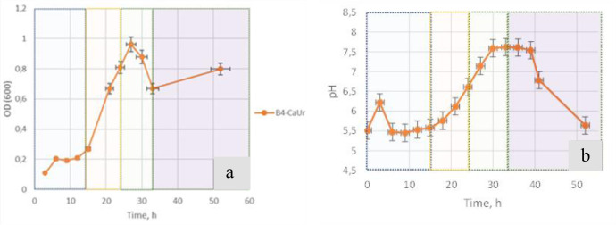
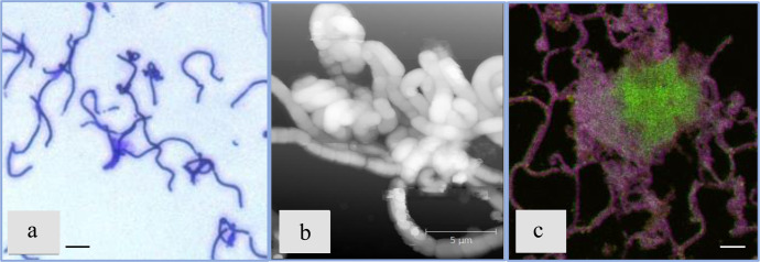
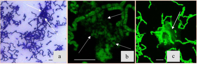
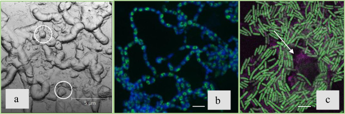
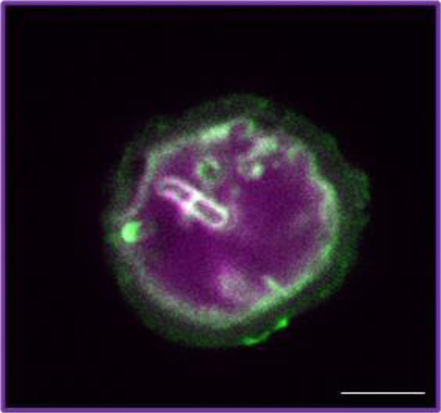
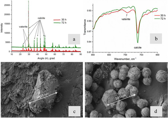
To date, the mechanisms of biomineralization induced by bacterial cells in the context of biofilm formation remain the subject of intensive studies. In this study, we analyzed the influence of the medium components on the induction of CaCO3 precipitation by the Bacillus cereus cells and composition of the extracellular matrix (ECM) formed in the submerged culture. While the accumulation of extracellular polysaccharides and amyloids appeared to be independent of the presence of calcium and urea during the growth, the accumulation of extracellular DNA (eDNA), as well as precipitation of calcium carbonate, required the presence of both ingredients in the medium. Removal of eDNA, which was sensitive to treatment by DNase, did not affect other matrix components but resulted in disruption of cell network formation and a sixfold decrease in the precipitate yield. An experiment with a cell-free system confirmed the acceleration of mineral formation after the addition of exogenous salmon sperm DNA. The observed pathway for the formation of CaCO3 minerals in B. cereus planktonic culture included a production of exopolysaccharides and negatively charged eDNA lattice promoting local Ca2+ supersaturation, which, together with an increase in the concentration of carbonate ions due to pH rise, resulted in the formation of an insoluble precipitate of calcium carbonate. Precipitation of amorphous CaCO3 on eDNA matrix was followed by crystal formation via the ACC-vaterite-calcite/aragonite pathway and further formation of larger mineral aggregates in complex with extracellular polymeric substances. Taken together, our data showed that DNA in extracellular matrix is an essential factor for triggering the biomineralization in B. cereus planktonic culture.


Conflict of interest statement

The authors declare no competing interests.

Figures

Fig. 1
Fig. 1. Biomass growth characteristics of B. cereus 4B in the B4_CaUr medium (with an excess of calcium and urea ions).
a Time-dependence of the bacterial biomass growth; b Time-dependence of the medium acidity during the incubation. Time stages are highlighted in color: 0–15 h in blue; 15–24 h in orange; 24–33 h in green; more than 33 h in lilac.
Fig. 2
Fig. 2. B. cereus 4B at the first stage of the growth (0–15 hours after inoculation).
a Light-microscopy image with crystal violet staining in the sample taken after 9 hours after the inoculation. b AFM microscopy image (after 15 h). c Confocal microscopy image with Sytox green staining (green color, specific for eDNA), Congo Red (pink color, amyloids and polysaccharides) and CaCO3 autofluorescence channel (yellow color); All scale bars correspond to 5 µm.
Fig. 3
Fig. 3. B. cereus at the second stage of the growth (15–24 h after inoculation).
a Light-microscopy image with crystal violet staining in the sample taken after 24 h after inoculation; white arrows show ECM thickening. b Confocal microscopy image with Sytox green staining (eDNA). c Confocal microscopy image with Thioflavin T staining (amyloid structures); All scale bars correspond to 5 µm.
Fig. 4
Fig. 4. B. cereus 4B at the third stage of the growth (24–33 h after inoculation).
a AFM image of the sample taken at 33 h after inoculation. b Confocal microscopy image with Sytox green (eDNA, green color) and Bromophenol blue (amyloid structures, blue color) staining. c Confocal microscopy image staining with Thioflavin T (amyloid structures, green color) and Congo red (amyloid structures and/or polysaccharides, pink color); All scale bars correspond to 5 µm.
Fig. 5
Fig. 5. B. cereus 4B at the fourth stage of the growth (45 h after inoculation).
a Confocal microscopy image with CaCO3 autofluorescence. b Confocal microscopy image with Sytox green (eDNA) staining. c Confocal microscopy image with Bromophenol blue staining (amyloid structures). d Confocal microscopy image with Sytox green (eDNA, green color), Bromophenol blue (amyloid structures, blue color) and CaCO3 autofluorescence (yellow color); All scale bars correspond to 5 µm.
Fig. 6
Fig. 6. Confocal microscopy image of B. cereus 4B at the fourth stage of the growth (52 h after inoculation).
DNA was stained with 7AAD (pink color) and amyloid structures were stained with Thioflavin T (green color). All scale bars correspond to 5 µm.
Fig. 7
Fig. 7. Mineral analysis of CaCO3 precipitates induced by the B. cereus during the growth in the B4-CaUr medium.
a X-ray diffraction patterns of the samples shows vaterite peak loss after 30 h. b FTIR spectra of precipitates at different times of growth shows polymorph transition from vaterite to calcite. c, d SEM images of CaCO3 particles induced by B. cereus at 24 and 72 h after inoculation, correspondingly.
Fig. 8
Fig. 8. Results of the eDNA removal from the extracellular matrix of the B. cereus planktonic culture.
Confocal microscopy images with Sytox green (eDNA, green color), Bromophenol blue (protein structures, blue color), and CaCO3 autofluorescence (yellow color) staining of B. cereus samples cultured in B4_CaUr medium without addition of DNase (I) (CONTROL a, c) and repeated addition of DNase (I) (EXPERIMENT b, d) for 26 h (a, b) and 44 h (c, d). e FTIR spectra of precipitates obtained during biomineralization of B. cereus in a medium with the addition of DNase (red line) and without DNase (black line). All scale bars correspond to 25 µm.
Fig. 9
Fig. 9. Confocal microscopy images of CaCO3 precipitates deposited in vitro.
a With exogenous DNA stained with Sytox green; b Without eDNA stained with Sytox green. All scale bars correspond to 25 µm.

References

    1. Dhami, N. K., Reddy, M. S. & Mukherjee, A. Application of calcifying bacteria for remediation of stones and cultural heritages, Front. Microbiol. 10.3389/fmicb.2014.00304 (2014). - PMC - PubMed
    1. le Métayer-Levrel G, Castanier S, Orial G, Loubière J-F, Perthuisot J-P. Applications of bacterial carbonatogenesis to the protection and regeneration of limestones in buildings and historic patrimony. Sediment Geol. 1999;126:25–34. doi: 10.1016/S0037-0738(99)00029-9. - DOI
    1. de Muynck W, de Belie N, Verstraete W. Microbial carbonate precipitation in construction materials: a review. Ecol. Eng. 2010;36:118–136. doi: 10.1016/j.ecoleng.2009.02.006. - DOI
    1. Yuan F, et al. Pathogenesis of Proteus mirabilis in catheter-associated urinary tract infections. Urol. Int. 2021;105:354–361. doi: 10.1159/000514097. - DOI - PubMed
    1. Ghosh T, Bhaduri S, Montemagno C, Kumar A. Sporosarcina pasteurii can form nanoscale calcium carbonate crystals on cell surface. PLoS ONE. 2019;14:e0210339. doi: 10.1371/journal.pone.0210339. - DOI - PMC - PubMed

Publication types

LinkOut - more resources