. 2023 Mar;58(3):185-195.
doi: 10.1007/s00535-022-01954-9. Epub 2023 Mar 1.

Diagnostic criteria and endoscopic and histological findings of autoimmune gastritis in Japan

Affiliations

Diagnostic criteria and endoscopic and histological findings of autoimmune gastritis in Japan

Tomoari Kamada et al. J Gastroenterol. 2023 Mar.

Abstract

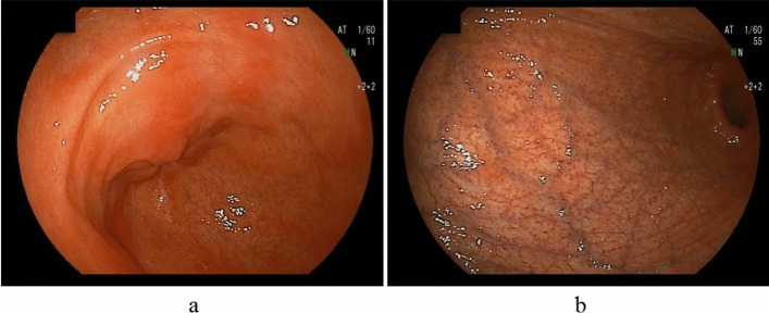
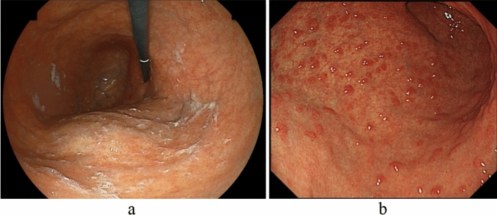
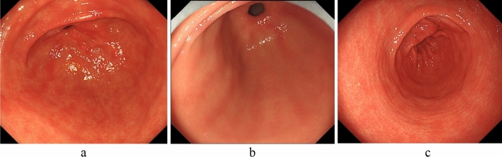
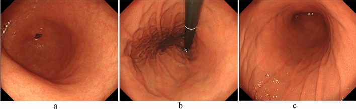
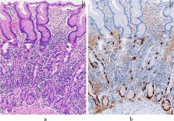
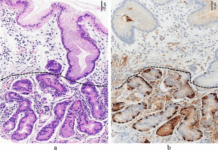
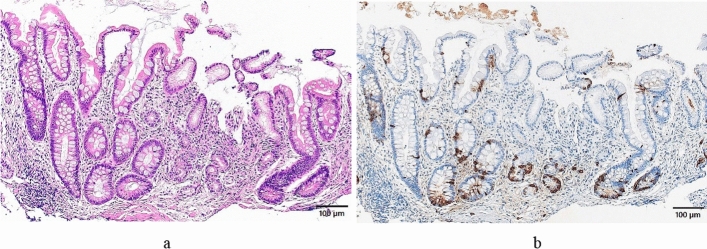
The Japanese diagnostic criteria for autoimmune gastritis (AIG) were established by the "Study Group on the establishment of diagnostic criteria for type A gastritis," which is related to a workshop associated with the Japan Gastroenterological Endoscopy Society (JGES) and the Committee of AIG Research Group (CARP). The criteria were set as follows: the cases of confirmed diagnosis are patients in whom either the endoscopic or histological findings, or both, meet the requirements for AIG and who are confirmed to be positive for gastric autoantibodies (either anti-parietal cell or anti-intrinsic factor antibodies, or both). The presentation of endoscopic findings of early-stage AIG in the diagnostic criteria was withheld owing to the need for further accumulation and characterization of endoscopic clinical data. Therefore, diagnosis of early-stage AIG only requires histological confirmation and gastric autoantibody positivity. Suspected cases are patients in whom either the endoscopic or histological findings, or both, meet only the requirements for AIG. Histological findings only meet the requirements for early stage. AIG has been underdiagnosed in the past, but our study group's newly proposed diagnostic criteria will enable a more accurate and early diagnosis of AIG. The criteria can be used to stratify patients into various high-risk groups for gastric tumors and pernicious anemia. They would allow the establishment of an appropriate surveillance system in the coming years. Nevertheless, issues such as establishing the endoscopic findings of early-stage AIG and obtaining Japanese insurance coverage for gastric autoantibody tests require attention.

Keywords: Autoimmune gastritis; Corpus-predominant atrophy; Diagnostic criteria; Early-stage AIG; Type A gastritis.


Conflict of interest statement

The authors declare that they have no conflict of interest.

Figures

Fig. 1
Fig. 1
Endoscopic findings of advanced AIG (primary findings). Absent or mild antral atrophy, severe atrophy predominantly in the corpus (uniform mucosal blood vessels are visible), and no atrophic border. a Antral area. b Greater curvature of the corpus
Fig. 2
Fig. 2
Endoscopic findings of advanced AIG (secondary findings). Sticky adherent dense mucus and residual fundic gland mucosa may be found in the corpus. a Sticky adherent dense mucus. b Remnant oxyntic mucosa (pseudopolyp-like)
Fig. 3
Fig. 3
Mucosal findings of AIG (secondary findings) in the antral area. a Patchy redness. b Red streaks. c Circular wrinkle-like pattern
Fig. 4
Fig. 4
Endoscopic findings of early-stage AIG. Reddened and mildly edematous gastric pits are diffusely observed in the corporal mucosa without RAC despite the absence of H. pylori infection. The folds of the greater curvature of the corpus are preserved. a Greater curvature of the antral area. b Gastric body (observation looking upward). c Gastric body (observation looking downward)
Fig. 5
Fig. 5
Histological features of early stage (greater curvature of the corpus). a The fundic gland mucosal structure that is almost similar to normal is preserved, although there is the elongation of the gastric pit (ratio of the gastric pit length to the gastric gland length: 1.0:1.8), and there is moderate chronic inflammatory cell infiltration between the fundic glands (stronger in the glandular area than in the gastric pit area). Numerous residual parietal cells exhibit pseudohypertrophy (degenerative swelling and cell protrusion). (HE staining). b Intraglandular hyperplasia of the ECL cells is observed (chromogranin A staining)
Fig. 6
Fig. 6
Histological features of advanced florid stage (greater curvature of the corpus). a The normal fundic gland mucosa structure has disappeared, the lengths of the gastric pit and glandular are 0.39 and 0.29 mm, respectively, (ratio: 1.0:0.7), and there is the elongation of the gastric pit and shortening of the gastric gland. The dashed line indicates the boundary between the gastric pit and gastric gland (HE staining). b Intraglandular linear hyperplasia of the ECL cells is observed (chromogranin A staining)
Fig. 7
Fig. 7
Histological features of advanced end stage (greater curvature of the corpus). a The normal fundic gland mucosa structure has disappeared, intestinal metaplasia has become severe, and a small amount of the pyloric gland mucosa remains at the center of this photo. The gastric pit length/the gastric gland duct length ratio is 0.34/0.08 mm (ratio: 4.2:1.0). (HE staining). b Intraglandular ECL cell hyperplasia has become clear. (chromogranin A staining)

References

    1. Strickland RG, Mackay IR. A reappraisal of the nature and significance of chronic atrophic gastritis. Am J Dig Dis. 1973;18:426–440. doi: 10.1007/BF01071995. - DOI - PubMed
    1. Notsu T, Adachi K, Mishiro T, et al. Prevalence of autoimmune gastritis in individuals undergoing medical checkups in Japan. Intern Med. 2019;58:1817–1823. doi: 10.2169/internalmedicine.2292-18. - DOI - PMC - PubMed
    1. Aoki R, Yasuda M, Shunto J, Haruma K. Type A gastritis detected during endoscopic examination. Stomach Intestine. 2019;54:1046–1052.
    1. Nishizawa T, Yoshida S, Watanabe H, et al. Clue of diagnosis for autoimmune gastritis. Digestion. 2021;102:903–910. doi: 10.1159/000516624. - DOI - PubMed
    1. Terao S, Tome M, Kure I, et al. Most cases categorized into the group D are not those with complete elimination of Helicobacter pylori infection because of advanced atrophy and intestinal metaplasia. Jpn J Helicobacter Res. 2013;14:5–14.