. 2023 Nov;26(6):514-523.
doi: 10.1111/vop.13075. Epub 2023 Feb 28.

Iris angiography in ADAMTS10 mutant dogs with open-angle glaucoma (ADAMTS10-OAG)

Affiliations

Iris angiography in ADAMTS10 mutant dogs with open-angle glaucoma (ADAMTS10-OAG)

Richard A Pytak 3rd et al. Vet Ophthalmol. 2023 Nov.

Abstract

Objective: To evaluate anterior segment angiographic findings in hypertensive ADAMTS10-open-angle glaucoma (ADAMTS10-OAG) eyes as compared to normotensive control eyes.

Animals studied: Nine ADAMTS10-OAG beagles and four wild-type control dogs.

Procedures: Anterior segment angiography was performed under general anesthesia following intravenous injection of indocyanine green (ICG; 1 mg/kg) and sodium fluorescein (SF; 20 mg/kg) using a Heidelberg Spectralis® confocal scanning laser ophthalmoscope. Time to onset of iridal angiographic phases and the presence/severity of dye leakage into the iris stromal and/or aqueous humor were recorded. Group findings were compared, and multiple linear regression analysis was performed to identify potential factor associations with disease status.

Results: Time to onset of all angiographic phases visualized using ICG was significantly prolonged while time to onset of SF leakage into the aqueous humor was significantly reduced in glaucomatous eyes compared to controls. Only glaucomatous eyes (n = 9) demonstrated evidence of SF stromal leakage. Mean intraocular pressure (IOP) and age were significantly higher, while mean cardiac pulse was significantly lower in glaucomatous eyes compared to controls. Blood pressure and ocular perfusion pressure were not significantly different between groups. Multiple linear regression analysis, controlling for age, IOP, and pulse demonstrated glaucoma, was not predictive of the time to onset of any angiographic phase, stromal, or aqueous humor leakage. However, pulse was a significant factor contributing to the severity of aqueous humor leakage.

Conclusions: A compromised vascular supply to the anterior segment exists in dogs with ADAMTS10-OAG. These observations warrant further exploration of what role altered perfusion and/or disruption to the blood-aqueous barrier may play.

Keywords: ADAMTS10-open-angle glaucoma (ADAMTS10-OAG); angiography; canine; uvea.


Conflict of interest statement

The authors declare no conflicts related to this study. Dr. Komáromy received research funding from PolyActiva Pty. Ltd. and CRISPR Therapeutics while the presented work was conducted. He also serves as a consultant for Reichert Technologies and Editor‐in‐Chief of Veterinary Ophthalmology.

Figures
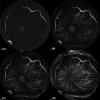
FIGURE 1
FIGURE 1
Representative anterior segment indocyanine green angiography (ASICGA) images of the left eye from an 8 month‐old intact male wild‐type control dog. After intravenous (IV) injection of indocyanine green (ICG), initial filling of the major arterial circle (A; 4 s) and early filling of radial iris arteries (B; 6 s) is observed during the arterial phase. Progressive filling of radial iris arteries and initial filling of terminal capillary loops within the peri‐pupillary region denotes the capillary phase (C; 8 s), and progressive filling of radial iris veins is observed during the venous phase D; 11 s).
FIGURE 2
FIGURE 2
Representative anterior segment sodium fluorescein angiography (ASSFA) images of the left eyes of two ADAMTS10‐OAG affected dogs (A; 4‐year‐old intact male, B; 4‐year‐old intact female) to illustrate the subjective grading scheme used to characterize the severity of dye leakage following intravenous (IV) injection of sodium fluorescein (SF). Images were acquired at 2 min after SF injection. Grade 0 corresponds to no SF leakage within the anterior chamber (not shown). Grade 1 corresponds to a mild degree of SF leakage within the anterior chamber (A). Grade 2 corresponds to a moderate amount of SF leakage (B). Grade 3 (not shown) corresponds to severe SF leakage within the anterior chamber.
FIGURE 3
FIGURE 3
Representative standard color (A), near‐infrared (B), anterior segment indocyanine green angiography (ASICGA) (C), and anterior segment sodium fluorescein angiography (ASSFA) (D–F) images of the left eye of an 8‐month‐old intact male wild‐type control beagle dog. Angiographic images were obtained at 20 s (C and D), 30 s (E), and 60 s (F) after intravenous (IV) injection of dye. There is no evidence of leakage of indocyanine green (ICG) (C) or SF (D–F).
FIGURE 4
FIGURE 4
Representative standard color (A), near‐infrared (B), anterior segment indocyanine green angiography (ASICGA) (C), and anterior segment sodium fluorescein angiography (ASSFA) (D–F) images of the left eye of a 4‐year‐old intact female ADAMTS10‐OAG‐affected beagle dog. Angiographic images were obtained at 20 s (C and D), 30 s (E), and 60 s (F) after intravenous (IV) injection of dye. There is no evidence of leakage of indocyanine green (ICG) (C). After IV injection of sodium fluorescein (SF), progressive leakage within the peripupillary region is apparent at 20 s (D), 30 s (E), and 60 s (F). Leakage of SF is evident in 4 quadrants of the peripupillary border (grade 2). There is marked leakage of SF into the aqueous humor (F; arrow). Extravasation of SF within the lower palpebral conjunctiva, due to outward rolling of the lower eyelid margin, is also noted (D–F).
FIGURE 5
FIGURE 5
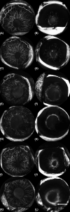
Representative anterior segment indocyanine green angiography (ASICGA) (A, C, E, G, I, and K), and anterior segment sodium fluorescein angiography (ASSFA) (B, D, F, H, J, and L) images from 6 ADAMTS10‐OAG‐affected beagle dogs. All images were obtained 20 s following intravenous (IV) dye injection. Images represent the left eye of a 4‐year‐old intact male (A and B), the right eye of a 3.3‐year‐old female (C and D), the right eye of a 5.5‐year‐old female (E and F), the left eye of a 4‐year‐old intact male (G and H), the right eye of a 3.3‐year‐old intact male (I and J), and the left eye of a 3.3‐year‐old female (K and L). No stromal leakage of indocyanine green (ICG) was observed (A, C, E, G, I, and K) Note that peripupillary leakage of SF in 3–4 quadrants (grade 2) of the iris stroma is readily apparent (B, D, F, H, J, and L). Early leakage of sodium fluorescein (SF) into the aqueous humor (L; arrow) is also noted 20 s following dye injection.

References

    1. Gelatt KN, Mackay EO. Prevalence of the breed‐related glaucomas in pure‐bred dog in North America. Vet Ophthalmol. 2004;7:97‐111. - PubMed
    1. Tham YC, Li X, Wong TY, Quigley HA, Aung T, Cheng CY. Global prevalence of glaucoma and projections of glaucoma burden through 2040: a systematic review and meta‐analysis. Ophthalmology. 2014;121(11):2081‐2090. - PubMed
    1. Miller PE, Bentley E. Clinical signs and diagnosis of the canine primary glaucomas. Vet Clin North Am Small Anim Pract. 2015;45(6):1183‐vi. doi:10.1016/j.cvsm.2015.06.006 - DOI - PMC - PubMed
    1. Miller PE. The glaucomas. In: Maggs DJ, Miller PE, Ofri R, eds. Slatter's Fundamentals of Veterinary Ophthalmology. 5th ed. Elsevier; 2013:247‐271.
    1. Kuchtey J, Olson LM, Rinkoski T, et al. Mapping of the disease locus and identification of ADAMTS10 as a candidate gene in a canine model of primary open angle glaucoma. PLoS Genet. 2011;7(2):e1001306. - PMC - PubMed

Substances