. 2023 Feb 28;23(1):86.
doi: 10.1186/s12883-023-03132-7.

Computed tomography derived cervical fat-free muscle fraction as an imaging-based outcome marker in patients with acute ischemic stroke: a pilot study

Affiliations

Computed tomography derived cervical fat-free muscle fraction as an imaging-based outcome marker in patients with acute ischemic stroke: a pilot study

Narine Mesropyan et al. BMC Neurol. .

Abstract

Background: Outcome assessment in stroke patients is essential for evidence-based stroke care planning. Computed tomography (CT) is the mainstay of diagnosis in acute stroke. This study aimed to investigate whether CT-derived cervical fat-free muscle fraction (FFMF) as a biomarker of muscle quality is associated with outcome parameters after acute ischemic stroke.

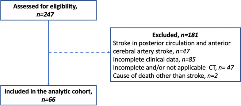
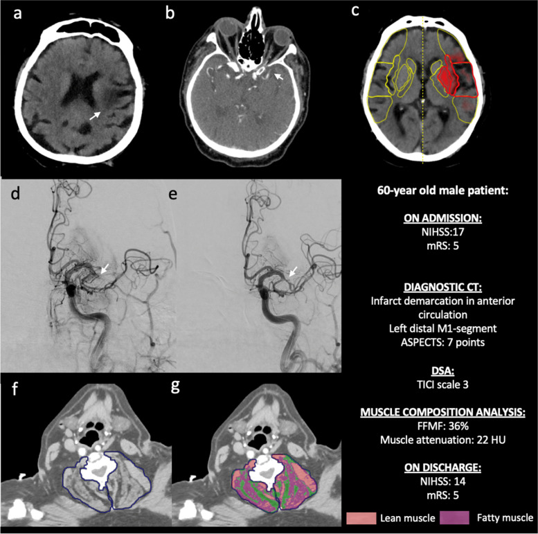
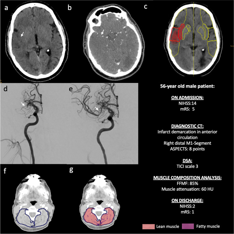
Methods: In this retrospective study, 66 patients (mean age: 76 ± 13 years, 30 female) with acute ischemic stroke in the anterior circulation who underwent CT, including CT-angiography, and endovascular mechanical thrombectomy of the middle cerebral artery between August 2016 and January 2020 were identified. Based on densitometric thresholds, cervical paraspinal muscles covered on CT-angiography were separated into areas of fatty and lean muscle and FFMF was calculated. The study cohort was binarized based on median FFMF (cutoff value: < 71.6%) to compare clinical variables and outcome data between two groups. Unpaired t test and Mann-Whitney U test were used for statistical analysis.

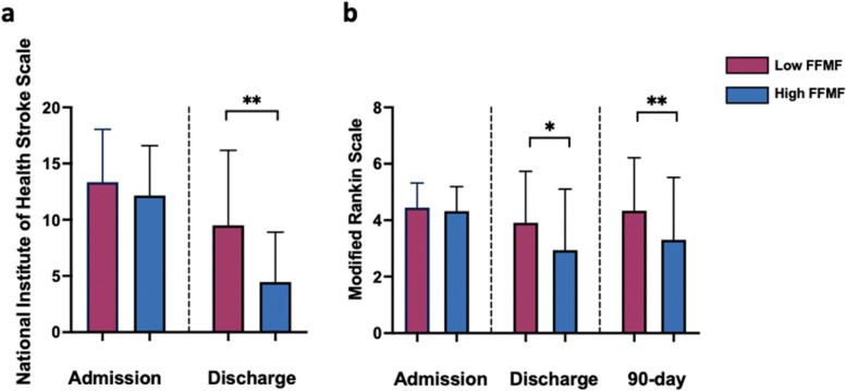
Results: National Institute of Health Stroke Scale (NIHSS) (12.2 ± 4.4 vs. 13.6 ± 4.5, P = 0.297) and modified Rankin scale (mRS) (4.3 ± 0.9 vs. 4.4 ± 0.9, P = 0.475) at admission, and pre-stroke mRS (1 ± 1.3 vs. 0.9 ± 1.4, P = 0.489) were similar between groups with high and low FFMF. NIHSS and mRS at discharge were significantly better in patients with high FFMF compared to patients with low FFMF (NIHSS: 4.5 ± 4.4 vs. 9.5 ± 6.7; P = 0.004 and mRS: 2.9 ± 2.1 vs.3.9 ± 1.8; P = 0.049). 90-day mRS was significantly better in patients with high FFMF compared to patients with low FFMF (3.3 ± 2.2 vs. 4.3 ± 1.9, P = 0.045).

Conclusion: Cervical FFMF obtained from routine clinical CT might be a new imaging-based muscle quality biomarker for outcome prediction in stroke patients.

Keywords: Acute ischemic stroke; Computed tomography; Fat-free muscle fraction; Muscle quality.


Conflict of interest statement

C.K. and J.R. are members of the European Reference Network on Rare Neuromuscular Diseases ERN EURO-NMD. ERN EURO-NMD had no influence on study conceptualization and design, collection and analysis of the data, manuscript preparation as well as the decision to publish. The remaining authors have no competing interests to declare that are relevant to the content of this article.

Figures

Fig. 1
Fig. 1
Flowchart illustrating included patients and patients which were excluded from analysis. CT: Computed tomography
Fig. 2
Fig. 2
Representative images with summary of skeletal muscle composition analysis. The paraspinal muscle area at the level of the intervertebral disc space C3/4 was separated into areas of lean and fatty muscle based on densitometric thresholds. Fat-free muscle fraction (FFMF) was calculated as a relation of fatty muscle area to the total skeletal muscle area. Opportunistic analysis of CT scans was performed using an in-house software written in MATLAB. CT: Computed tomography; FFMF: Fat-free muscle fraction
Fig. 3
Fig. 3
Column graphs showing the National Institute of Health Stroke (a) and the modified Rankin Scale (b) in the group of patients with low and high fat-free muscle fraction (FFMF) on admission, discharge and after 3 months. Data are presented as mean with standard deviation error bars. *, ** represents significance levels of pairwise comparisons with P values of ≤0.05 and ≤ 0.01, respectively. P values were obtained using U Mann-Whitney test. FFMF: Fat-free muscle fraction
Fig. 4
Fig. 4
Representative images of a 60-year-old male patient with a low fat-free muscle fraction of 36% and ischemic stroke by left middle cerebral artery occlusion. Non-enhanced CT scan (a) demonstrates beginning infarct area demarcation (white arrow) in the anterior circulation due to occlusion of the distal M1 segment of the left middle cerebral artery (white arrow, b). Additional analysis of computer-assisted early ischemic changes revealed an ASPECT score of 7 (c). The patient underwent systemic thrombolysis and successful endovascular intervention with mechanical thrombectomy and complete recanalization (TICI scale 3) (white arrows, d and e). Images f and g visualize the assessment of skeletal muscle fat infiltration as previously described
Fig. 5
Fig. 5
Representative images of a 56-year-old male patient with a high fat-free muscle fraction of 85% and ischemic stroke by right cerebral artery occlusion. Non-enhanced CT scan (a) demonstrates beginning infarct area demarcation (white arrow) in the anterior circulation due to occlusion of the distal M1 segment of the right middle cerebral artery (white arrow, b). Additional analysis of computer-assisted early ischemic changes revealed an ASPECT score of 8 (c). The patient underwent systemic thrombolysis and successful endovascular intervention with mechanical thrombectomy with complete recanalization (TICI scale 3) (white arrows, d and e). Images f and g visualize the assessment of skeletal muscle fat infiltration as previously described

References

    1. Feigin VL, Stark BA, Johnson CO, et al. Global, regional, and national burden of stroke and its risk factors, 1990–2019: a systematic analysis for the global burden of disease study 2019. Lancet Neurol. 2021;20:795–820. doi: 10.1016/S1474-4422(21)00252-0. - DOI - PMC - PubMed
    1. Navis A, Garcia-Santibanez R, Skliut M. Epidemiology and outcomes of ischemic stroke and transient ischemic attack in the adult and geriatric population. J Stroke Cerebrovasc Dis. 2019;28:84–89. doi: 10.1016/j.jstrokecerebrovasdis.2018.09.013. - DOI - PubMed
    1. Banks JL, Marotta CA. Outcomes validity and reliability of the modified Rankin scale: implications for stroke clinical trials: a literature review and synthesis. Stroke. 2007;38:1091–1096. doi: 10.1161/01.STR.0000258355.23810.c6. - DOI - PubMed
    1. Falk-Delgado A, Kuntze Söderqvist Å, Fransén J, et al. Improved clinical outcome 3 months after endovascular treatment, including thrombectomy, in patients with acute ischemic stroke: a meta-analysis. J Neurointerv Surg. 2016;8:665–670. doi: 10.1136/neurintsurg-2015-011835. - DOI - PMC - PubMed
    1. Faron A, Kreyer S, Sprinkart AM, et al. CT fatty muscle fraction as a new parameter for muscle quality assessment predicts outcome in venovenous extracorporeal membrane oxygenation. Sci Rep. 2020;10:22391. doi: 10.1038/s41598-020-79495-5. - DOI - PMC - PubMed