

A rare case of H3K27-altered diffuse midline glioma with multiple osseous and spinal metastases at the time of diagnosis
- PMID: 36855102
- PMCID: PMC9972747
- DOI: 10.1186/s12883-023-03135-4
A rare case of H3K27-altered diffuse midline glioma with multiple osseous and spinal metastases at the time of diagnosis
Abstract
Background: H3K27-altered diffuse midline gliomas are uncommon central nervous system tumors with extremely poor prognoses.
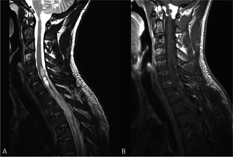
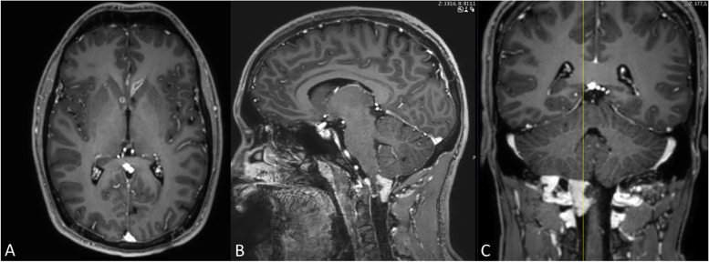
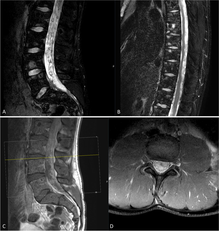
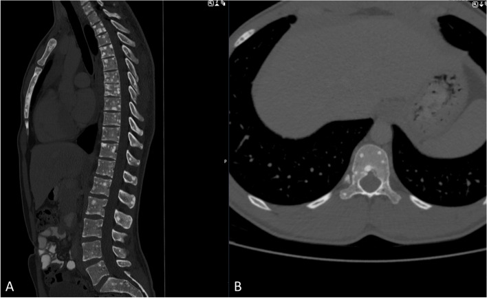
Case presentation: We report the case of a 24-year-old man patient with multiple, inter alia osseous metastases who presented with back pain, hemi-hypoesthesia, and hemi-hyperhidrosis. The patient underwent combined radio-chemotherapy and demonstrated temporary improvement before deteriorating.
Conclusions: H3K27-altered diffuse midline glioma presents an infrequent but crucial differential diagnosis and should be considered in cases with rapid neurological deterioration and multiple intracranial and intramedullary tumor lesions in children and young adults. Combined radio-chemotherapy delayed the neurological deterioration, but unfortunately, progression occurred three months after the diagnosis.
Keywords: Diffuse midline Glioma; Extraneural metastases; H3K27M; Neuro-oncology.
© 2023. The Author(s).
Conflict of interest statement
JG and BM work as consultants for Brainlab (Brainlab AG, Feldkirchen). In addition, BM works as a consultant for Medtronic, Spineart, Icotec, Relievant and Depuy/Synthes. In these firms, BM acts as a member of the advisory board. Furthermore, BM reports a financial relationship with Medtronic, Ulrich Medical, Brainlab, Spineart, Icotec, Relievant and Depuy/Synthes. He received personal fees and research grants for clinical studies from Medtronic, Ulrich Medical, Brainlab, Icotec and Relievant. All this happened independently of the submitted work. BM holds the royalties/patent for Spineart. All named potential conflicts of interest are unrelated to this study. There are no further conflicts of interest regarding the other authors.
Figures
Similar articles
-
Pediatric diffuse midline glioma H3K27- altered: A complex clinical and biological landscape behind a neatly defined tumor type.Front Oncol. 2023 Jan 16;12:1082062. doi: 10.3389/fonc.2022.1082062. eCollection 2022. Front Oncol. 2023. PMID: 36727064 Free PMC article. Review.
-
Spinal intramedullary H3K27M mutant glioma with vertebral metastasis: a case report.Childs Nerv Syst. 2021 Dec;37(12):3933-3937. doi: 10.1007/s00381-021-05119-6. Epub 2021 Mar 19. Childs Nerv Syst. 2021. PMID: 33742289
-
Adult H3K27M-mutant diffuse midline glioma with gliomatosis cerebri growth pattern: Case report and review of the literature.Int J Surg Case Rep. 2020;68:124-128. doi: 10.1016/j.ijscr.2020.02.046. Epub 2020 Feb 28. Int J Surg Case Rep. 2020. PMID: 32145563 Free PMC article.
-
Spinal cord diffuse midline glioma with postoperative acute swelling: A case report and review of literature.Surg Neurol Int. 2023 Oct 13;14:360. doi: 10.25259/SNI_636_2023. eCollection 2023. Surg Neurol Int. 2023. PMID: 37941612 Free PMC article.
-
Case series of diffuse extraneural metastasis in H3F3A mutant high-grade gliomas: Clinical, molecular phenotype and literature review.J Clin Neurosci. 2021 Jul;89:405-411. doi: 10.1016/j.jocn.2021.05.033. Epub 2021 May 27. J Clin Neurosci. 2021. PMID: 34053821 Review.
Cited by
-
Retinol dehydrogenase 10 promotes epithelial-mesenchymal transition in spinal cord gliomas via PI3K/AKT pathway.Int J Immunopathol Pharmacol. 2024 Jan-Dec;38:3946320241276336. doi: 10.1177/03946320241276336. Int J Immunopathol Pharmacol. 2024. PMID: 39180753 Free PMC article.
-
Extra-neural metastases in pediatric diffuse midline gliomas, H3 K27-altered: presentation of two cases and literature review.Front Mol Neurosci. 2023 Jul 20;16:1152430. doi: 10.3389/fnmol.2023.1152430. eCollection 2023. Front Mol Neurosci. 2023. PMID: 37547920 Free PMC article.
-
Diffuse bone-marrow metastasis of grade 4 isocitrate dehydrogenase-mutant astrocytoma associated with hematological abnormalities: Gliomatosis of the bone marrow.Surg Neurol Int. 2025 May 23;16:201. doi: 10.25259/SNI_49_2025. eCollection 2025. Surg Neurol Int. 2025. PMID: 40469347 Free PMC article.
-
Diffuse midline glioma H3K27-altered with thoracic epidural metastasis: illustrative case.J Neurosurg Case Lessons. 2025 Jul 14;10(2):CASE25249. doi: 10.3171/CASE25249. Print 2025 Jul 14. J Neurosurg Case Lessons. 2025. PMID: 40658994 Free PMC article.
-
Pediatric-type diffuse high-grade glioma with systemic metastasis: A case report.Neurooncol Adv. 2025 Apr 3;7(1):vdaf069. doi: 10.1093/noajnl/vdaf069. eCollection 2025 Jan-Dec. Neurooncol Adv. 2025. PMID: 40351828 Free PMC article. No abstract available.
References
-
- Raco A, Esposito V, Lenzi J, Piccirilli M, Delfini R, Cantore G. Long-term follow-up of intramedullary spinal cord tumors: a series of 202 cases. Neurosurgery 2005;56(5):972-81; discussion -81. - PubMed
Publication types
MeSH terms
LinkOut - more resources
Full Text Sources
